All posts by peferguson

Endometrial Adenocarcinoma

Endocervical vs. Endometrial Adenocarcinoma
Endocervical and endometrial adenocarcinomas may be morphologically indistinguishable morphologically.  The question often comes up as to whether a case represents an endometrial adenocarcinoma involving the cervix, or and endocervical adenocarcinoma involving the endometrium.  The following antibodies may be helpful in such circumstances:
 
 
Endocervical
Adenocarcinoma
Endometrial
Adenocarcinoma
Negative (7-8%+)
Positive (70-93%)
Positive (65-95%)
Usually Negative
Negative (4-20%+, 38% weak)
Strong Positive (67-90%)
Strong & Diffuse Positive (90-100%)
Patchy Positive cells (~35%)
HPV
Positive (67%)
Negative
References
AJSP 2002;26:998
 
“Endocervical vs. Endometrial Adenocarcinoma:  Update on Useful Immunohistochemical Markers.”  RT Miller,The Focus ProPath Immunohistochemistry.”   April 2003.
 

Cervical Adenocarcinoma

Endocervical vs. Endometrial Adenocarcinoma
Endocervical and endometrial adenocarcinomas may be morphologically indistinguishable morphologically.  The question often comes as to whether a case represents an endometrial adenocarcinoma involving the cervix, or and endocervical adenocarcinoma involving the endometrium.  The following antibodies may be helpful in such circumstances:


Endocervical
Adenocarcinoma
Negative (7-8%+)
Positive (70-93%)
Positive (65-95%)
Usually Negative
Negative (4-20%+, 38% weak)
Strong Positive (67-90%)
Strong & Diffuse Positive (90-100%)
Patchy Positive cells (~35%)
HPV
Positive (67%)
Negative
References
AJSP 2002;26:998
 
“Endocervical vs. Endometrial Adenocarcinoma:  Update on Useful Immunohistochemical Markers.”  RT Miller,The Focus ProPath Immunohistochemistry.”   April 2003.

Mature B-Cell Neoplasms

CD61

CD61 is a marker of megakaryocytes/platelets.  The antigen is glycoprotein IIIa, and any cellular proliferation with megakaryocytic differentiation may express CD61.  This marker has been found useful in bone marrow biopsies looking for identifying highlighting abnormal and micromegakaryocytes, which is found in some cases of myelodysplasia (MDS) and myeloproliferative neoplasms (MPN).  
 
Some data suggests that at least 25% of the staining megakaryocytes should be micromegakaryocytes to have specificity for MDS (normal bone marrow <10% micromegakaryocytes).  The opinion of some expert hematopathologists is that a significant proportion for MDS is >50% micromegakaryocytes.
Photomicrographs
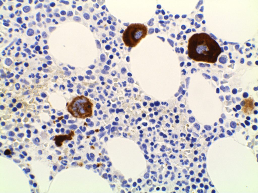
CD61 - Megakaryocytes
CD61 expression in a normal bone marrow highlighting megakaryocytes.
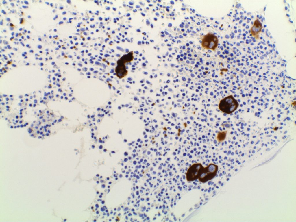
CD61 - Megakaryocytes
CD61 expression in a normal bone marrow highlighting megakaryocytes.
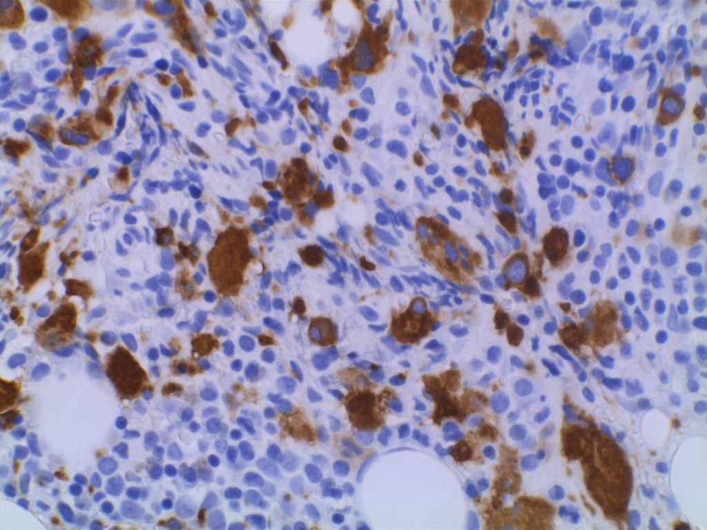
CD61 - RAEB
CD61 expression in MDS (RAEB) with increased numbers of small and hypolobated megakaryocytes.
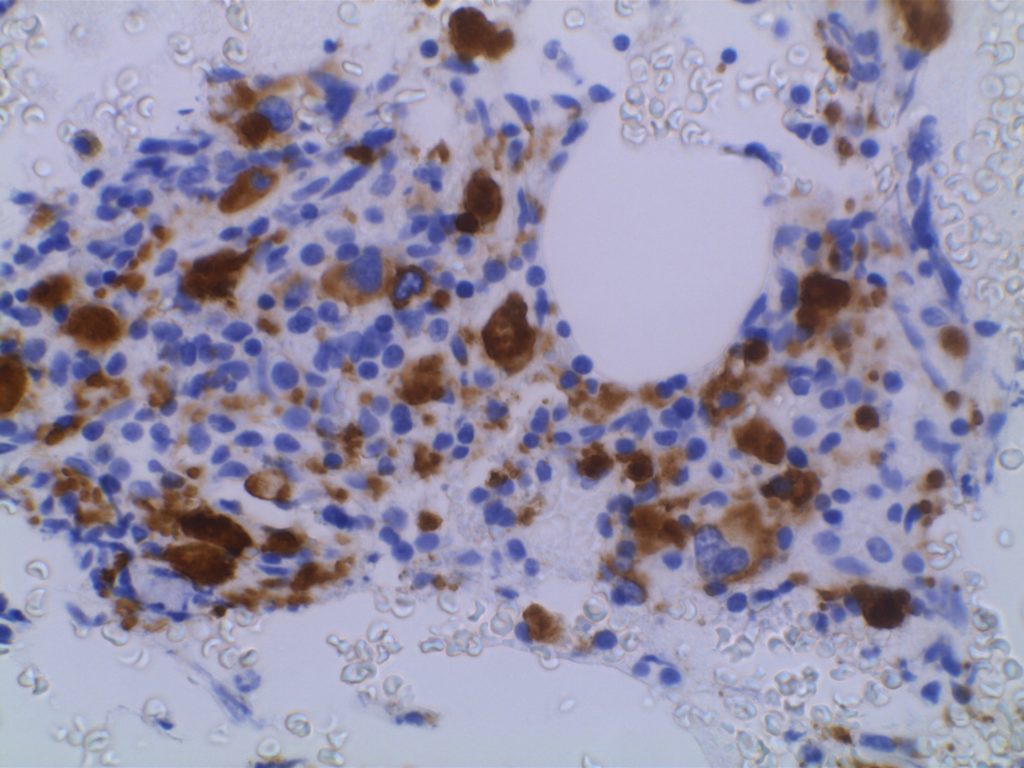
CD61 - RAEB
CD61 expression in MDS (RAEB) with increased numbers of small and hypolobated megakaryocytes.

References
Quitmann H, Wagner S, Fischer R. Dysmegakaryopoiesis in myelodysplastic syndromes (MDS): an immunomorphometric study of bone marrow trephine biopsy specimens. Journal of Clinical Pathology. 1991.
 
Chuang SS, Li CY. Useful Panel of Antibodies for the Classification of Acute Leukemia by lmmunohistochemical Methods in Bone Marrow Trephine Biopsy Specimens. Am J Clin Pathol. 1997.
 
Jawad MD, Go RS, Reichard KK, Shi M. Increased Multinucleated Megakaryocytes as an Isolated Finding in Bone Marrow:  A Rare Finding and Its Clinical Significance. Am J Clin Pathol. 2016;146: 561–566. doi:10.1093/ajcp/aqw144
 
FOX, S.B., LORENZEN, J., HERYET, A., JONES, M., GATTER, K.C. and MASON, D.Y. (1990), Megakaryocytes in myelodysplasia: an immunohistochemical study on bone marrow trephines. Histopathology, 17: 69–74. doi:10.1111/j.1365-2559.1990.tb00665.x
 
Bone Marrow IHC.  Torlakovic, EE, et. al. American Society for Clinical Pathology Pathology Press © 2009.  pp. 109-113.