All posts by peferguson

WT-1 (Wilms tumor gene product)

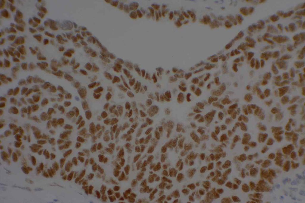
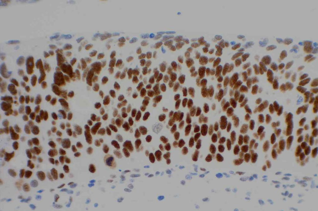
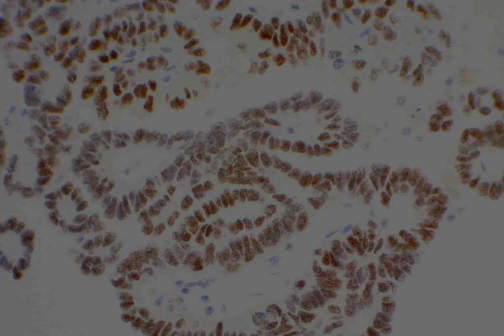
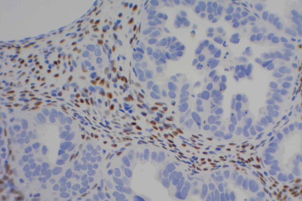
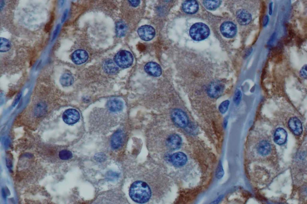
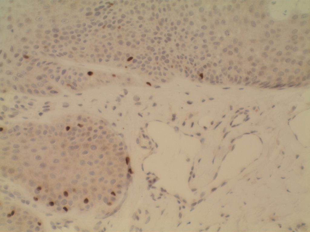
WT-1 (Wilms tumor gene product) is a marker most commonly used to identify OVARIAN SEROUS CARCINOMAS.  It is also commonly used in a panel to differentiate mesothelioma (positive) from adenocarcinoma (negative).  WT-1 has a normal expression distribution in adult tissues, which includes Sertoli cells, ovarian stromal and surface epithelium, and mesothelium.  The 6F-H2 clone has shown better sensitivity compared to other WT-1 antibodies (Ordonez).
 
Tumors / lesions expressing WT-1:
  • Mesothelioma (>75% of epithelial mesotheliomas, usually negative in sarcomatoid variants)
  • Ovarian Serous Carcinomas
  • Wilms tumor
  • Desmoplastic Small Round Cell Tumors
  • Metanephric Adenomas
 
Tumor
Expression (%)
Ovarian Serous Carcinoma
93%
Ovarian Mucinous Carcinoma
0%
Pancreatobiliary Carcinoma
0%
Breast Carcinoma
0%
Lung Carcinoma
0%
Colon Adenocarcinoma
0%
Renal Cell Carcinoma
0%
Thyroid Carcinoma
0%
Prostate Adenocarcinoma
0%
 
WT-1 expression in differentiating mesothelioma from adenocarcinoma (Marchevsky).
Tumor
Expression (%)
Epithelioid Mesothelioma
77%
Sarcomatoid Mesothelioma
13%
Adenocarcnioma
4%
Photomicrographs
WT-1 - Wilms Tumor
WT-1 – Wilms Tumor
WT-1 - Mesothelioma
WT-1 – Mesothelioma
WT-1 - Ovarian Serous Carcinoma
WT-1 – Ovarian Serous Carcinoma
WT-1 - Stromal Cells
WT-1 expression in stromal cells.

 
References:
Muir, T. E., Cheville, J. C., & Lager, D. J. (2001). Metanephric adenoma, nephrogenic rests, and Wilms’ tumor: a histologic and immunophenotypic comparison. The American Journal of Surgical Pathology, 25(10), 1290–1296.
 
Marchevsky, A. M. (2008). Application of immunohistochemistry to the diagnosis of malignant mesothelioma. Archives of Pathology & Laboratory Medicine, 132(3), 397–401.
 
Ordóñez, N. G. (2005). Immunohistochemical diagnosis of epithelioid mesothelioma: an update. Archives of Pathology & Laboratory Medicine, 129(11), 1407–1414.  
  
Hadi, AIMM Annual Meeting, “Carcinomas of Unknown Primary”, presentation, 2011.

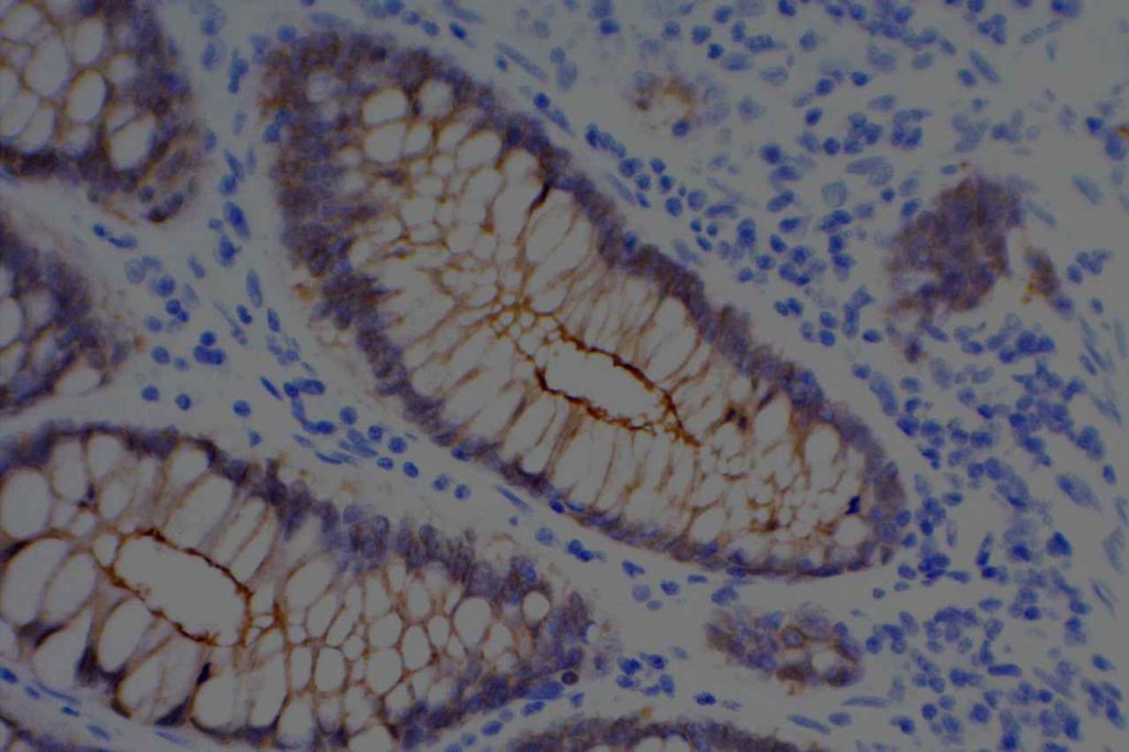
Villin

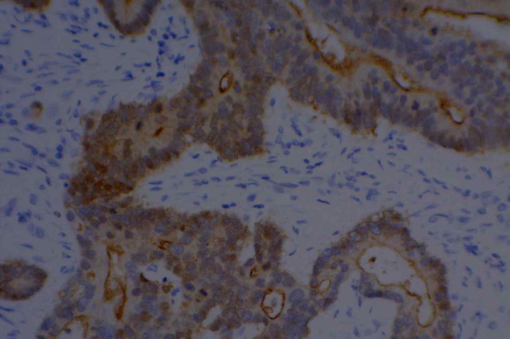
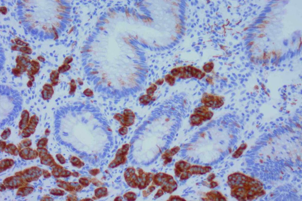
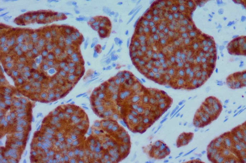
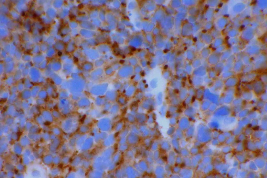
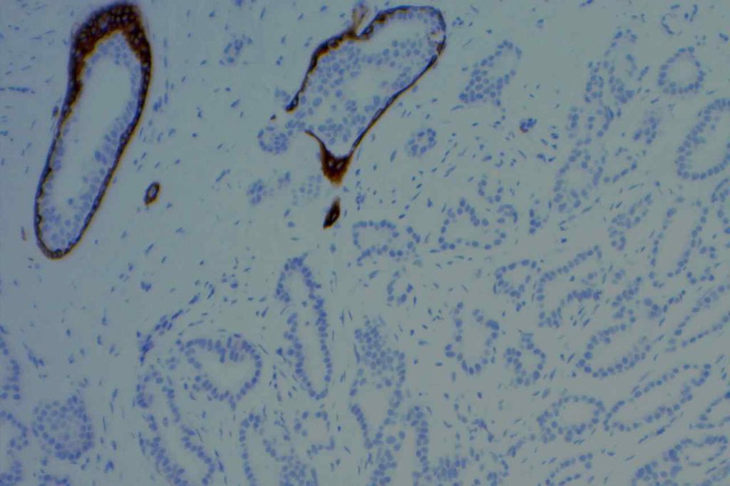
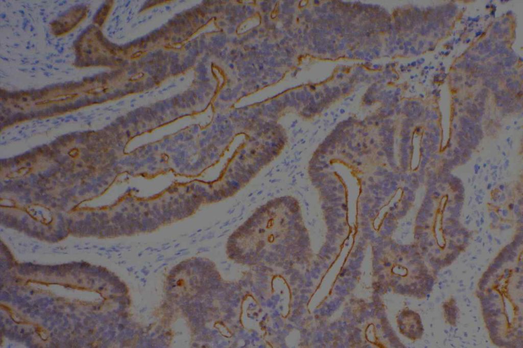
Villin is an actin-binding protein found in microvilli.  It’s expression pattern is cytoplasmic and generally restricted to epithelial cells in the GI tract.  It is a sensitive marker for colon adenocarcinomas, but not entirely specific.  It also stains renal proximal tubules and hepatic bile ducts.
 
CDX-2 and Villin expression in human tumors (2-3+ expression) (Werling, R.W., et. al.)
Tumor Type
No.
CDX2
Villin
G.I. Tract


Colon Adenocarcinoma
75
99%
82% (n=73)
Duodenal Adenoma
10
100%
100%
Gastric Adenocarcinoma
24
70%
42%
Esophageal Adenocarcinoma
9
67%
78%
Pancreatic Adenocarcinoma
22
32%
40%
Cholangiocarcinoma/GB
16
25%
60%
Hepatocellular Carcinoma
12
0%
0%
Carcinoid Tumors
12
42%
34%
Ovary


Mucinous Adenocarcinoma
14
64%
64%
Mucinous Cystadenoma
13
8%
0%
Mucinous Borderline Tumor
4
25%
0%
Non-Mucinous
36
0%
0% (n=31)
Genitourinary Tract


Urothelial Carcinoma
21
0%
0%
Adenocarcinoma
2
100%
100%
Urachal Caricinoma
1
100%
100%
Renal Cell Carcinoma
7
0%
0% (n=3)
Prostate Adenocarcinoma
27
4%
0% (n=24)
Breast Carcinoma
34
0%
0%
Lung (45 primary; 18 met)
63
0%
5%
Adenocarcinoma
11
0%
0%
Squamous Cell Carcinoma
11
0%
0%
Non-Small Cell Carcinoma NOS
33
0
9%
Mucinous Carcinoma
2
0%
0%
Small Cell Carcinoma
4
0%
0%
Mesothelioma
7
0%
0%
Head and Neck


Thyroid
36
3%
0%
Papillary Carcinoma
11
9%
0%
Follicular Adenoma/Carcinoma
25
0%
0%
Salivary Gland
12
0%
0%
Mixed Tumor
6
0%
0%
Low Grade Carcinoma
6
0%
0%
Squamous Cell Carcinoma
13
0%
0% (n=11)
Photomicrographs
Villin - Colon Adenocarcinoma
Villin – Colon Adenocarcinoma
Villin - Colon
Villin – Colon
Villin - Colon Adenocarcinoma
Villin – Colon Adenocarcinoma

References
Gastroenterol94:343-52, 1988. 
 
Hadi, AIMM Annual Meeting, “Carcinomas of Unknown Primary”, presentation, 2011.
 
Werling, R. W., Yaziji, H., Bacchi, C. E., & Gown, A. M. (2003). CDX2, a highly sensitive and specific marker of adenocarcinomas of intestinal origin: an immunohistochemical survey of 476 primary and metastatic carcinomas. The American Journal of Surgical Pathology, 27(3), 303–310.  

TTF-1

TTF-1 is a nuclear transcription factor that is expressed in thyroid and respiratory epithelium.  It is a useful marker for lung adenocarcinomas and thyroid malignancies.  In the setting of neuroendocrine carcinomas, TTF-1 expression is not specific as to the site of origin, except that Merkel cell carcinomas of the skin do not usually express TTF-1 (some have reported focal expression).  The expression pattern of TTF-1, like other transcription markers, is generally strong and diffuse.  Lower levels of positivity should bring caution to the interpretation.
 
TTF-1 use has become more important to sub-classify lung tumors on small biopsy specimens.  It is generally considered the most sensitive and specific individual marker for lung adenocarcinomas, but is often used in combination with Napsin A to maximize sensitivity and specificity for the determination of primary lung adenocarcinomas.  Current recommendations are to test non-small cell lung carcinomas (non-squamous cell carcinoma) for ALK, ROS-1 and EGFR mutations, as targeted therapies are available (this list is ever expanding and should be verified with current medical literature).

Continue reading TTF-1

Thyroglobulin

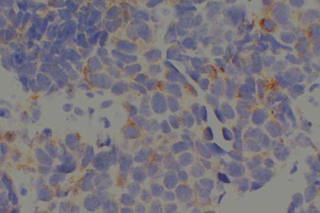
Thyroglobulin is a glycoprotein expressed by thyroid follicular cells.  It is a specific marker for thyroid derivation, and may be useful in the work-up of carcinomas of unknown primary.  It may be less sensitive in less differentiated thyroid tumors, as anapestic thyroid carcinoma is notorious for not staining with about anything.
 
Pitfalls
  • Medullary thyroid carcinoma does not express thyroglobulin.
  • Thyroglobulin can have a lot of artifactual staining, which may cause difficultly diagnosing tumors like medullary carcinoma (expected to be thyroglobulin negative).
  • Dedifferentiated thyroid tumors may not express thyroglobulin.
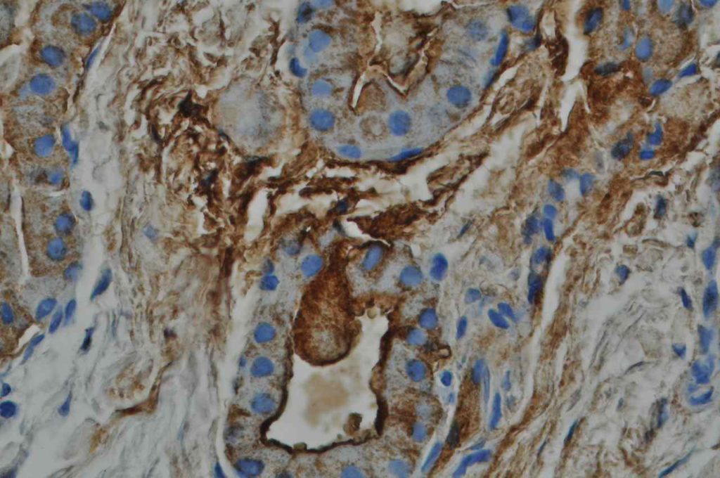
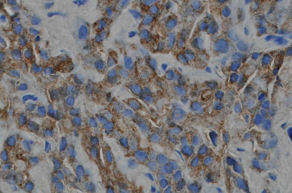
Photomicrographs
Thyroglobulin - Follicular Carcinoma of Thyroid
Thyroglobulin – Follicular Carcinoma of Thyroid
Thyroglobulin - Follicular Carcinoma of Thyroid
Thyroglobulin – Follicular Carcinoma of Thyroid

References:
 
Fischer, S., & Asa, S. L. (2008). Application of immunohistochemistry to thyroid neoplasms. Archives of Pathology & Laboratory Medicine, 132(3), 359–372. 

Terminal Deoxynucleotiyl Transferase (TdT)

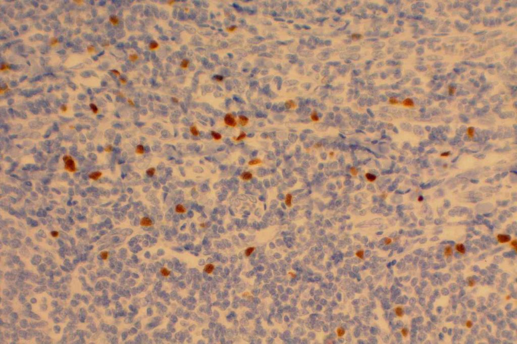
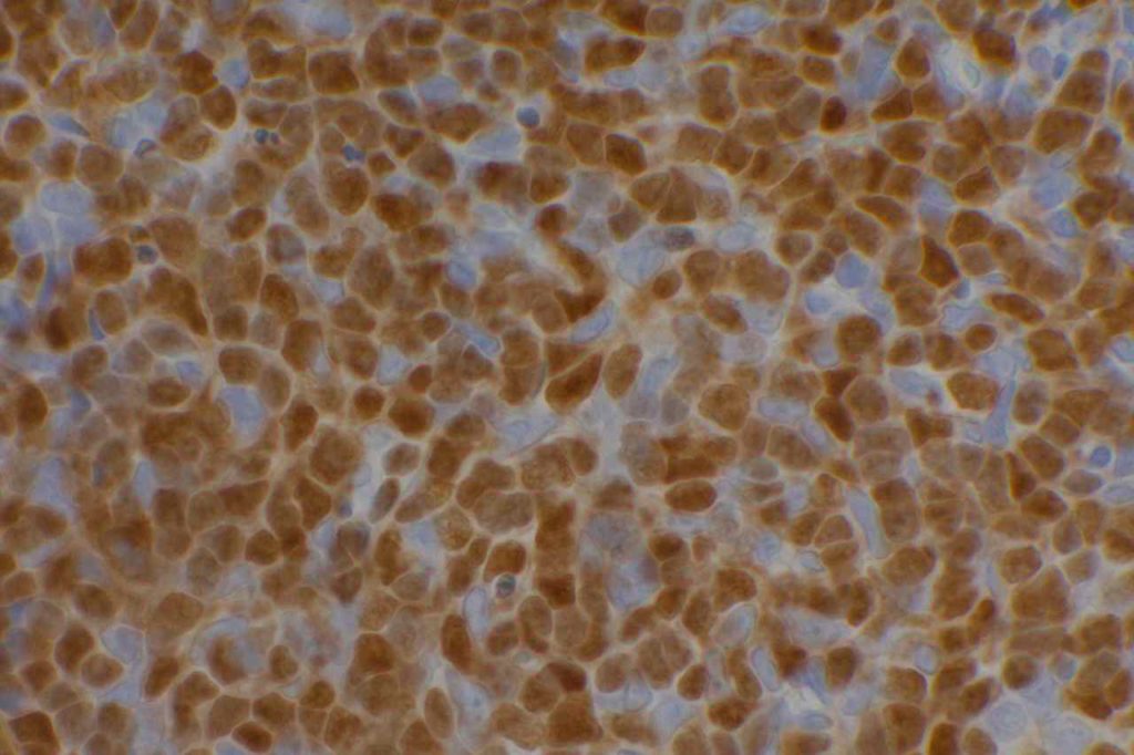
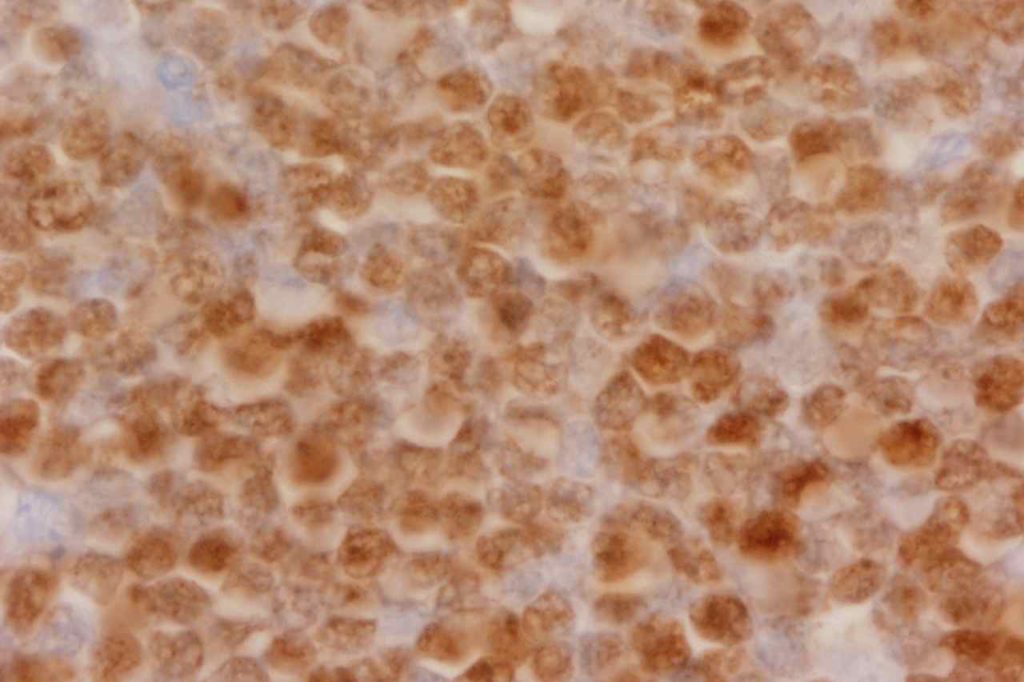
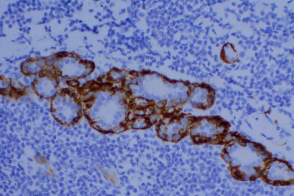
Terminal deoxynucleotiyl transferase (TdT) is a nuclear transcription factor expressed in B and T-cell precursors and cortical thymocytes.  It is expressed in acute lymphoblastic lymphoma (95%), AML (<25%), Merkel cell carcinoma (~50%), hematogones, and thymomas.  It is negative in Burkitt lymphoma.
 
Scattered TdT positive cells in benign lymphoid tissue (e.g. tonsil) is a common finding.
 
TdT Expression Pattern

Photomicrographs
TdT - Benign Tonsil
Scattered staining TdT positive cells in normal tonsil tissue.
TdT - Thymoma
TdT – Thymoma
TdT - T-ALL
TdT – T-ALL

References:
Bone Marrow IHC.  Torlakovic, EE, et. al. American Society for Clinical Pathology Pathology Press © 2009.  pp. 167.
 
Sidiropoulos, M., Hanna, W., Raphael, S. J., & Ghorab, Z. (2011). Expression of TdT in Merkel cell carcinoma and small cell lung carcinoma. American Journal of Clinical Pathology, 135(6), 831–838. doi:10.1309/AJCPLCB2Q9QXDZAA 
 
Onciu, M., Lorsbach, R. B., Henry, E. C., & Behm, F. G. (2002). Terminal deoxynucleotidyl transferase-positive lymphoid cells in reactive lymph nodes from children with malignant tumors: incidence, distribution pattern, and immunophenotype in 26 patients. American Journal of Clinical Pathology, 118(2), 248–254. doi:10.1309/L3UW-3AE7-L4LB-3QX3
 
Strauchen, J. A., & Miller, L. K. (2001). Terminal deoxynucleotidyl transferase-positive cells in human tonsils. American Journal of Clinical Pathology, 116(1), 12–16. doi:10.1309/M8V2-DWPB-DVX1-UBPC  
 
Hurford, M. T., Altman, A. J., Digiuseppe, J. A., Sherburne, B. J., & Rezuke, W. N. (2008). Unique Pattern of Nuclear TdT Immunofluorescence Distinguishes Normal Precursor B Cells (Hematogones) From Lymphoblasts of Precursor B-Lymphoblastic Leukemia. American Journal of Clinical Pathology, 129(5), 700–705. doi:10.1309/ANERT51H38TUEC45 

TFE3 (transcription factor E3)

TFE3 (transcription factor E3) is a gene located on chromosome Xp11.2, and is a member of the microphthalmia-associated transcription factor (MITF) family.  Translocations with this gene are associated with different partners in  alveolar soft part sarcoma and a subset of renal cell carcinomas (Xp11.2 translocation RCC).  TFE3 protein over expression can be detected by IHC in cases with a TFE3 translocation, and the expression is nuclear.  The normal gene product (non-TFE3 translocation cases) is not detectable by IHC.
 
In a large case set (1476 cases) by Argani, et. al., TFE3 expression was demonstrated in 1/2 high-grade myxofibrosarcomas, 3/60 adrenal cortical carcinomas, and 1/15 distal bile duct carcinomas.  TFE3 is a sensitive and specific marker for alveolar soft part sarcoma and Xp11.2 RCC, as it appears to be expressed in >90% of Xp11.2 translocation associated renal cell carcinomas and alveolar soft part sarcoma.
 
Xp11.2 RCC cases tend to express E-cadherin and CD10, while NOT expressing CK7 and EMA.  Conventional clear cell RCC tend not to express E-cadherin while expressing EMA.

Photomicrographs
Xp11.2 Renal Cell Carcinoma
Xp11.2 Renal Cell Carcinoma – H&E section
TFE3 - Xp11.2 Renal Cell Carcinoma
TFE3 – Xp11.2 Renal Cell Carcinoma
TFE3 - Xp11.2 Renal Cell Carcinoma
TFE3 – Xp11.2 Renal Cell Carcinoma

References:

Camparo, P., Vasiliu, V., Molinie, V., Couturier, J., Dykema, K. J., Petillo, D., et al. (2008). Renal translocation carcinomas: clinicopathologic, immunohistochemical, and gene expression profiling analysis of 31 cases with a review of the literature. The American Journal of Surgical Pathology, 32(5), 656–670. doi:10.1097/PAS.0b013e3181609914
 
Argani, P., Lal, P., Hutchinson, B., Lui, M. Y., Reuter, V. E., & Ladanyi, M. (2003). Aberrant nuclear immunoreactivity for TFE3 in neoplasms with TFE3 gene fusions: a sensitive and specific immunohistochemical assay. The American Journal of Surgical Pathology, 27(6), 750–761.
 
Argani, P., Olgac, S., Tickoo, S. K., Goldfischer, M., Moch, H., Chan, D. Y., et al. (2007). Xp11 translocation renal cell carcinoma in adults: expanded clinical, pathologic, and genetic spectrum. The American Journal of Surgical Pathology, 31(8), 1149–1160. doi:10.1097/PAS.0b013e318031ffff
 
Argani, P., Antonescu, C. R., Couturier, J., Fournet, J.-C., Sciot, R., Debiec-Rychter, M., et al. (2002). PRCC-TFE3 renal carcinomas: morphologic, immunohistochemical, ultrastructural, and molecular analysis of an entity associated with the t(X;1)(p11.2;q21). The American Journal of Surgical Pathology, 26(12), 1553–1566.
 
Argani, P., Lal, P., Hutchinson, B., Lui, M. Y., Reuter, V. E., & Ladanyi, M. (2003). Aberrant nuclear immunoreactivity for TFE3 in neoplasms with TFE3 gene fusions: a sensitive and specific immunohistochemical assay. The American Journal of Surgical Pathology, 27(6), 750–761.
 
Armah, H. B., & Parwani, A. V. (2010). Xp11.2 translocation renal cell carcinoma. Archives of Pathology & Laboratory Medicine, 134(1), 124–129. doi:10.1043/2008-0391-RSR.1

Synaptophysin

Synaptophysin reacts with the integral membrane glycoprotein of presynaptic vesicles.  It is less specific than chromogranin, and it may mark neurofilaments and epithelial filaments.  Some neuroendocrine neoplasms will mark with synaptophysin and not chromogranin A.   The converse can also be true.  Therefore, chromogranin A and synaptophysin should be used in tandem to identify neuroendocrine differentiation (Wick, MR).  CD56 is often considered the most sensitive neuroendocrine marker, but it is not as specific as chromogranin and synaptophysin (Bahrami, A, et al).
 
Neuroendocrine differentiation may not be readily recognized until it is revealed by immunohistochemistry, but may be important as these tumors may be responsive to similar therapy used for small cell carcinoma (Bahrami, A, et al).
Microscopic Images
Synaptophysin - Carcinoid
Synaptophysin expression in a gastric carcinoid.
Synaptophysin - Pheochromocytoma
Synaptophysin expression in a pheochromocytoma.
Synaptophysin - Carcinoid
Synaptophysin expression in a carcinoid tumor.
Synaptophysin - Merkel Cell Carcinoma
Synaptophysin expression in a Merkel cell carcinoma.
Synaptophysin - Lung
Synaptophysin expression in a lung small cell carcinoma.

 References
Wick, M. R. (2008). Immunohistochemical approaches to the diagnosis of undifferentiated malignant tumors. Annals of Diagnostic Pathology, 12(1), 72–84. doi:10.1016/j.anndiagpath.2007.10.003 
 
Bahrami, A., Truong, L. D., & Ro, J. Y. (2008). Undifferentiated tumor: true identity by immunohistochemistry. Archives of Pathology & Laboratory Medicine, 132(3), 326–348. 

SOX10

SOX10 (member of the SOX family of transcription factors and located at chromosome 22q13.1) is a transcription factor (Sry-related HMg-Box gene 10) protein, which functions as a nucleocytoplasmic shuttle protein, and is important in neural and melanocytic development. Expression has been identified in benign tissues (melanocytes, breast, ganglia, and optic vesicle) and in tumors (melanoma, malignant peripheral nerve sheath tumors, gliomas, breast carcinoma, and schwannoma).
 
Studies have shown SOX10 to be an excellent marker for melanoma (>95% sensitivity), but is not specific in differentiating nevi from melanoma as benign nevi/melanocytes also express SOX10.  Specificity with respect to other tumor types appears relatively good with the exceptions described above.  However, current data sets appear limited, and more study is needed to better define specificity.  
 
SOX10 may be a helpful marker in the diagnosis of melanoma, especially when used with a panel of other markers like S-100, MART-1, or HMB-45.  A head to head comparison with S-100 might be helpful to further characterize the antibodies usefulness as a screening tool for melanomas.  
 
Expression of SOX10 is nuclear, as one would expect with transcription based markers.
 
In breast carcinoma, SOX10 is more commonly expressed in basal-like, triple negative, and metaplastic subtypes.
 
SOX10 Expression statistics using BC34 clone (Tacha, et. al)
Lesion
Percent +
Number
Melanoma (all subtypes)
92.6%
257
– Spindle Cell / Desmoplastic Melanoma
98%
51
Nevi
100%
20
Schwannomas
100%
28
Invasive Ductal (Breast) Carcinoma
16.5%
109
CNS Neoplasms
48%
52
– Astrocytomas
53%
41
Various Sarcomas
4%
99
Microscopic Images
SOX10 - Benign Skin
SOX10 highlighting scattered melanocytes in the skin epidermis.

References
Mohamed A, Gonzalez RS, Lawson D, Wang J, Cohen C. SOX10 expression in malignant melanoma, carcinoma, and normal tissues. Appl Immunohistochem Mol Morphol. 2013;21: 506–510. doi:10.1097/PAI.0b013e318279bc0a
 
Tacha D, Qi W, Ra S, Bremer R, Yu C, Chu J, et al. A Newly Developed Mouse Monoclonal SOX10 Antibody Is a Highly Sensitive and Specific Marker for Malignant Melanoma, Including Spindle Cell and Desmoplastic Melanomas. Arch Pathol Lab Med. 2015;139: 530–536. doi:10.5858/arpa.2014-0077-OA
 
Chiu, K., Ionescu, D. N. & Hayes, M. SOX10 expression in mammary invasive ductal carcinomas and benign breast tissue. Virchows Arch 474, 667–672 (2019).

Smooth Muscle Myosin Heavy Chain (SMM-HC)

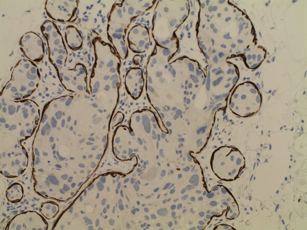
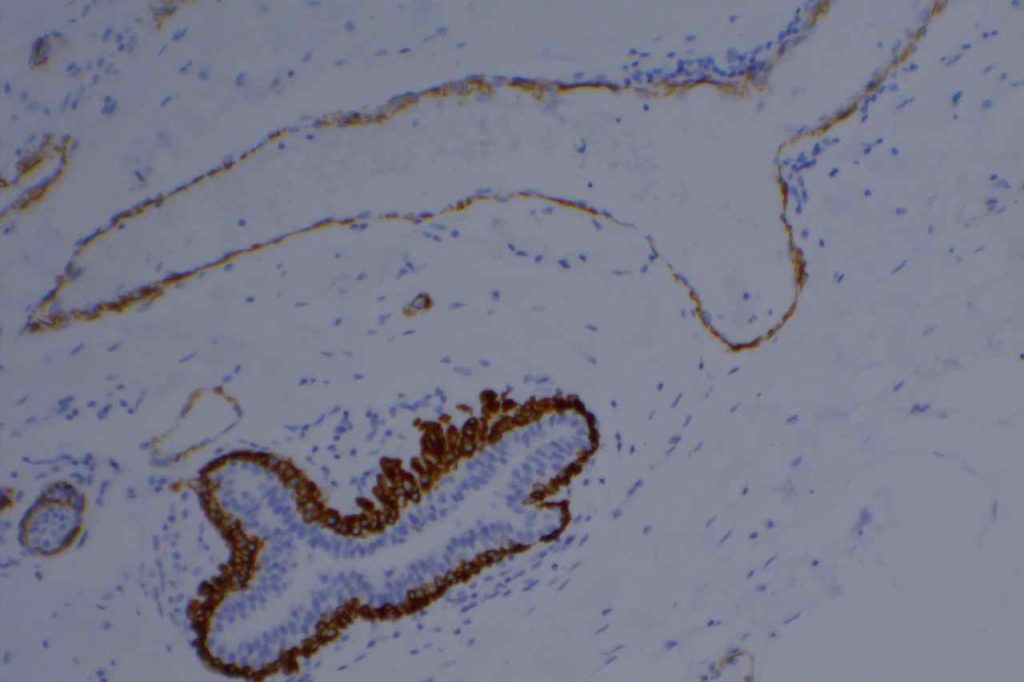
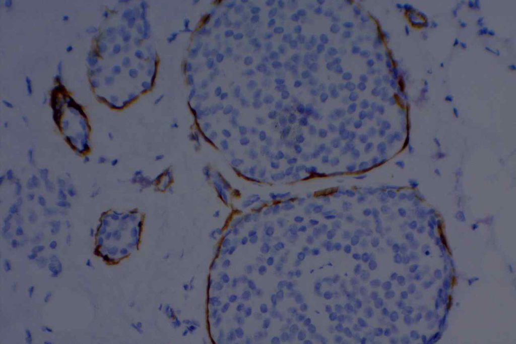
Smooth Muscle Myosin Heavy Chain (SMM-HC) is an antibody to the cytoplasmic structural protein, which is a component of the contractile apparatus in smooth muscle cells.  SMM-HC is expressed in myoepithelial cells of the breast, which makes it a very useful stain in the differentiation of in situ and invasive lesions.
 
One of the big problems in the breast with smooth muscle markers is that myofibroblasts may have background expression, which can occasionally mimic myoepithelial cells.  SMM-HC is one of the best markers (comparatively speaking) not to have a lot background expression in myofibroblasts.  Calponin and smooth muscle actin have higher background staining of myofibroblasts.
 
SMM-HC is a more sensitive and specific marker in the differentiation of in situ and invasive breast carcinoma compared to CD10 [J Clin Pathol. 2007 Aug;60(8):958-9].  Another study noted that SMM-HC stained myofibroblasts in ~8% of cases compared to ~76% of cases for calponin.  p63 is also a useful marker for breast myoepithelial cells, but the staining may not be as continuous as SMM-HC and occasional tumor cells may be positive for p63 (although the patter is recognizably different) [Am J Surg Pathol 2003 Jan;27(1):82-90]. SMM-HC combined with p63 as a dual stain may have synergistic and complementary effects.
 
Usefulness may be decreased in cases of DCIS, in which myoepithelial cells have been reported negative in up to 16% of DCIS cases (especially high grade DCIS). (Dewar, et al)
 
SMM-HC is also expressed in vascular smooth muscle cells.
Microscopic Images
SMM-HC - High Grade DCIS
SMM-HC – High Grade DCIS. Intact myoepithelial cell layer highlighted by smooth muscle myosin-heavy chain.
Smooth Muscle Myosin (SMM-HC) Breast
Smooth Muscle Myosin (SMM-HC) demonstrating loss of the myoepithelial cell layer in the invasive component.
Smooth Muscle Myosin (SMM-HC) Breast
SMM-HC highlighting the myoepithelial layer in benign breast epithelium.
Smooth Muscle Myosin (SMM-HC) Breast
SMM-HC highlighting vascular smooth muscle and benign duct structures.
Smooth Muscle Myosin (SMM-HC) Breast
SMM-HC highlighting expanded duct structures.
References
Kalof AN, et. al. “Immunostaining patterns of myoepithelial cells in breast lesions: a comparison of CD10 and smooth muscle myosin heavy chain.”  J Clin Pathol. 2004 vol. 57(6) pp. 625-629.  doi:10.1136/jcp.2003.013227
 
Werling RW, et. al. “Immunohistochemical distinction of invasive from noninvasive breast lesions: a comparative study of p63 versus calponin and smooth muscle myosin heavy chain.” Am J Surg Pathol 2003 Jan;27(1):82-90.
 
Dewar, R., Fadare, O., Gilmore, H., & Gown, A. M. (2011). Best practices in diagnostic immunohistochemistry: myoepithelial markers in breast pathology. Archives of pathology & laboratory medicine, 135(4), 422–429. doi:10.1043/2010-0336-CP.1