All posts by peferguson

CDX-2

CDX-2 is a nuclear transcription marker that regulates differentiation and proliferation of intestinal epithelial cells, and also serves as tumor suppressor gene.  CDX-2 is normally expressed in small and large intestine epithelium.  It is a sensitive marker for colorectal adenocarcinomas (especially with strong diffuse expression).  It can also mark non-colorectal GI adenocarcinomas, columnar cell variant of papillary thyroid carcinoma (and other variants of papillary thyroid carcinoma, classic and modular variant),  ovarian mutinous carcinomas, bladder adenocarcinomas, goblet cells in Barrett’s esophagus, along with other tumors (data in tables below). Practically speaking, if a particular tumor can have any type of intestinal differentiation, then it most likely can have CDX-2 expression.
Pitfalls
It is important to note that antibody sensitivity can have significant inter laboratory variation due to both clone selection and antibody optimization (Borrisholt, et. al.).  Each laboratory should have a good understanding of the performance characteristics of their CDX-2 clone and optimization.
 
IHC Survey of Primary and Metastatic Caricnomas (Werling, R. W., et. al.)
Tumor
Expression (%)
Gastric
  Adencarcinoma
70% (N=24)
Pancreatic
  Carcinoma
32% (N=22)
Cholangiocarcinoma
25% (N=16)
Carcinoid Tumor
42% (N=12)
Ovarian Carcinoma
  (Mucinous)
64% (N=14)
Bladder
  Adenocarcinoma
100% (N=2)
 
Common expression patterns in carcinoma (Dennis, J. L, et. al.)
Tumor
Expression (%)
Breast
0%
Colon
>90%
Lung
<5%
Pancreas
0-32%
Stomach
20-70%
Prostate
<5%
 
(Enriquez, M.L., et. al.)
Tumor
Expression (%)
Columnar Cell Variant
  Papillary Thyroid Carcnioma
55% (N=11)
 
CDX-2 and Villin expression in human tumors (2-3+ expression) (Werling, R.W., et. al.)
Tumor Type
No.
CDX2
Villin
G.I. Tract


Colon Adenocarcinoma
75
99%
82% (n=73)
Duodenal Adenoma
10
100%
100%
Gastric Adenocarcinoma
24
70%
42%
Esophageal Adenocarcinoma
9
67%
78%
Pancreatic Adenocarcinoma
22
32%
40%
Cholangiocarcinoma/GB
16
25%
60%
Hepatocellular Carcinoma
12
0%
0%
Carcinoid Tumors
12
42%
34%
Ovary


Mucinous Adenocarcinoma
14
64%
64%
Mucinous Cystadenoma
13
8%
0%
Mucinous Borderline Tumor
4
25%
0%
Non-Mucinous
36
0%
0% (n=31)
Genitourinary Tract


Urothelial Carcinoma
21
0%
0%
Adenocarcinoma
2
100%
100%
Urachal Caricinoma
1
100%
100%
Renal Cell Carcinoma
7
0%
0% (n=3)
Prostate Adenocarcinoma
27
4%
0% (n=24)
Breast Carcinoma
34
0%
0%
Lung (45 primary; 18 met)
63
0%
5%
Adenocarcinoma
11
0%
0%
Squamous Cell Carcinoma
11
0%
0%
Non-Small Cell Carcinoma NOS
33
0
9%
Mucinous Carcinoma
2
0%
0%
Small Cell Carcinoma
4
0%
0%
Mesothelioma
7
0%
0%
Head and Neck


Thyroid
36
3%
0%
Papillary Carcinoma
11
9%
0%
Follicular Adenoma/Carcinoma
25
0%
0%
Salivary Gland
12
0%
0%
Mixed Tumor
6
0%
0%
Low Grade Carcinoma
6
0%
0%
Squamous Cell Carcinoma
13
0%
0% (n=11)
Photomicrograph
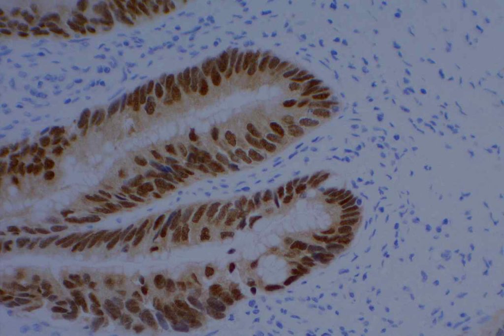
CDX-2 Benign Colon
CDX-2 expression in benign colon epithelium.
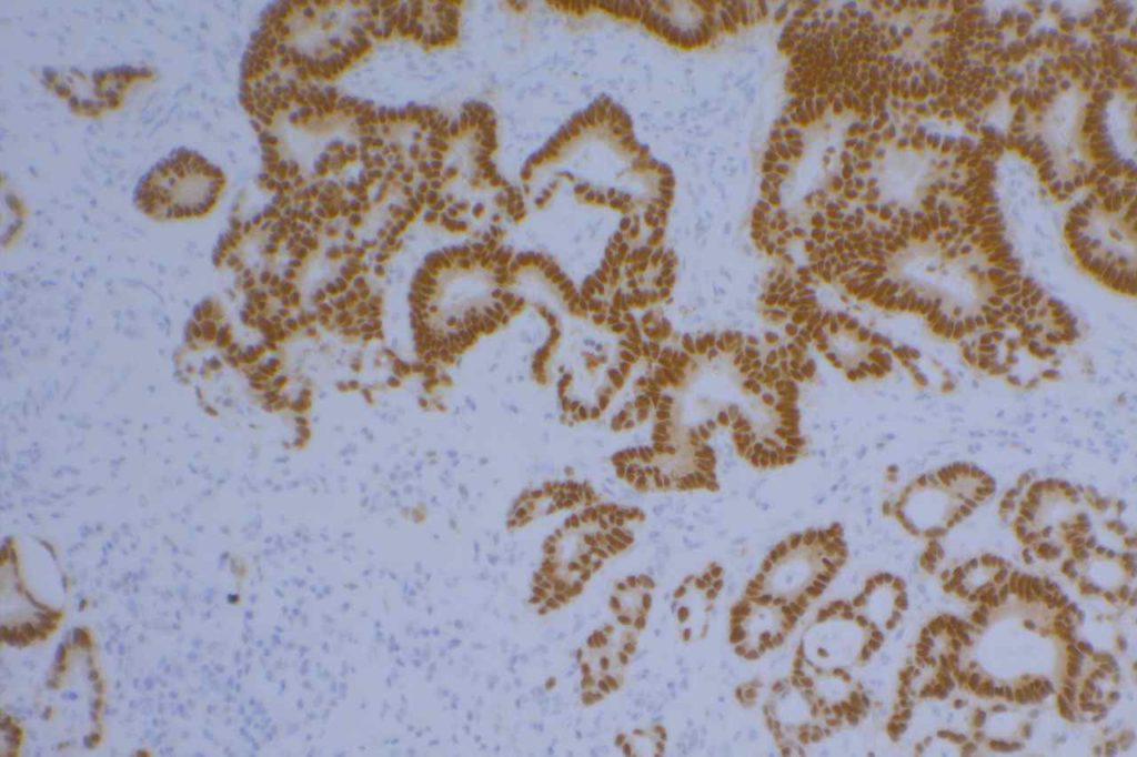
CDX-2 Colon Adenocarcinoma
CDX-2 staining in colon adenocarcinoma.
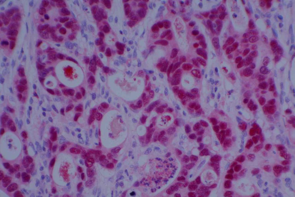
CDX-2 Colon Adenocarcinoma
Colon adenocarcinoma stained with CDX-2 using a red alkaline phosphatase staining kit. Brown (DAB) staining tends to be easier to interpret with nuclear markers (red is good with cytoplasmic markers).
References
Werling, R. W., Yaziji, H., Bacchi, C. E., & Gown, A. M. (2003). CDX2, a highly sensitive and specific marker of adenocarcinomas of intestinal origin: an immunohistochemical survey of 476 primary and metastatic carcinomas. The American Journal of Surgical Pathology, 27(3), 303–310.  Link: http://www.ncbi.nlm.nih.gov/pubmed/?term=American+Journal+of+Surgical+Pathology%2C+27(3)%2C+303%E2%80%93310. 
 
Dennis, J. L., Hvidsten, T. R., Wit, E. C., Komorowski, J., Bell, A. K., Downie, I., et al. (2005). Markers of adenocarcinoma characteristic of the site of origin: development of a diagnostic algorithm. Clinical Cancer Research : an Official Journal of the American Association for Cancer Research, 11(10), 3766–3772. doi:10.1158/1078-0432.CCR-04-2236  Link:  http://www.ncbi.nlm.nih.gov/pubmed/?term=Markers+of+adenocarcinoma+characteristic+of+the+site+of+origin%3A+development+of+a+diagnostic+algorithm 
 
Enriquez, M. L., Baloch, Z. W., Montone, K. T., Zhang, P. J., & LiVolsi, V. A. (2012). CDX2 expression in columnar cell variant of papillary thyroid carcinoma. American Journal of Clinical Pathology, 137(5), 722–726. doi:10.1309/AJCPXE3PUBWVZCGZ Link:  http://www.ncbi.nlm.nih.gov/pubmed/22523209
 
Cameselle-Teijeiro, J., Alberte-Lista, L., Peteiro-Gonzalez, D., Abdulkader-Nallib, I., Reyes-Santias, R., Soares, P., et al. (2012). CDX2 Expression in Some Variants of Papillary Thyroid Carcinoma. American Journal of Clinical Pathology, 138(6), 907–910. doi:10.1309/AJCP1BGCA6MFCNKH  Link:  http://www.ncbi.nlm.nih.gov/pubmed/23161723 
 
Borrisholt, M., Nielsen, S., & Vyberg, M. (2013). Demonstration of CDX2 is highly antibody dependant. Applied Immunohistochemistry & Molecular Morphology : AIMM / Official Publication of the Society for Applied Immunohistochemistry, 21(1), 64–72. doi:10.1097/PAI.0b013e318257f8aa Link: http://www.ncbi.nlm.nih.gov/pubmed/?term=Demonstration+of+CDX2+is+highly+antibody+dependant 
 
Werling, R. W., Yaziji, H., Bacchi, C. E., & Gown, A. M. (2003). CDX2, a highly sensitive and specific marker of adenocarcinomas of intestinal origin: an immunohistochemical survey of 476 primary and metastatic carcinomas. The American Journal of Surgical Pathology, 27(3), 303–310. Link: http://www.ncbi.nlm.nih.gov/pubmed/?term=CDX2%2C+a+highly+sensitive+and+specific+marker+of+adenocarcinomas+of+intestinal+origin%3A+an+immunohistochemical+survey+of+476+primary+and+metastatic+carcinomas 

Calponin

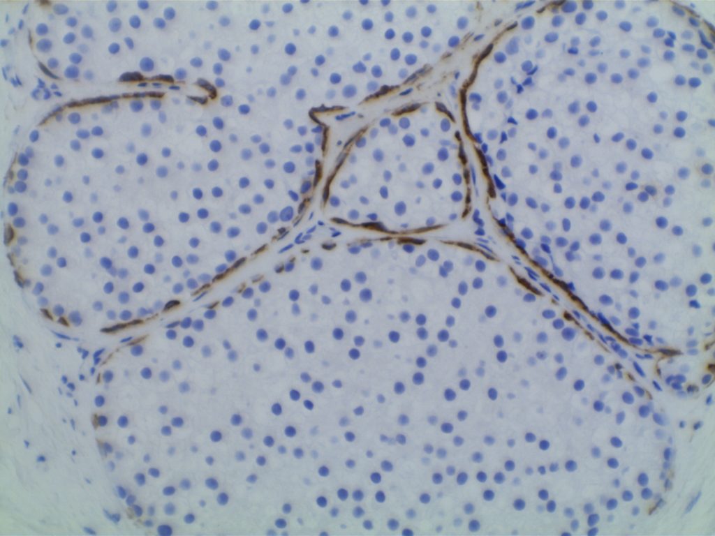
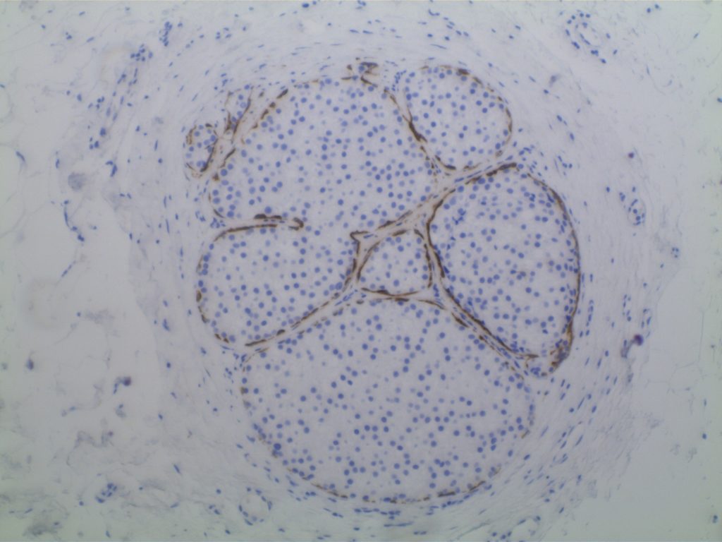
Calponin is an actin filament and is expressed in smooth muscle.  It is often used individually or combined with other IHC markers to identify the myoepithelial layer in breast epithelium.  It is important to remember, like other smooth muscle markers, that myofibroblasts may also express calponin.  Therefore, care should be taken not to mistake myofibroblast positivity for myoepithelial positivity. 
 
It is also expressed in cases of collagenous spherulosis (positive), but not adenoid cystic carcinoma of the breast.  Calponin is not a specific stain, and has been identified in numbers pathologic disease processes.  It is recommended to refer to specific medical literature for the IHC performance based on the differential diagnosis.
 
Positive Expression:
  • Atypical fibroxanthoma (30%) [Virchows Arch 200;437:58]
  • Benign Fibrous Histiocytoma (65%)
  • Collagenous Spherulosis[Mod Path 2006;19:1351]
  • DFSP (40%)
  • Fibromatosis [Am J Dermatopathol 2006;28:105]
  • Fibrosarcoma (60%)
  • Glomus Tumor [AJSP 2002;26:301]
  • Leiomyoma
  • Leiomyosarcoma
  • MFH of bone (47%) [J Clin Pathol 2002;55:853]
  • MPNST (40%)
  • Myoepithelioma-skin
  • Solitary fibrous tumor (70%)
  • Synovial Sarcoma [Histopathology 2003;42:588]
  • Myofibroblastic lesions
Photomicorgraphs
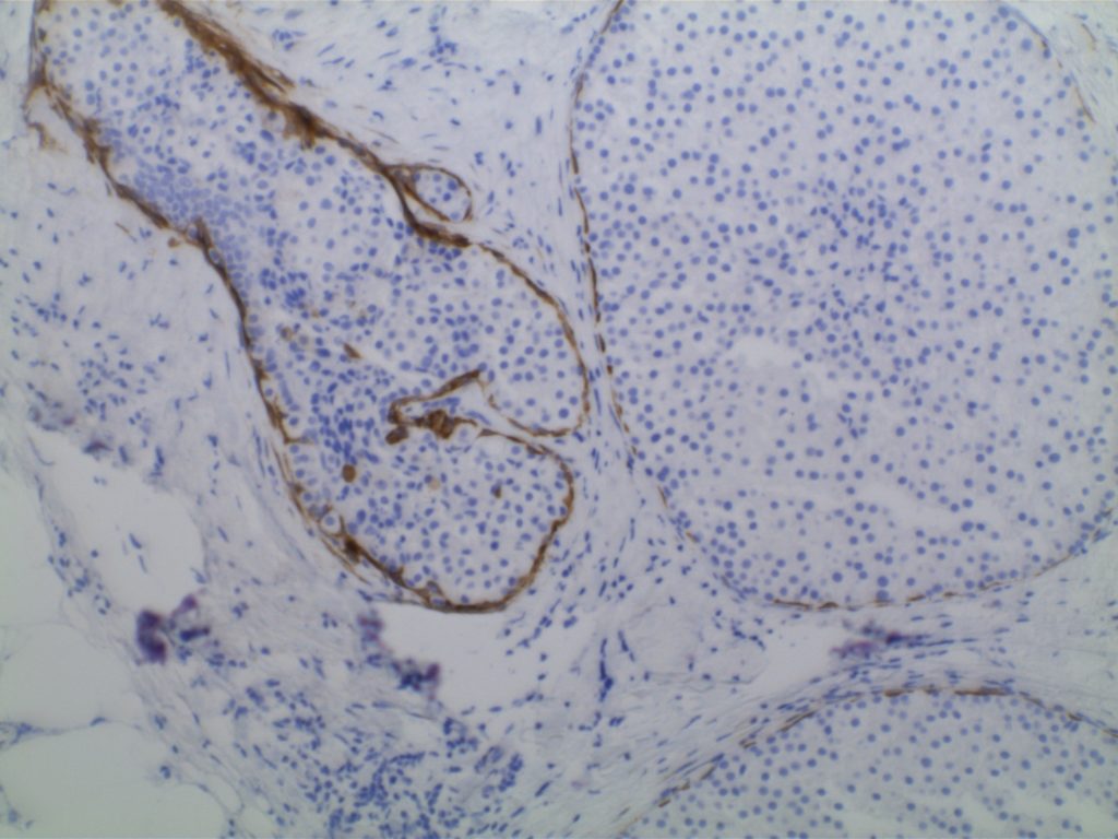
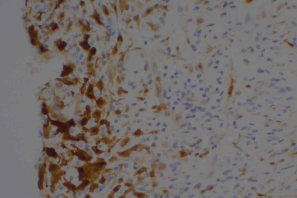
Calponin - Pleomorphic Lobular Carcinoma of Breast
Calponin – Pleomorphic Lobular Carcinoma of Breast
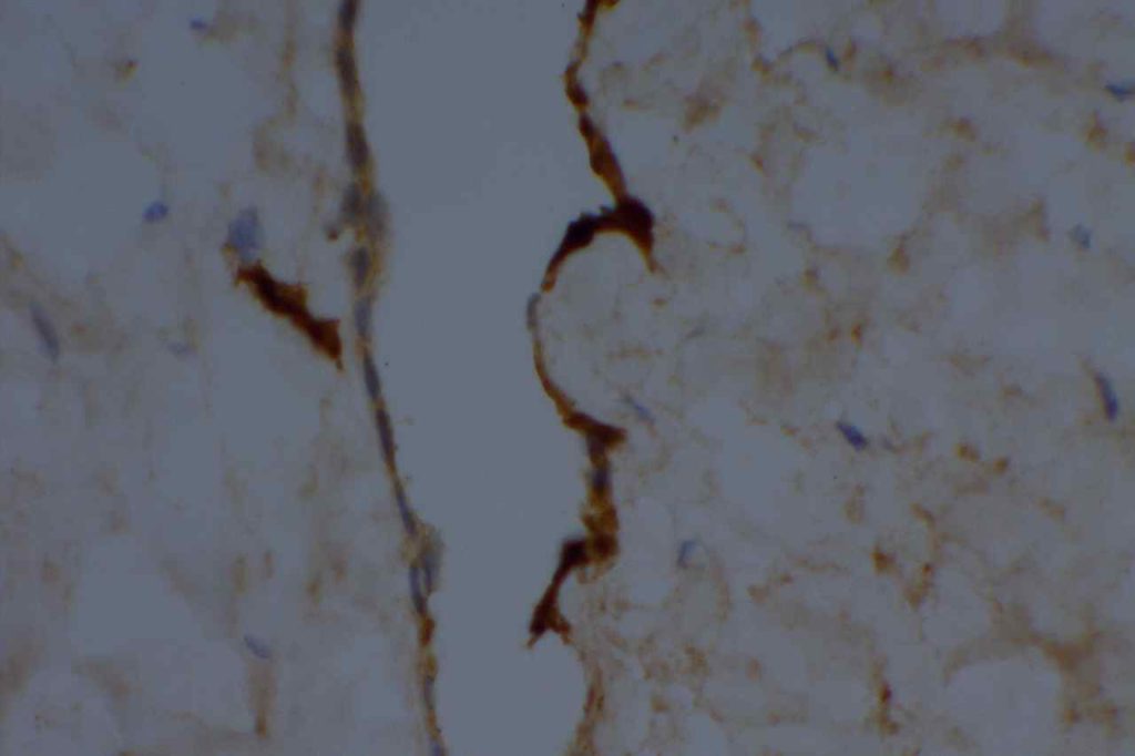
Calponin - Pleomorphic Lobular Carcinoma of Breast
Calponin – Pleomorphic Lobular Carcinoma of Breast
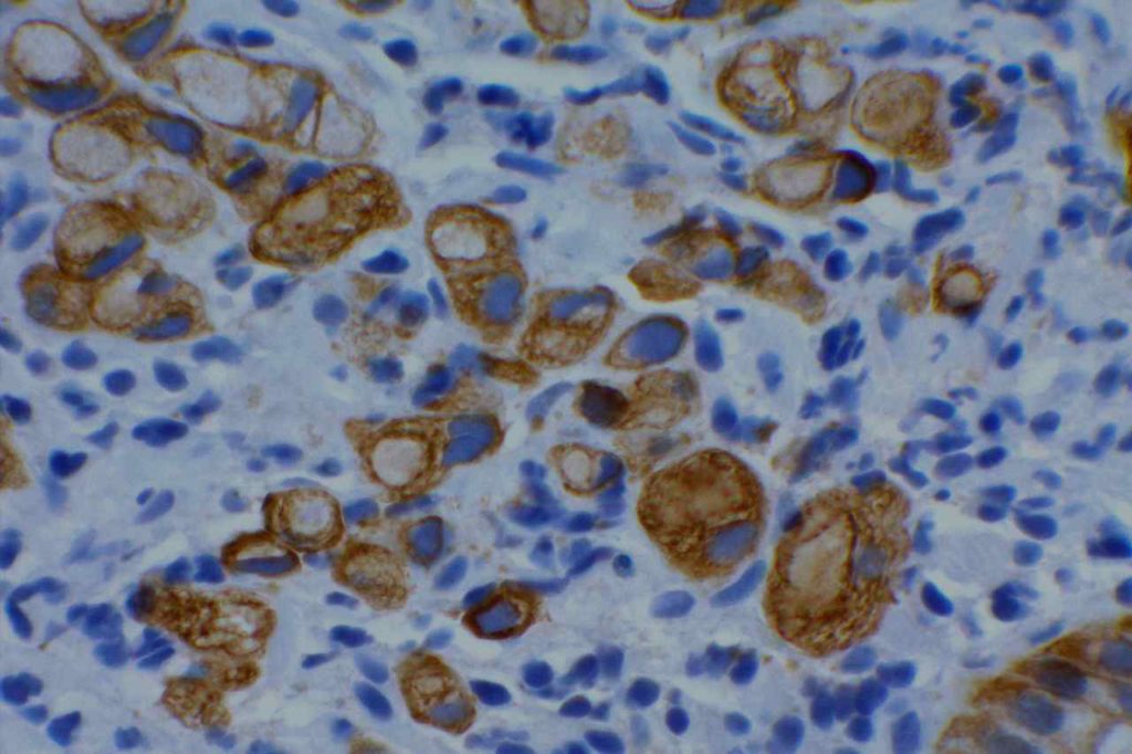
Calponin - Pleomorphic Lobular Carcinoma of Breast
Calponin – Pleomorphic Lobular Carcinoma of Breast
References
Zhao L, Yang X, Khan A, Kandil D. Diagnostic role of immunohistochemistry in the evaluation of breast pathology specimens. Arch Pathol Lab Med. 2014;138: 16–24. doi:10.5858/arpa.2012-0440-RA
 
Dewar R, Fadare O, Gilmore H, Gown AM. Best practices in diagnostic immunohistochemistry: myoepithelial markers in breast pathology. Arch Pathol Lab Med. 2011;135: 422–429. doi:10.1043/2010-0336-CP.1
 
Werling RW, Hwang H, Yaziji H, Gown AM. Immunohistochemical distinction of invasive from noninvasive breast lesions: a comparative study of p63 versus calponin and smooth muscle myosin heavy chain. Am J Surg Pathol. 2003;27: 82–90. 
 
Liu H. Application of immunohistochemistry in breast pathology: a review and update. Arch Pathol Lab Med. 2014;138: 1629–1642. doi:10.5858/arpa.2014-0094-RA

CA-125

Common expression patterns in carcinoma [Clin Cancer Res 2005;11(10) May 15, 2005]
Tumor
Expression (%)
Breast
10-25%
Colon
0-15%
Lung
20-40%
Ovary
60-95%
Pancreas
50%
Stomach
~10%
Prostate
<5%
References
Dennis, J. L., Hvidsten, T. R., Wit, E. C., Komorowski, J., Bell, A. K., Downie, I., et al. (2005). Markers of adenocarcinoma characteristic of the site of origin: development of a diagnostic algorithm. Clinical Cancer Research : an Official Journal of the American Association for Cancer Research, 11(10), 3766–3772. doi:10.1158/1078-0432.CCR-04-2236 

CAM5.2

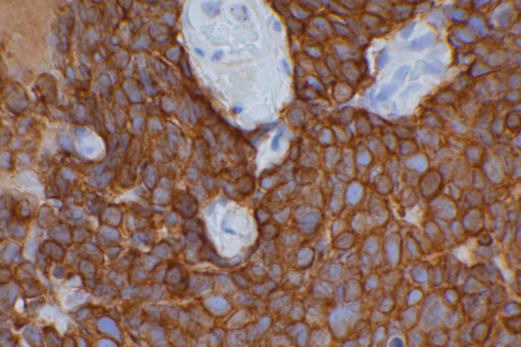
CAM5.2 is an antibody clone manufactured by Becton Dickinson (BD), which shows reactivity to CK8 primarily and CK7 to a less extent.  It does not demonstrate reactivity to CK18 or CK19.  Sometimes CAM5.2 has been used interchangeably with CK8/18 in the medical literature, which is not correct (Hsu, JD, et al).  It is an excellent screening marker for poorly differentiated carcinomas, micro-metastasis of carcinoma in lymph nodes, and for characteristic peri-nuclear dot-like reactivity in Merkel cell carcinoma.
 
AE1/AE3 is generally the most common screening cytokeratin marker used, which has both high and low molecular weight cytokeratins.  Rarely AE1/AE3 may not stain a poorly differentiated carcinoma, which will mark with CAM5.2.
 
CAM5.2 may occasionally stain myofibroblasts and smooth muscle cells, which may express cytokeratins in certain situations (Moll, RT, et al).
 
Moll, RT, et al.  Cytokeratin expression in various tumors.
Tumor
CK8/CK18
CK19
CK7
CK20
CK5
Hepatocellular Ca.
+
+/-
+/-
+/-
=
Colorectal ACA
+
+
+/-
+
=
Stomach ACA
+
+
+/-
+/-
=
Pancreas Ductal ACA
+
+
+
+/-
+/-
Lung ACA
+
+
+
=
=
Breast Inv. Ductal
+
+
+
=
+/-
Endometrium ACA
+
+
+
=
+/-
Ovary ACA 
+
+
+
=
=
RCC, Clear Cell Type
+
+/-
=
=
=
RCC, Papillary Type
+
+
+
=
=
RCC, Chromophobe
+
+/-
+
=
=
Mesothelioma
+
+
+/-
=
+
Lung, Small Cell Ca.
+
+/-
=
=
=
Merkel Cell Ca.
+
+
=
+
=
Urothelial Carcinoma
+
+
+
+/-
+/-
Squamous Cell Ca.
+/-
+/-
=
=
+
Key:  “+/-“, focal staining in some cases. “=“, negative, “+”, positive.
Microscopic Images
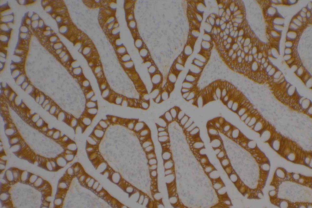
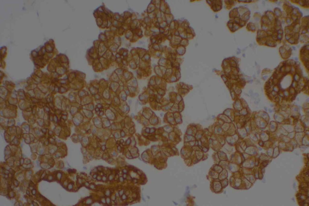
CAM5.2 - Colon Adenocarcinoma
Poorly differentiated sigmoid colon adenocarcinoma with CAM5.2 expression.
CAM5.2 - Lung Small Cell Carcinoma
Small cell carcinoma of the lung stained with CAM5.2.
CAM5.2 - Small Intestine
Normal small bowel epithelium stained with CAM5.2.
CAM5.2 - Salivary Gland
Salivary gland epithelium stained with CAM5.2.
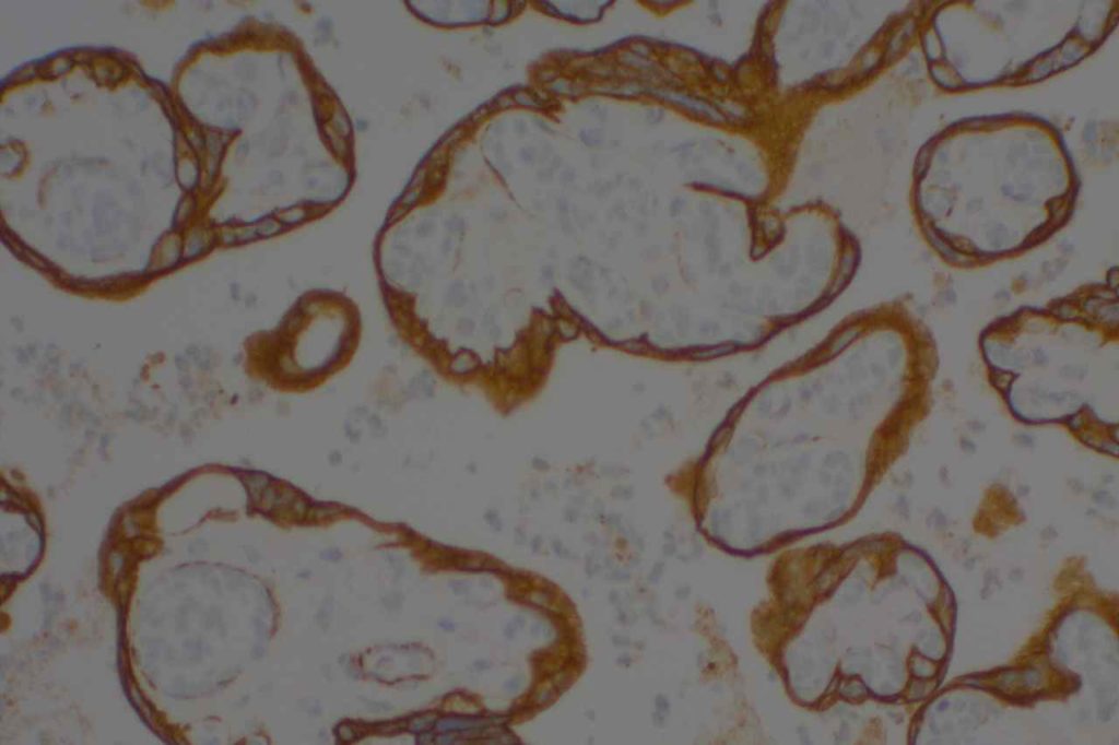
CAM5.2 - Placenta
CAM5.2 expression in placental tissue.
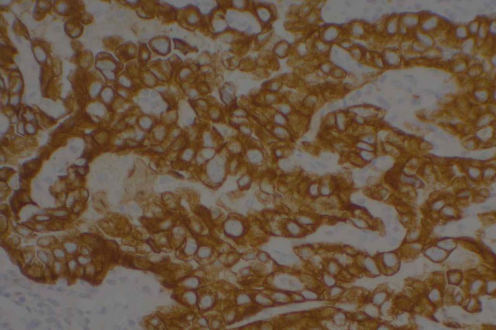
CAM5.2 - Lung Adenocarcinoma
Lung adenocarcinoma stained with CAM5.2.

References
Tamas, EF, Epstein, JI. (2007).  Detection of residual tumor cells in bladder biopsy specimens:  pitfalls in the interpretation of cytokeratin stains.  American Journal of Surgical Pathology, Mar;31(3):390-7.
 
Moll, R., Divo, M., & Langbein, L. (2008). The human keratins: biology and pathology. Histochemistry and Cell Biology, 129(6), 705–733. doi:10.1007/s00418-008-0435-6 
 
Hsu, J.-D., Yao, C.-C., Han, L.-W., & Han, C.-P. (2010). CAM5.2 is not identical to cytokeratins 8 and 18. American Journal of Clinical Pathology, 133(3), 514. doi:10.1309/AJCPAKB6JEBVMX5U  

Calretinin

Calretinin is a very sensitive marker used most commonly as a positive marker for mesothelioma.  It is expressed in >80% of epithelial and up to 70% of sarcomatous subtypes, staining in a cytoplasmic and nuclear pattern.  It is recommended to be used as part of a panel (2 mesothelial markers and 2 adenocarcinoma markers) since there are no sensitive and specific markers to differentiate mesothelioma from adenocarcinoma.
Other tumors expressing Calretinin
  • Sex-cord stromal tumors
  • Adrenal cortical tumors
  • Rare adenocarcinomas
  • Rare squamous cell carcinomas
Photomicrographs
Calretinin - Mesothelium
Chronically inflamed mesothelium highlighted by calretinin.
Calretinin - Mesothelium
Benign mesothelium highlighted by calretinin.
References 
Arch Pathol Lab Med- Vol. 134, February 2010 p. 208.
Wick, MR. “Immunohistochemical approaches to the diagnosis of undifferentiated malignant tumor.”Annals of Diagnostic Pathology12(2008):72-84.

BRAF

BRAF mutations (mainly V600E) are seen in many different tumors (see table below), and the presence of such mutations have therapeutic predictive and diagnostic information for the patient based on the type of tumor implicated.  For example, BRAF mutated colorectal carcinomas, like KRAS mutated tumors do not respond to EGFR inhibitors, but BRAF mutated melanomas may respond to the BRAF kinase inhibitor, vemurafenib.

Continue reading BRAF

Beta-Catinin

β-catinin is a protein that forms cytoplasmic/membranous complexes with E-cadherin (involved by zonula adherens assembly). It is part of the Wnt signaling pathway, and plays an important role in embryogenesis and neoplasia.  The expression may be membraneous, cytoplasmic, or nuclear.  The pattern of expression may be important dependent upon what the differential diagnosis one is considering.  Expression of beta-catinin is closely regulated by the APC gene.  APC mutation associated tumor may have accumulation of protein in the nucleus, and it is the nuclear pattern of expression that is significant in this circumstance.  This stain is best used as part of a panel of markers, and it is important to understand the uses and limitations for a given differential diagnosis.
 
Breast – β-catinin has limited usefulness in stromal proliferations of the breast.  While there is documented variability of expression between fibroadenomas (FA) and phyllodes tumors (PT), the specificity of expression pattern limits this stain’s usefulness. (Yang et al.)
 
Pancreas – nuclear expression of β-catinin is found in pancreatic acing cell carcinoma and solid and pseudo papillary neoplasm, but is negative in pancreatic ductal carcinomas and pancreatic endocrine tumors.  Pancreatoblastoma may have focal expression. 
 
Soft Tissue – Cases of fibrzomatosis have been shown to demonstrate nuclear expression of β-catinin (may be focal, e.g. breast biopsies), compared with nodular fascitis, myofibroma, scar, fibrosarcomas, and low-grade fibromyxoid sarcomas.  There is still limited data on many entities, which may be in the differential diagnosis of soft tissue lesions.  It is important that one understands the usefulness and limitations of this marker.
 
Thyroid – β catinin shows strong to weak membraneous expression in follicular adenomas and well-differentiated carcinomas.  There is nuclear and cytoplasmic expression in poorly differentiated and anapestic carcinomas, and loss of membraneous staining (adverse prognostic indicator).
 
Uterus – nuclear β-catinin expression may be seen in approximately 50% of low-grade endometrial stromal sarcomas, but not in smooth muscle tumors. About half of high grade endometriod carcinomas will express nuclear beta-catinin, but not in high grade serous carcinomas. 
 
References:
Yang X, Kandil D, Cosar EF, Khan A. Fibroepithelial tumors of the breast: pathologic and immunohistochemical features and molecular mechanisms. Arch Pathol Lab Med. 2014;138: 25–36. doi:10.5858/arpa.2012-0443-RA 
 
Fischer S, Asa SL. Application of immunohistochemistry to thyroid neoplasms. Arch Pathol Lab Med. 2008;132: 359–372.
 
Folpe AL, Montgomery EA. The Diagnostic Value of Beta-Catinin Immunihistochemistry. Adv Anat Pathol. 2012;12. doi:10.1186/s13613-016-0144-6 

Bcl-6

Bcl-6 is a nuclear transcription marker that is normally expressed on follicular (germinal center) B-cells and a subset of intrafollicular T-cells.  It is usually expressed in cases of follicular lymphoma, and a subset of diffuse large B-cell lymphomas (40%).
Follicular Lymphoma
In follicular lymphoma, the expression intensity of bcd-6 is an independent prognostic marker.  In general, the stronger the bcd-6 expression, the better the overall and disease free survival.  Overall, at least 88% of cases show expression.
DLBCL
In cases of diffuse large B-cell lymphoma, bcd-6 is used as part of a panel, including CD10 and MUM-1, to sub classify cases as either germinal center or non-germinal center immunophenotypes (Hans’ classifier).  Germinal center immunophenotypes are associated with better survival compared to a non-germinal immunophenotype. 
Reporting
Bcl-6 is generally interpreted and reported in a semi-quantitative manner with regard to intensity (e.g. 1+, 2+, or 3+) and distribution (e.g. >50% of the lymphoma cells)
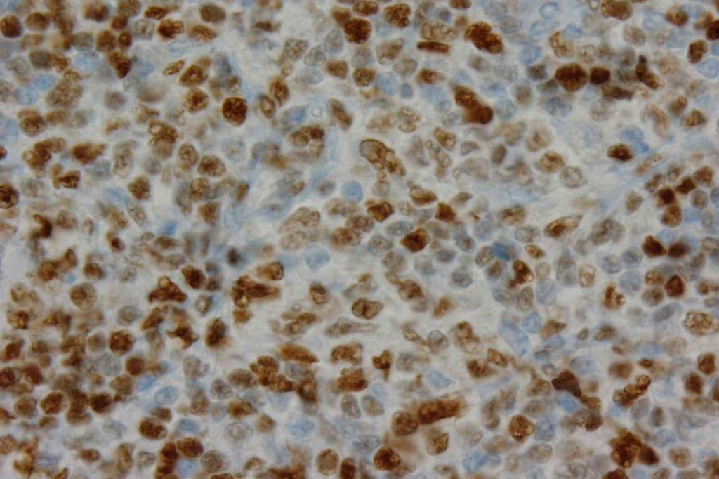
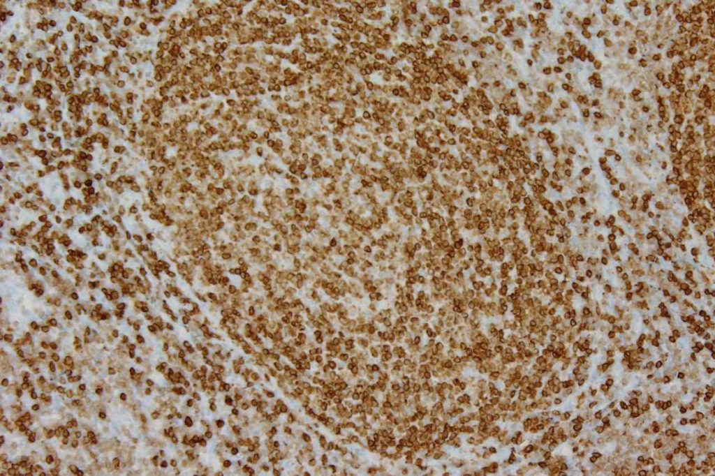
Bcl-6 Expression Pattern
Photomicrographs
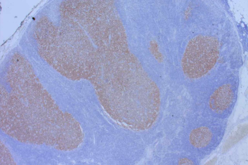
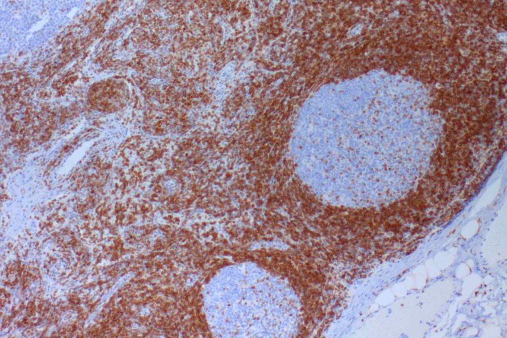
Bcl-6 Reactive Lymph Node
Bcl-6 expression in a reactive lymph node.
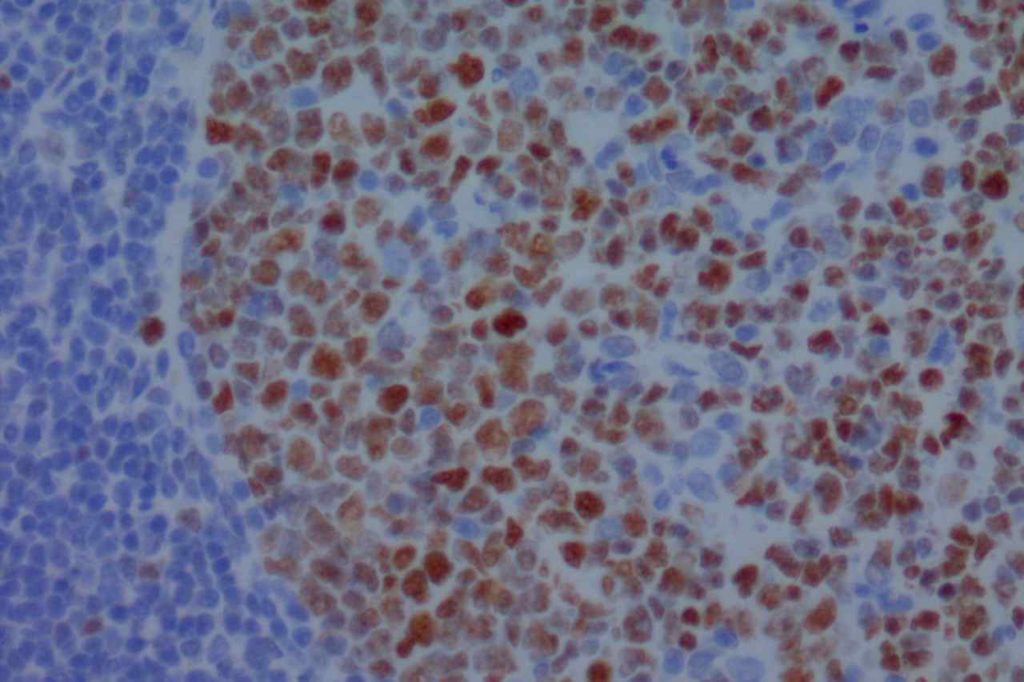
Bcl-6 Reactive Lymph Node
Bcl-6 expression in a reactive lymph node.
Bcl-6 Follicular Lymphoma
Bcl-6 expression in follicular lymphoma.

References
Boyd SD, Natkunam Y, Allen JR, Warnke RA.Selective immunophenotyping for diagnosis of B-cell neoplasms: immunohistochemistry and flow cytometry strategies and results. Appl Immunohistochem Mol Morphol. 2013;21: 116–131. doi:10.1097/PAI.0b013e31825d550a
 
Bilalovic N, Blystad AK, Golouh R, Nesland JM, Selak I, Trinh D, et al. Expression of bcl-6 and CD10 Protein Is Associated With Longer Overall Survival and Time to Treatment Failure in Follicular Lymphoma. Am J Clin Pathol. 2004;121: 34–42. doi:10.1309/TNKL7GDC66R9WPV5
 
Arch Pathol Lab Med.Vol. 130, December 2006:1819-1824.
 
Bone Marrow IHC.  Torlakovic, EE, et. al. American Society for Clinical Pathology Pathology Press © 2009.  pp. 226.
 
Berglund, M., Thunberg, U., Amini, R.-M., Book, M., Roos, G., Erlanson, M., et al. (2005). Evaluation of immunophenotype in diffuse large B-cell lymphoma and its impact on prognosis. Modern Pathology : an Official Journal of the United States and Canadian Academy of Pathology, Inc, 18(8), 1113–1120. doi:10.1038/modpathol.3800396 
 
Herbeck R, Teodorescu Brînzeu D, Giubelan M, Lazăr E, Dema A, Ioniţă H. B-cell transcription factors Pax-5, Oct-2, BOB.1, Bcl-6, and MUM1 are useful markers for the diagnosis of nodular lymphocyte predominant Hodgkin lymphoma. Rom J Morphol Embryol. 2011;52: 69–74.
 
Khokhar FA, Payne WD, Talwalkar SS, Jorgensen JL, Bueso-Ramos CE, Medeiros LJ, et al. Angioimmunoblastic T-cell lymphoma in bone marrow: a morphologic and immunophenotypic study. Hum Pathol. 2010;41: 79–87. doi:10.1016/j.humpath.2009.06.016
 
Hans CP, Weisenburger DD, Greiner TC, Gascoyne RD, Delabie J, Ott G, et al. Confirmation of the molecular classification of diffuse large B-cell lymphoma by immunohistochemistry using a tissue microarray. Blood. 2004;103: 275–282. doi:10.1182/blood-2003-05-1545

Bcl-2

Bcl-2 (B-cell leukemia/lymphoma 2) is a mitochondrial membrane protein involved in blocking apoptotic death of certain cells.  Over expression is associated with multiple lymphomas.  Follicular Lymphoma (FL) is the most important diagnostically.  The vast majority of FLs express bcl-2.  In contrast, benign germinal centers are negative for bcl-2 (except for small follicular T-cells). 
 
In small lymphoid cells, there is little to no specificity to support a benign or malignant diagnosis with bcl-2, and the stain is not helpful in that setting.
Bcl-2 Expression Patterns
  • Follicular Lymphoma (bcl-2 expressed), some high grade tumors may be negative
    • Grade 1 = 87% +
    • Grade 2 = 81% +
    • Grade 3 = 73% +
  • Normal B-cells (non-germinal centers)
  • Normal T-cells
  • Burkitt Lymphoma (NEGATIVE)
  • DLBCL (independent adverse prognostic indicator)
Skin
Bcl-2 is expressed in most basal cell carcinomas of the skin, compared to squamous cell carcinomas, which are negative.  Bcl-2, like BerEP4 may be helpful in differentiating basal cell carcinoma from squamous cell carcinoma.
Microscopic Images
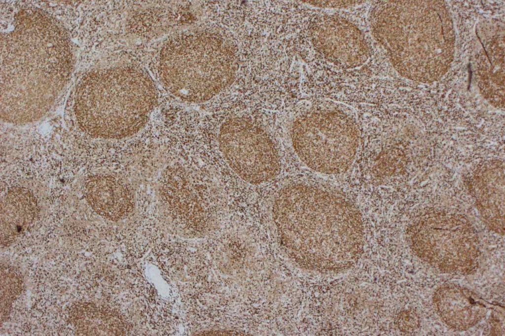
Bcl-2 Follicular Lymphoma
Bcl-2 expression in follicular lymphoma (high power).
Bcl-2 Tonsil
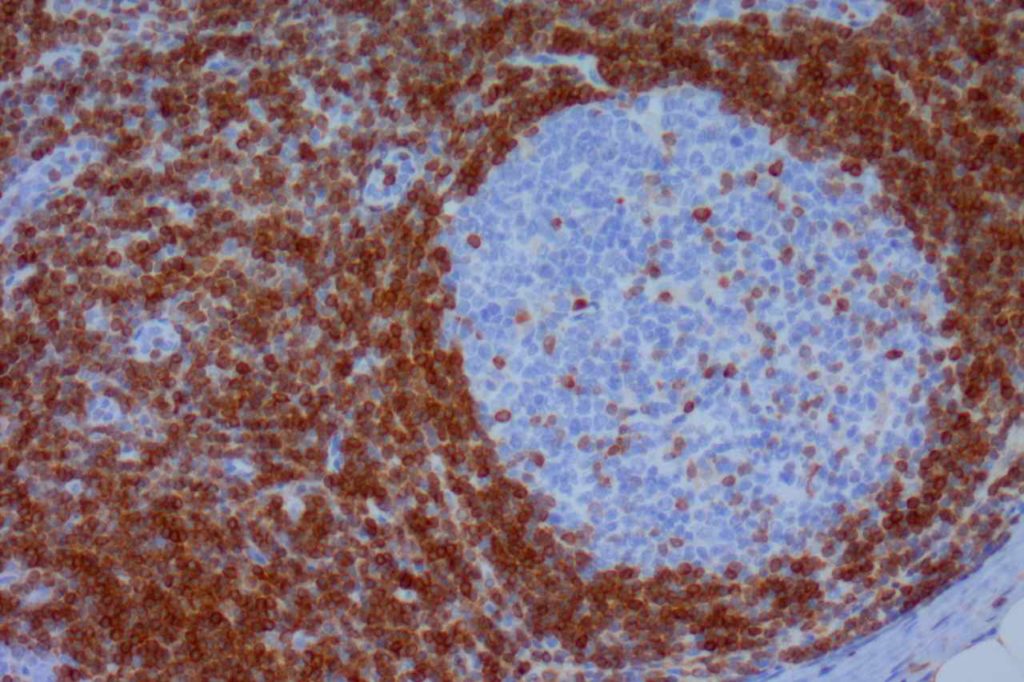
Bcl-2 expression (high power) in reactive tonsil.
Bcl-2
Bcl-2 expression in a benign tonsil.
Bcl-2
Bcl-2 expression in a benign tonsil.
Bcl-2 Follicular Lymphoma
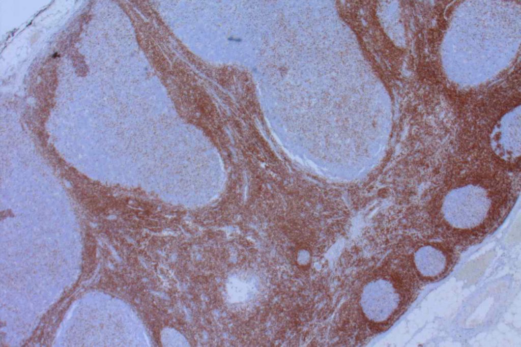
Low power view of Bcl-2 expression in follicular lymphoma.

References
Chuang S-S, Ye H, Du M-Q, Lu C-L, Dogan A, Hsieh P-P, et al. Histopathology and immunohistochemistry in distinguishing Burkitt lymphoma from diffuse large B-cell lymphoma with very high proliferation index and with or without a starry-sky pattern: a comparative study with EBER and FISH. Am J Clin Pathol. 2007;128: 558–564. doi:10.1309/EQJR3D3V0CCQGP04
 
Boyd SD, Natkunam Y, Allen JR, Warnke RA. Selective immunophenotyping for diagnosis of B-cell neoplasms: immunohistochemistry and flow cytometry strategies and results. Appl Immunohistochem Mol Morphol. 2013;21: 116–131. doi:10.1097/PAI.0b013e31825d550a
 
Chan JKC, Ip YT, Cheuk W. The Utility of Immunohistochemistry for Providing Genetic Information on Tumors. International Journal of Surgical Pathology. 2013;21: 455–475. doi:10.1177/1066896913502529
 
Bone Marrow IHC.  Torlakovic, EE, et. al. American Society for Clinical Pathology Pathology Press © 2009.  pp. 226.
 
Tellechea, O., Reis, J.P., Domingues, J.C., Baptista, A.P. (1993).  Monoclonal antibody Ber EP4 distinguishes basal-cell carcinoma from squamous-cell carcinoma of the skin.  American Journal of Dermatopathology.  Oct;15(5):452-5. 
 
Berglund, M., Thunberg, U., Amini, R.-M., Book, M., Roos, G., Erlanson, M., et al. (2005). Evaluation of immunophenotype in diffuse large B-cell lymphoma and its impact on prognosis. Modern Pathology : an Official Journal of the United States and Canadian Academy of Pathology, Inc, 18(8), 1113–1120. doi:10.1038/modpathol.3800396 

BerEP4

BerEP4 is expressed on the surface and cytoplasm of epithelial cells.  It does not mark superficial layers of squamous epithelium, mesothelial cells, or hepatocytes.  It does mark basal layers of squamous epithelium.  BerEP4 is an antibody to cell membrane glycoproteins (not cytokeratins).
 
BerEP4 is most often used as an adenocarcinoma marker for adenocarcinoma (80-100%) in the differential with mesothelioma (0-18%).  [100% sensitive, 91% specific.  AJSP 2001;25:43]  As is the case in examining multiple sources in the medical literature, one must have a clear understanding of how “positive” is defined.  A general take home point is that most adenocarcinomas show strong/diffuse expression compared to more dim/focal expression when found (rare) in mesotheliomas.  BerEP4 is a valuable tool to help differentiate adenocarcinomas from mesothelioma, and is best used as part of a larger panel chosen for the specific differential diagnosis at hand.
 
Summary of multiple studies describing BerEP4 expression in various tumors (Ordones, NG)
Tumor
No.
Expression
(%)
Mesothelioma
14
0%
“Various Carcinomas”
144
99%
Adenocarcinoma of “various” origin
120
86%
Mesothelioma
49
20%
Renal Cell Carcinoma
 
35-50%
Skin
Basal cell carcinomas have been found to express BerEP4, compared to squamous cell carcinomas, which do not express BerEP4.  Bcl-2 also has a similar expression pattern, like BerEP4, in basal cell carcinomas and squamous cell carcinomas.
General Points
Photomicrographs
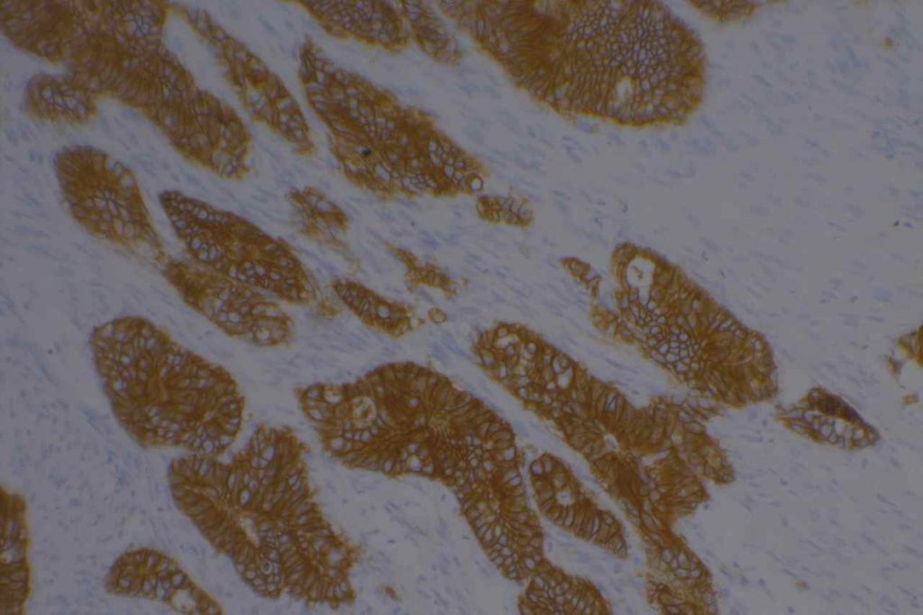
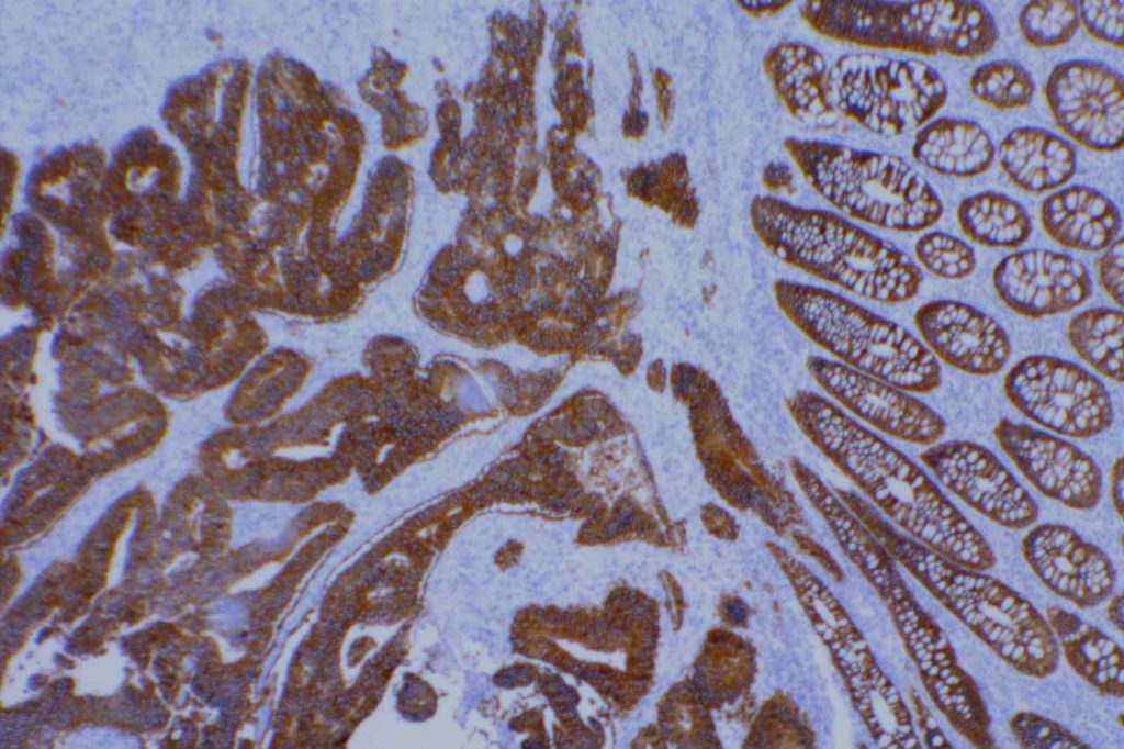
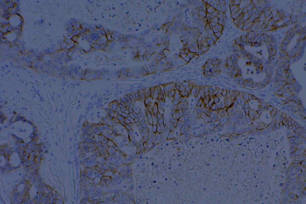
Ber-EP4 Colon Adenocarcinoma
Ber-EP4 expression in colon adenocarcinoma.
Ber-EP4 Colon Adenocarcinoma
Ber-EP4 expression in colon adenocarcinoma.
Ber-EP4 Breast Carcinoma
Ber-EP4 expression in breast carcinoma.
References
Ordóñez, N. G. (2005). Immunohistochemical diagnosis of epithelioid mesothelioma: an update. Archives of Pathology & Laboratory Medicine, 129(11), 1407–1414.  
 
Tellechea, O., Reis, J.P., Domingues, J.C., Baptista, A.P. (1993).  Monoclonal antibody Ber EP4 distinguishes basal-cell carcinoma from squamous-cell carcinoma of the skin.  American Journal of Dermatopathology.  Oct;15(5):452-5.
 
Marchevsky AM. Application of immunohistochemistry to the diagnosis of malignant mesothelioma. Arch Pathol Lab Med. 2008;132: 397–401.
 
Allende D, Yerian L. Immunohistochemical Markers in the Diagnosis of Hepatocellular Carcinoma. Pathology Case Reviews. Vol. 14: 40–46.
 
Sandeck HP, Røe OD, Kjærheim K, Willén H, Larsson E. Re-evaluation of histological diagnoses of malignant mesothelioma by immunohistochemistry. Diagnostic pathology. 2010;5: 47. doi:10.1186/1746-1596-5-47