Non-neoplastic luminal proliferation of ductal epithelial and myoepithelial cells, which can fill and expand ductal strucutures. The morphology shows a mixed cellularity pattern with overlapping nuclei, which often have a “streaming” appearance. In challenging cases immunohistochemistry can help differentiated these lesions from ADH or DCIS.
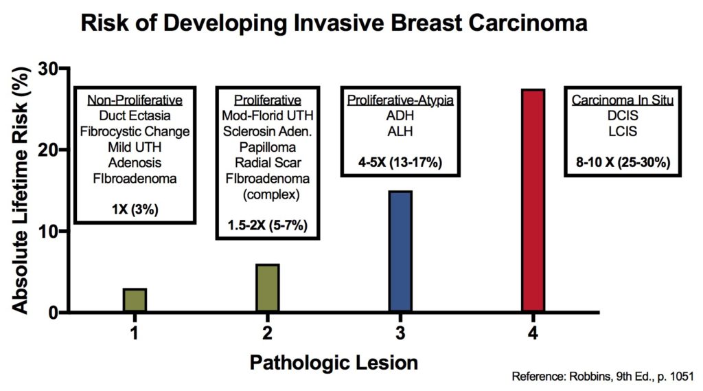
If present only focally, there is not an increased risk of breast carcinoma. However, moderate to florid usual type hyperplasia is associated with a 1.5-2X increased relative risk of breast carcinoma.

Breast lesions and risk of developing an invasive carcinoma

References
Kumar, Vinay, Abul K. Abbas, and Jon C. Aster. Robbins and Cotran Pathologic Basis of Disease. Ninth edition. Philadelphia, PA: Elsevier/Saunders, 2015.
