Category Archives: A – F Antibodies

Beta-Catinin

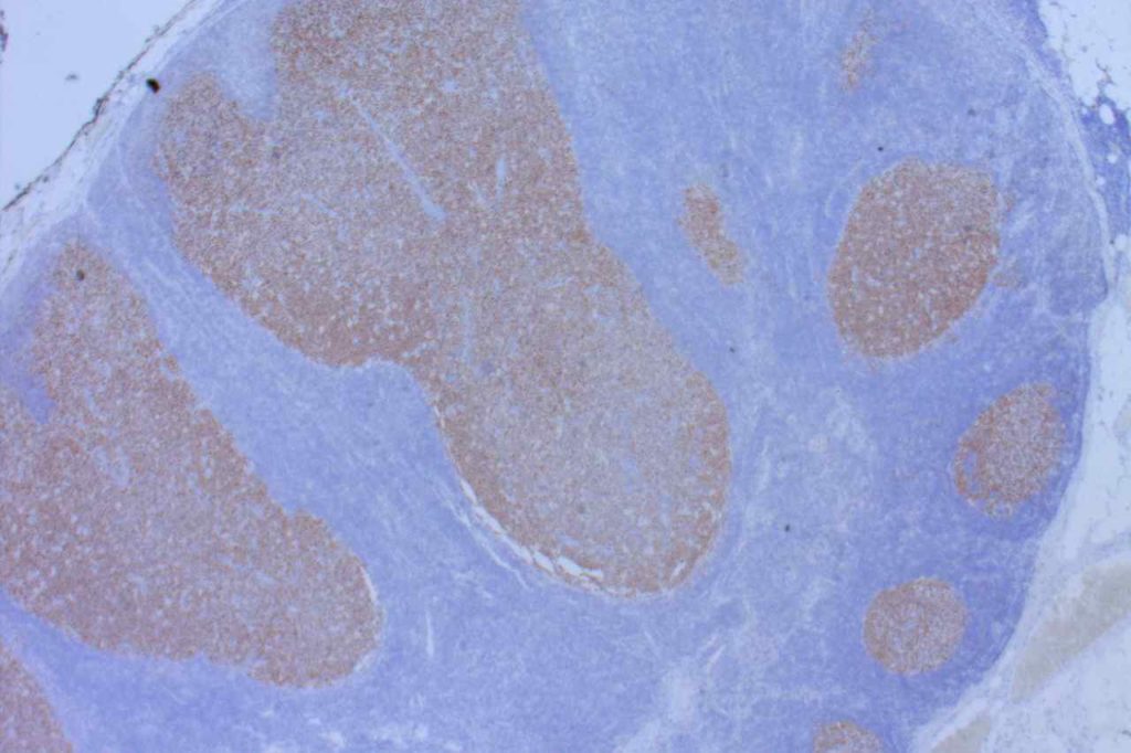
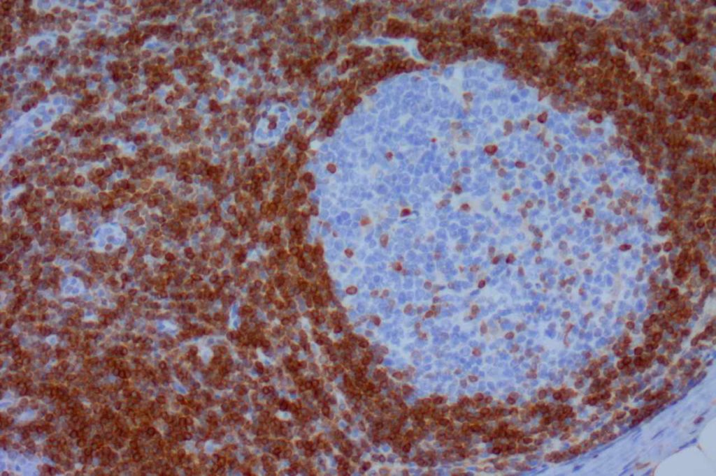
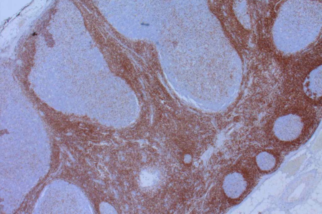
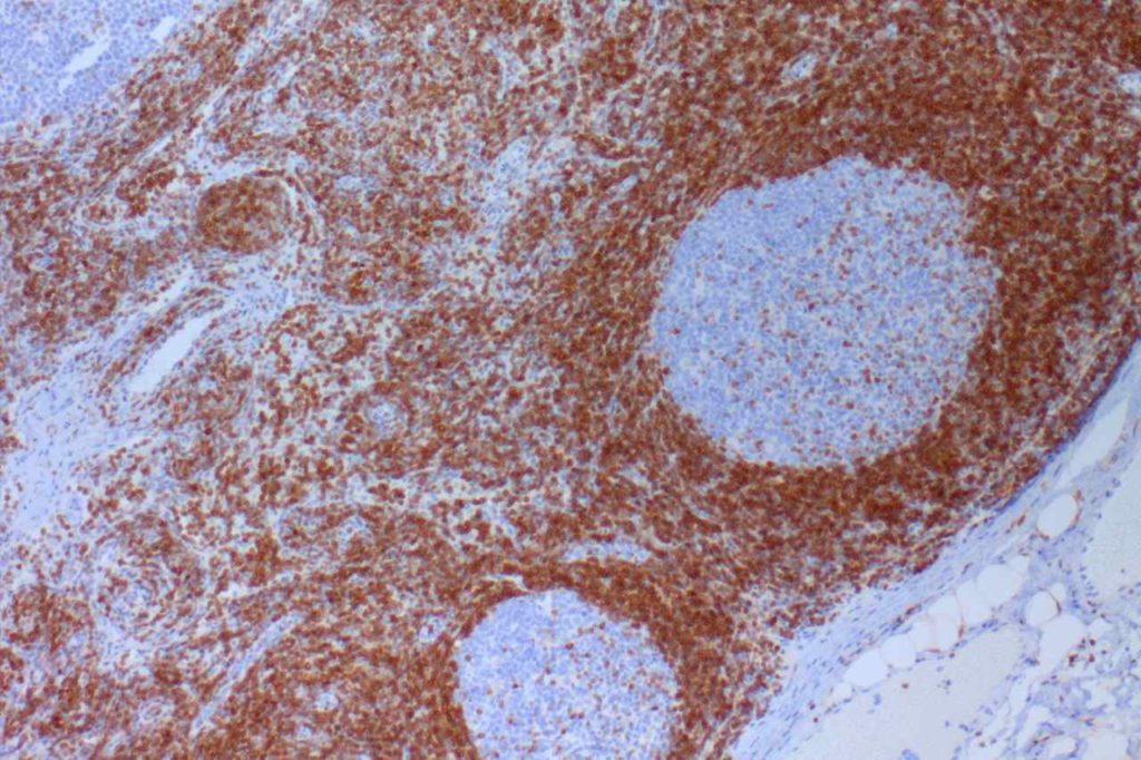
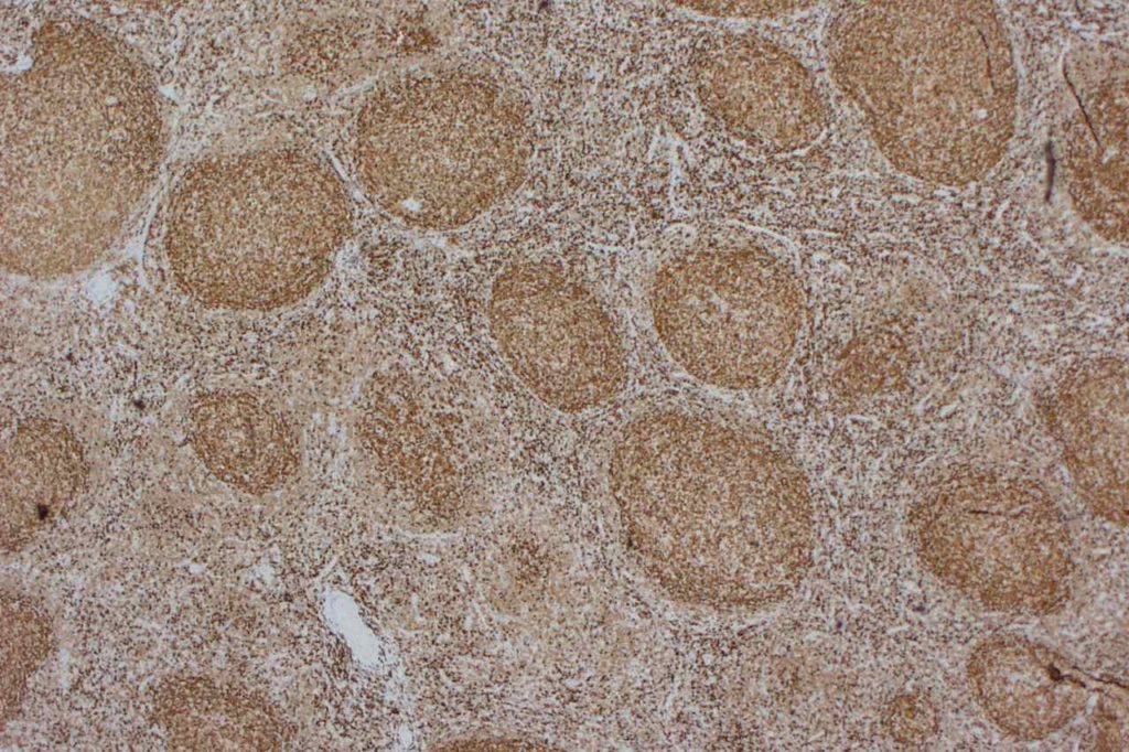
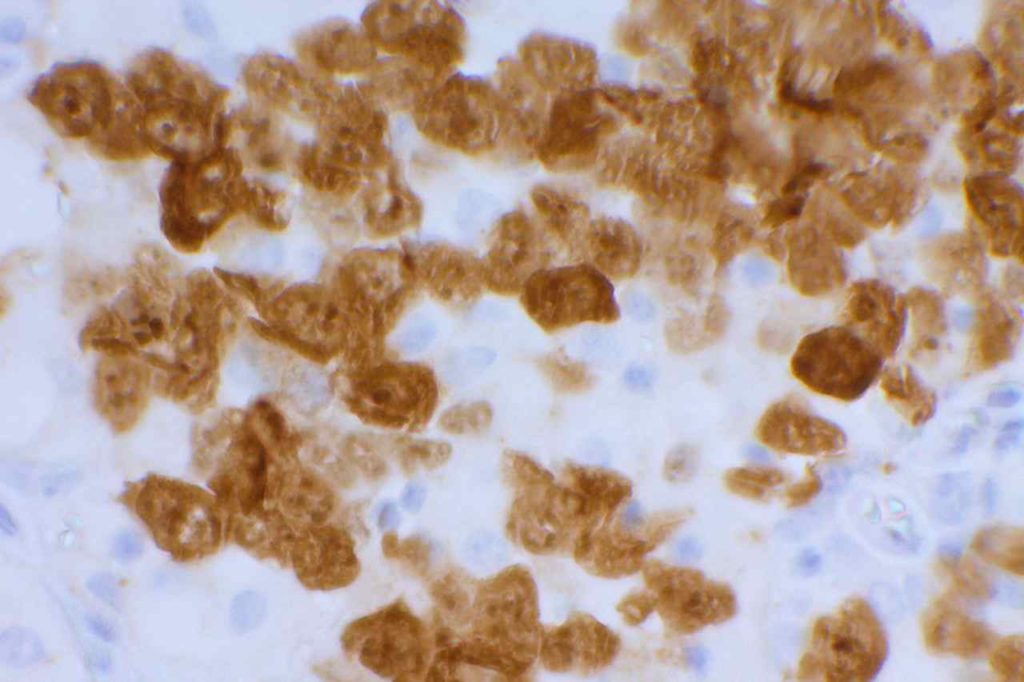
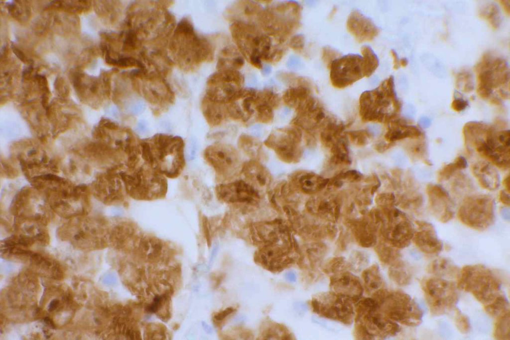
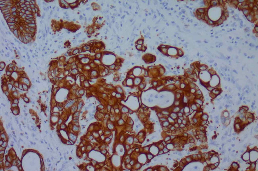
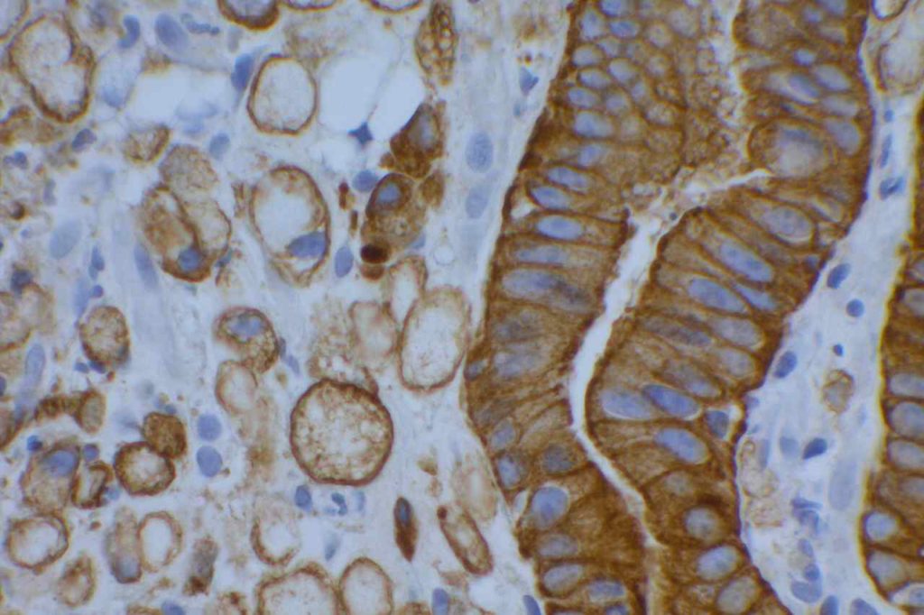
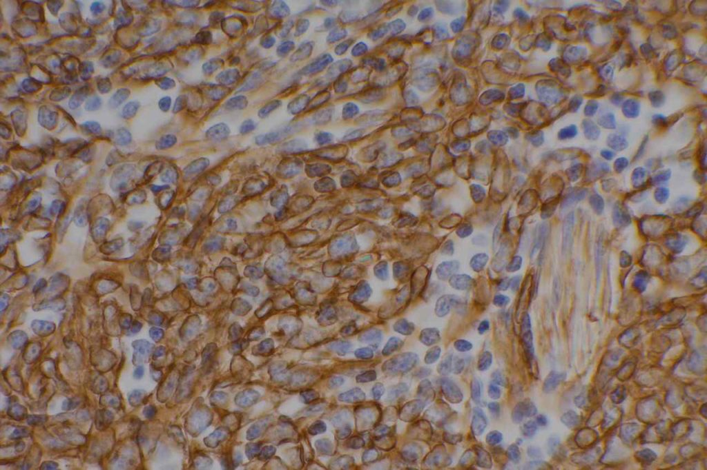
β-catinin is a protein that forms cytoplasmic/membranous complexes with E-cadherin (involved by zonula adherens assembly). It is part of the Wnt signaling pathway, and plays an important role in embryogenesis and neoplasia.  The expression may be membraneous, cytoplasmic, or nuclear.  The pattern of expression may be important dependent upon what the differential diagnosis one is considering.  Expression of beta-catinin is closely regulated by the APC gene.  APC mutation associated tumor may have accumulation of protein in the nucleus, and it is the nuclear pattern of expression that is significant in this circumstance.  This stain is best used as part of a panel of markers, and it is important to understand the uses and limitations for a given differential diagnosis.
 
Breast – β-catinin has limited usefulness in stromal proliferations of the breast.  While there is documented variability of expression between fibroadenomas (FA) and phyllodes tumors (PT), the specificity of expression pattern limits this stain’s usefulness. (Yang et al.)
 
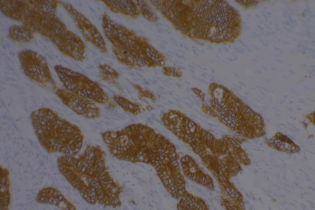
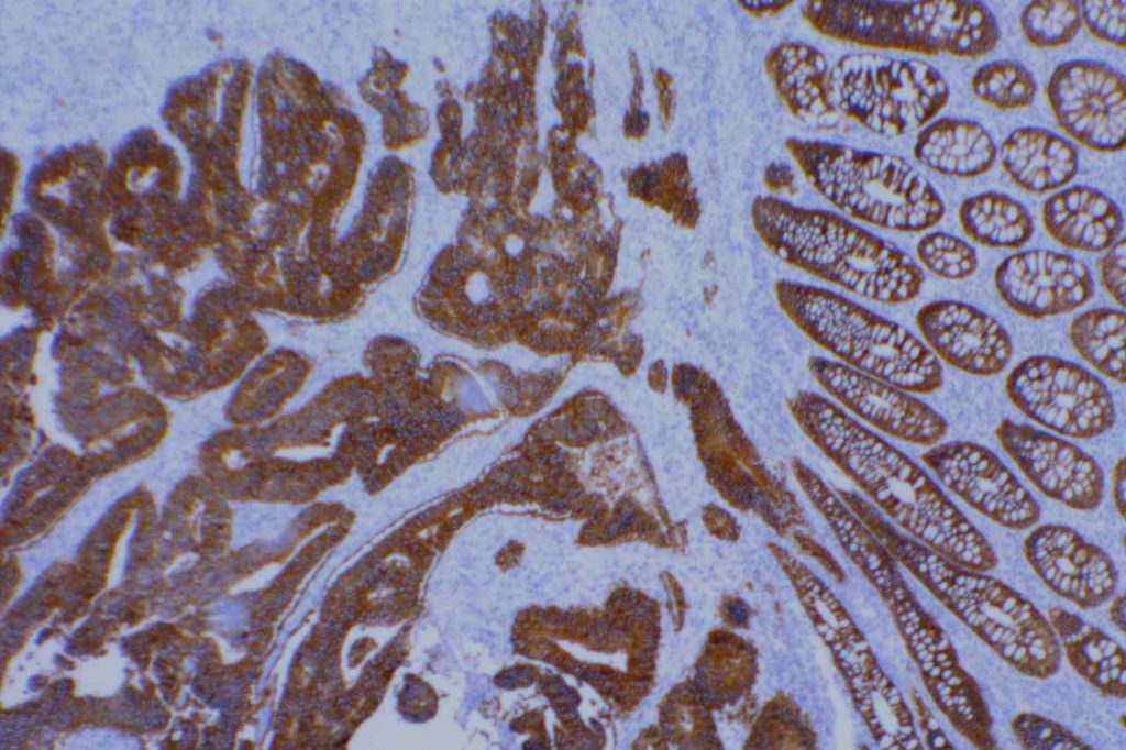
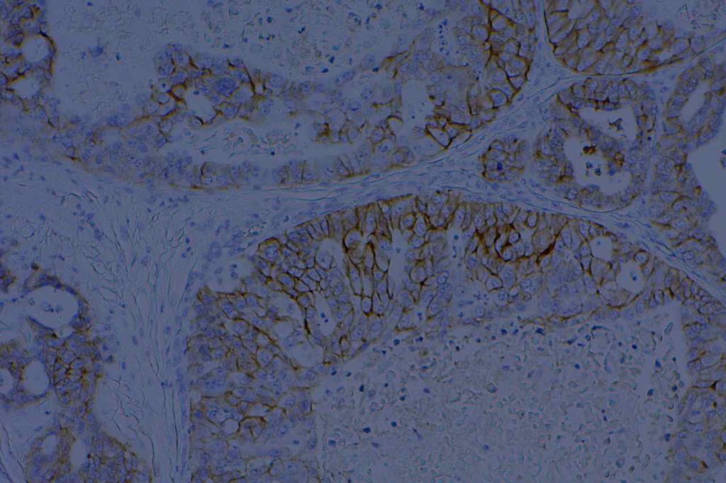
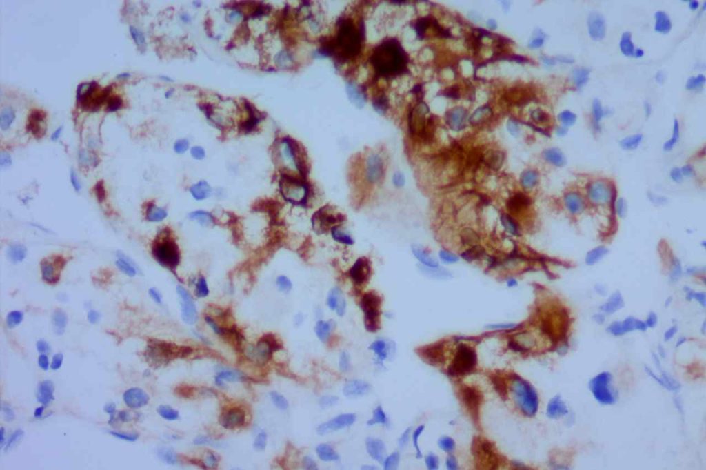
Pancreas – nuclear expression of β-catinin is found in pancreatic acing cell carcinoma and solid and pseudo papillary neoplasm, but is negative in pancreatic ductal carcinomas and pancreatic endocrine tumors.  Pancreatoblastoma may have focal expression. 
 
Soft Tissue – Cases of fibrzomatosis have been shown to demonstrate nuclear expression of β-catinin (may be focal, e.g. breast biopsies), compared with nodular fascitis, myofibroma, scar, fibrosarcomas, and low-grade fibromyxoid sarcomas.  There is still limited data on many entities, which may be in the differential diagnosis of soft tissue lesions.  It is important that one understands the usefulness and limitations of this marker.
 
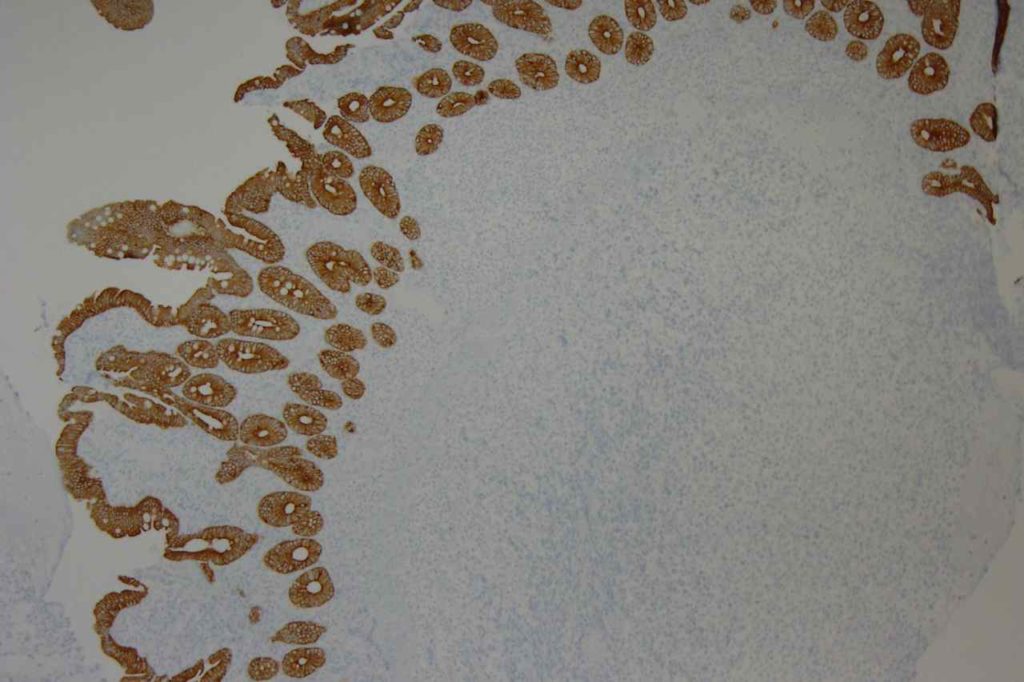
Thyroid – β catinin shows strong to weak membraneous expression in follicular adenomas and well-differentiated carcinomas.  There is nuclear and cytoplasmic expression in poorly differentiated and anapestic carcinomas, and loss of membraneous staining (adverse prognostic indicator).
 
Uterus – nuclear β-catinin expression may be seen in approximately 50% of low-grade endometrial stromal sarcomas, but not in smooth muscle tumors. About half of high grade endometriod carcinomas will express nuclear beta-catinin, but not in high grade serous carcinomas. 
 
References:
Yang X, Kandil D, Cosar EF, Khan A. Fibroepithelial tumors of the breast: pathologic and immunohistochemical features and molecular mechanisms. Arch Pathol Lab Med. 2014;138: 25–36. doi:10.5858/arpa.2012-0443-RA 
 
Fischer S, Asa SL. Application of immunohistochemistry to thyroid neoplasms. Arch Pathol Lab Med. 2008;132: 359–372.
 
Folpe AL, Montgomery EA. The Diagnostic Value of Beta-Catinin Immunihistochemistry. Adv Anat Pathol. 2012;12. doi:10.1186/s13613-016-0144-6 

Bcl-6

Bcl-6 is a nuclear transcription marker that is normally expressed on follicular (germinal center) B-cells and a subset of intrafollicular T-cells.  It is usually expressed in cases of follicular lymphoma, and a subset of diffuse large B-cell lymphomas (40%).
Follicular Lymphoma
In follicular lymphoma, the expression intensity of bcd-6 is an independent prognostic marker.  In general, the stronger the bcd-6 expression, the better the overall and disease free survival.  Overall, at least 88% of cases show expression.
DLBCL
In cases of diffuse large B-cell lymphoma, bcd-6 is used as part of a panel, including CD10 and MUM-1, to sub classify cases as either germinal center or non-germinal center immunophenotypes (Hans’ classifier).  Germinal center immunophenotypes are associated with better survival compared to a non-germinal immunophenotype. 
Reporting
Bcl-6 is generally interpreted and reported in a semi-quantitative manner with regard to intensity (e.g. 1+, 2+, or 3+) and distribution (e.g. >50% of the lymphoma cells)
Bcl-6 Expression Pattern
Photomicrographs
Bcl-6 Reactive Lymph Node
Bcl-6 expression in a reactive lymph node.
Bcl-6 Reactive Lymph Node
Bcl-6 expression in a reactive lymph node.
Bcl-6 Follicular Lymphoma
Bcl-6 expression in follicular lymphoma.

References
Boyd SD, Natkunam Y, Allen JR, Warnke RA.Selective immunophenotyping for diagnosis of B-cell neoplasms: immunohistochemistry and flow cytometry strategies and results. Appl Immunohistochem Mol Morphol. 2013;21: 116–131. doi:10.1097/PAI.0b013e31825d550a
 
Bilalovic N, Blystad AK, Golouh R, Nesland JM, Selak I, Trinh D, et al. Expression of bcl-6 and CD10 Protein Is Associated With Longer Overall Survival and Time to Treatment Failure in Follicular Lymphoma. Am J Clin Pathol. 2004;121: 34–42. doi:10.1309/TNKL7GDC66R9WPV5
 
Arch Pathol Lab Med.Vol. 130, December 2006:1819-1824.
 
Bone Marrow IHC.  Torlakovic, EE, et. al. American Society for Clinical Pathology Pathology Press © 2009.  pp. 226.
 
Berglund, M., Thunberg, U., Amini, R.-M., Book, M., Roos, G., Erlanson, M., et al. (2005). Evaluation of immunophenotype in diffuse large B-cell lymphoma and its impact on prognosis. Modern Pathology : an Official Journal of the United States and Canadian Academy of Pathology, Inc, 18(8), 1113–1120. doi:10.1038/modpathol.3800396 
 
Herbeck R, Teodorescu Brînzeu D, Giubelan M, Lazăr E, Dema A, Ioniţă H. B-cell transcription factors Pax-5, Oct-2, BOB.1, Bcl-6, and MUM1 are useful markers for the diagnosis of nodular lymphocyte predominant Hodgkin lymphoma. Rom J Morphol Embryol. 2011;52: 69–74.
 
Khokhar FA, Payne WD, Talwalkar SS, Jorgensen JL, Bueso-Ramos CE, Medeiros LJ, et al. Angioimmunoblastic T-cell lymphoma in bone marrow: a morphologic and immunophenotypic study. Hum Pathol. 2010;41: 79–87. doi:10.1016/j.humpath.2009.06.016
 
Hans CP, Weisenburger DD, Greiner TC, Gascoyne RD, Delabie J, Ott G, et al. Confirmation of the molecular classification of diffuse large B-cell lymphoma by immunohistochemistry using a tissue microarray. Blood. 2004;103: 275–282. doi:10.1182/blood-2003-05-1545

Bcl-2

Bcl-2 (B-cell leukemia/lymphoma 2) is a mitochondrial membrane protein involved in blocking apoptotic death of certain cells.  Over expression is associated with multiple lymphomas.  Follicular Lymphoma (FL) is the most important diagnostically.  The vast majority of FLs express bcl-2.  In contrast, benign germinal centers are negative for bcl-2 (except for small follicular T-cells). 
 
In small lymphoid cells, there is little to no specificity to support a benign or malignant diagnosis with bcl-2, and the stain is not helpful in that setting.
Bcl-2 Expression Patterns
  • Follicular Lymphoma (bcl-2 expressed), some high grade tumors may be negative
    • Grade 1 = 87% +
    • Grade 2 = 81% +
    • Grade 3 = 73% +
  • Normal B-cells (non-germinal centers)
  • Normal T-cells
  • Burkitt Lymphoma (NEGATIVE)
  • DLBCL (independent adverse prognostic indicator)
Skin
Bcl-2 is expressed in most basal cell carcinomas of the skin, compared to squamous cell carcinomas, which are negative.  Bcl-2, like BerEP4 may be helpful in differentiating basal cell carcinoma from squamous cell carcinoma.
Microscopic Images
Bcl-2 Follicular Lymphoma
Bcl-2 expression in follicular lymphoma (high power).
Bcl-2 Tonsil
Bcl-2 expression (high power) in reactive tonsil.
Bcl-2
Bcl-2 expression in a benign tonsil.
Bcl-2
Bcl-2 expression in a benign tonsil.
Bcl-2 Follicular Lymphoma
Low power view of Bcl-2 expression in follicular lymphoma.

References
Chuang S-S, Ye H, Du M-Q, Lu C-L, Dogan A, Hsieh P-P, et al. Histopathology and immunohistochemistry in distinguishing Burkitt lymphoma from diffuse large B-cell lymphoma with very high proliferation index and with or without a starry-sky pattern: a comparative study with EBER and FISH. Am J Clin Pathol. 2007;128: 558–564. doi:10.1309/EQJR3D3V0CCQGP04
 
Boyd SD, Natkunam Y, Allen JR, Warnke RA. Selective immunophenotyping for diagnosis of B-cell neoplasms: immunohistochemistry and flow cytometry strategies and results. Appl Immunohistochem Mol Morphol. 2013;21: 116–131. doi:10.1097/PAI.0b013e31825d550a
 
Chan JKC, Ip YT, Cheuk W. The Utility of Immunohistochemistry for Providing Genetic Information on Tumors. International Journal of Surgical Pathology. 2013;21: 455–475. doi:10.1177/1066896913502529
 
Bone Marrow IHC.  Torlakovic, EE, et. al. American Society for Clinical Pathology Pathology Press © 2009.  pp. 226.
 
Tellechea, O., Reis, J.P., Domingues, J.C., Baptista, A.P. (1993).  Monoclonal antibody Ber EP4 distinguishes basal-cell carcinoma from squamous-cell carcinoma of the skin.  American Journal of Dermatopathology.  Oct;15(5):452-5. 
 
Berglund, M., Thunberg, U., Amini, R.-M., Book, M., Roos, G., Erlanson, M., et al. (2005). Evaluation of immunophenotype in diffuse large B-cell lymphoma and its impact on prognosis. Modern Pathology : an Official Journal of the United States and Canadian Academy of Pathology, Inc, 18(8), 1113–1120. doi:10.1038/modpathol.3800396 

BerEP4

BerEP4 is expressed on the surface and cytoplasm of epithelial cells.  It does not mark superficial layers of squamous epithelium, mesothelial cells, or hepatocytes.  It does mark basal layers of squamous epithelium.  BerEP4 is an antibody to cell membrane glycoproteins (not cytokeratins).
 
BerEP4 is most often used as an adenocarcinoma marker for adenocarcinoma (80-100%) in the differential with mesothelioma (0-18%).  [100% sensitive, 91% specific.  AJSP 2001;25:43]  As is the case in examining multiple sources in the medical literature, one must have a clear understanding of how “positive” is defined.  A general take home point is that most adenocarcinomas show strong/diffuse expression compared to more dim/focal expression when found (rare) in mesotheliomas.  BerEP4 is a valuable tool to help differentiate adenocarcinomas from mesothelioma, and is best used as part of a larger panel chosen for the specific differential diagnosis at hand.
 
Summary of multiple studies describing BerEP4 expression in various tumors (Ordones, NG)
Tumor
No.
Expression
(%)
Mesothelioma
14
0%
“Various Carcinomas”
144
99%
Adenocarcinoma of “various” origin
120
86%
Mesothelioma
49
20%
Renal Cell Carcinoma
 
35-50%
Skin
Basal cell carcinomas have been found to express BerEP4, compared to squamous cell carcinomas, which do not express BerEP4.  Bcl-2 also has a similar expression pattern, like BerEP4, in basal cell carcinomas and squamous cell carcinomas.
General Points
Photomicrographs
Ber-EP4 Colon Adenocarcinoma
Ber-EP4 expression in colon adenocarcinoma.
Ber-EP4 Colon Adenocarcinoma
Ber-EP4 expression in colon adenocarcinoma.
Ber-EP4 Breast Carcinoma
Ber-EP4 expression in breast carcinoma.
References
Ordóñez, N. G. (2005). Immunohistochemical diagnosis of epithelioid mesothelioma: an update. Archives of Pathology & Laboratory Medicine, 129(11), 1407–1414.  
 
Tellechea, O., Reis, J.P., Domingues, J.C., Baptista, A.P. (1993).  Monoclonal antibody Ber EP4 distinguishes basal-cell carcinoma from squamous-cell carcinoma of the skin.  American Journal of Dermatopathology.  Oct;15(5):452-5.
 
Marchevsky AM. Application of immunohistochemistry to the diagnosis of malignant mesothelioma. Arch Pathol Lab Med. 2008;132: 397–401.
 
Allende D, Yerian L. Immunohistochemical Markers in the Diagnosis of Hepatocellular Carcinoma. Pathology Case Reviews. Vol. 14: 40–46.
 
Sandeck HP, Røe OD, Kjærheim K, Willén H, Larsson E. Re-evaluation of histological diagnoses of malignant mesothelioma by immunohistochemistry. Diagnostic pathology. 2010;5: 47. doi:10.1186/1746-1596-5-47

B72.3

B72.3 (monoclonal antibody) is also known as TAG-72, as it reacts with the so-named tumor associated protein.  It is expressed on many carcinoma cells, and is sometimes part of a panel to differentiate lung adenocarcinoma (+) from mesothelioma (=).  It has also been used in cytology effusion specimens.  B72.3 stains in a membraneous and cytoplasmic pattern.  B72.3 is expressed in high percentage of adenocarcinomas compared to extremely rare expression in mesotheliomas.
 
Ordonez, NG discusses the expression pattern of B72.3 in the context of multiple large studies with the expression characteristics highlighted in the table below.  It is important to evaluate the expression pattern in the setting of the differential diagnosis.  As example, renal cell carcinomas do not react with B72.3, and this antibody would not be helpful in a strategy to differentiate a mesothelioma from renal cell carcinoma.
 
Tumor
No.
Expression (%+)
Epitheliod Mesothelioma
57
3.5%
Mesothelioma
175
2%
Adenocarcinomas (various origins)
211
80.5%
  Pulmonary Adenocarcinoma
110
81%
  Ovarian Serous Carcinoma
45
87%
  Renal Cell Carcinomas
 
0%
Reference
Riera, J.R., Astengo-Osuna, C., Longmate, J.A., Barrifora, H.,  The immunohistochemical diagnostic panel for epithelial mesothelioma: a reevaluation after heat-induced epitope retrieval.  Am J Surg Pathol.  1997;21:1409.
 
Ordóñez, N. G. (2005). Immunohistochemical diagnosis of epithelioid mesothelioma: an update. Archives of Pathology & Laboratory Medicine, 129(11), 1407–1414. 

BOB.1

BOB.1 (B-cell Oct-binding protein 1) is a nuclear marker whose expression is primarily restricted to B-cells (germinal center, mantle zone, and plasma cells).  Cases of Nodular Lymphocyte Predominant Hodgkin Lymphoma (NLPHL) typically expressed both OCT2 and BOB.1.  The vast majority of cases of Classical Hodgkin Lymphoma (CHL) will be negative for both markers, and a small minority (~20%) will express with one of the two markers.  BOB.1 has also been found helpful to differentiate between Mediastinal Large B-Cell Lymphoma and CHL. 

Continue reading BOB.1

Annexin A1

Annexin A1 is antibody to the ANXA1 gene product (cell membrane), and is normally expressed in T-cells, macrophages, and myeloid cells.  It is a useful marker for Hairy Cell Leukemia.  This marker is better than the TRAcP antibody, and helps to distinguish HCL from other mimics (splenic lymphoma with villous lymphocytes and marginal zone lymphoma).
 
It should be noted that with the identification of BRAF mutations in hairy cell leukemia and other markers (and flow cytometry) this antibody is not utilized widely.
 
Clone:  29
Titer:  1:4,000

AMACR (p504s)

AMACR (alpha-methylacyl coenzyme) is a racemese most commonly used in the diagnosis of prostate cancer.  AMACR shows excellent (though not perfect) sensitivity and specificity for prostate adenocarcinoma in prostate tissue.  There are several pitfalls to be aware of with AMACR in the non-prostate gland setting.  Expression is seen in normal hepatocytes, proximal renal tubule epithelium, and bronchial epithelium.  Metanephric adenoma of the bladder has also been reported to be positive for AMACR.
 
From a renal carcinoma standpoint, AMACR is expressed in a high percentage of papillary RCCs and translocation associated RCC (TFE3) (Shen, et al., Wilkerson, et al. & Al-Ghawi et al.), but is rarely expressed by other subtypes of RCC.  Many other tumor types have been shown to express AMACR (see table below).
 
AMACR over expression (moderate to strong staining) in various tissue sites/types (Zhou, et. al.)
Tumor / Tissue
Expression (%)
Colorectal Adenocarcinoma
92%
Prostate Adenocarcinoma
83%
Breast Carcinoma
44%
High Grade PIN
64%
Colon Adenomas
75%
Ovarian
~60%
Melanoma
~40%
Lung
~35%
Urothelial
~30%
Renal Cell Carcinoma
~30%
Lymphoma
~30%
 
Carcinoma in situ of the urothelium has been shown to express AMACR in 50-78% of cases by Aron, et. al.  AMACR has also been shown to have increasing expression levels in Barrett’s esophagus with dysplasia and esophageal adenocarinoma.  Although it is unclear if anyone uses AMACR on a routine basis in cases of Barrett’s esophagus.
 
Pitfalls
An important point is to understand that AMACR is very helpful (especially as part of a panel with basal markers) in differentiating benign prostate glands from prostate adenocarcinoma, but AMACR is not a specific marker of prostate gland differentiation.
Photomicrographs
PIN-4 Prostate Adenocarcinoma
PIN-4 staining in prostate adenocarcinoma. AMACR (red) expression in neoplastic cells.
References
Shen, SS. “Role of Immunohistochemistry in Diagnosing Renal Neoplams: When Is It Really Useful?”Arch Pathol Lab Med, Vol. 136, April 2012.  pp. 410-417. 
 
Jiang, Z., Li, C., Fischer, A., Dresser, K., & Woda, B. A. (2005). Using an AMACR (P504S)/34bE12/p63 Cocktail for the Detection of Small Focal Prostate Carcinoma in Needle Biopsy Specimens. American Journal of Clinical Pathology, 123(2), 231–236. doi:10.1309/1G1NK9DBGFNB792L
 
Yang, X. J., Wu, C.-L., Woda, B. A., Dresser, K., Tretiakova, M., Fanger, G. R., & Jiang, Z. (2002). Expression of alpha-Methylacyl-CoA racemase (P504S) in atypical adenomatous hyperplasia of the prostate. The American Journal of Surgical Pathology, 26(7), 921–925. doi:10.1097/01.PAS.0000017328.13364.17
 
Aron, M., Luthringer, D. J., McKenney, J. K., Hansel, D. E., Westfall, D. E., Parakh, R., et al. (2013). Utility of a Triple Antibody Cocktail Intraurothelial Neoplasm-3 (IUN-3-CK20/CD44s/p53) and α-Methylacyl-CoA Racemase (AMACR) in the Distinction of Urothelial Carcinoma In Situ (CIS) and Reactive Urothelial Atypia. The American Journal of Surgical Pathology, 37(12), 1815–1823. doi:10.1097/PAS.0000000000000114 
 
Al-Ghawi, H., Asojo, O. A., Truong, L. D., Ro, J. Y., Ayala, A. G., & Zhai, Q. J. (2010). Application of Immunohistochemistry to the Diagnosis of Kidney Tumors. Pathology Case Reviews, 15(1), 25–34. doi:10.1097/PCR.0b013e3181d51c70 
 
Shi, X. Y., Bhagwandeen, B., & Leong, A. S.-Y. (2008). p16, cyclin D1, Ki-67, and AMACR as markers for dysplasia in Barrett esophagus. Applied Immunohistochemistry & Molecular Morphology : AIMM / Official Publication of the Society for Applied Immunohistochemistry, 16(5), 447–452. doi:10.1097/PAI.0b013e318168598b 
 
Wilkerson ML, Lin F, Liu H, Cheng L. The application of immunohistochemical biomarkers in urologic surgical pathology. Arch Pathol Lab Med. 2014;138(12):1643–1665. doi:10.5858/arpa.2014-0078-RA.

ALK

ALK (Anaplastic Lymphoma Kinase) is a transmembrane molecule that is only normally expressed in some neural tissues.  It has characteristic expression in a significant proportion of Anaplastic Large Cell Lymphoma (ALCL) cases.  It has also been expressed in cases of pleomorphic liposarcoma, inflammatory myofibroblastic tumor, Merkel cell carcinoma, and a small subset of diffuse large B-cell lymphomas.
 
A subset (1-5%) of non-small cell lung carcinomas (usually adenocarcinomas) have an ELM4-ALK mutation, which is often sensitive to the tyrosine kinase inhibitor (TKI) crizotinib (Pfizer).  Newer TKIs, including ceritinib (Novartis) and alectinib (Hoffmann-La Roche), have also been found to be effective.  
 
Historically, ALK translocations have been identified by FISH analysis.  IHC is also an accepted method with an FDA approved test (IHC CDx Assay).  PCR is being studied as an alternative.  ALK antibody clones 5A4 (Novocastra, Leica Biosystems, Buffalo Grove, Illinois), ALK1 (Dako, Santa Clara, California), 1A4 (Origene, Rockville, Maryland) and D5F3 (Cell signaling Technology, Danvers, Massachusetts) have been successfully used to identify ALK mutated lung tumors, with the 5A4 & D5F3 having equivalent sensitivity. 
 
The ALK1 clone is not as sensitive and the 1A4 clone lacks specificity compared to other antibodies.  Please consult the current medical literature for FDA approved tests for ALK translocation identification in non-small cell lung carcinomas.
Normal Expression
  • Neural Tissue
Abnormal Expression

Interpretation
ALK may stain in a cytoplasmic and/or nuclear pattern.  In ALCL the combined pattern of cytoplasmic and nuclear staining is associated with the t(2;5).
 

In lung ALK staining/expression is cytoplasmic.  Like other markers (e.g. Napsin A), staining may be present in macrophages.  Necrotic tumor, extracellular mucin, and cells of neural origin may also stain.


Photo Gallery
ALK - ALCL
ALK expression in anaplastic large cell lymphoma
ALK - ALCL
ALK expression in anaplastic large cell lymphoma

References
Thunnissen E, Allen TC, Adam J, Aisner DL, Beasley MB, Borczuk AC, et al. Immunohistochemistry of Pulmonary Biomarkers: A Perspective From Members of the Pulmonary Pathology Society. Arch Pathol Lab Med. 2018;142: 408–419. doi:10.5858/arpa.2017-0106-SA
 
Chan, J. K. C., Ip, Y. T., & Cheuk, W. (2013). The Utility of Immunohistochemistry for Providing Genetic Information on Tumors. International Journal of Surgical Pathology, 21(5), 455–475. doi:10.1177/1066896913502529
 
Chan, J. K. C. (2013). Newly Available Antibodies With Practical Applications in Surgical Pathology. International Journal of Surgical Pathology, 21(6), 553–572. doi:10.1177/1066896913507601
 
Paik, J. H., Choe, G., Kim, H., Choe, J.-Y., Lee, H. J., Lee, C.-T., et al. (2011). Screening of Anaplastic Lymphoma Kinase Rearrangement by Immunohistochemistry in Non-small Cell Lung Cancer: Correlation with Fluorescence In Situ Hybridization. Journal of Thoracic Oncology : Official Publication of the International Association for the Study of Lung Cancer, 6(3), 466–472. doi:10.1097/JTO.0b013e31820b82e8
 
Yi, E. S., Boland, J. M., Maleszewski, J. J., Roden, A. C., Oliveira, A. M., Aubry, M.-C., et al. (2011). Correlation of IHC and FISH for ALK Gene Rearrangement in Non-small Cell Lung Carcinoma: IHC Score Algorithm for FISH. Journal of Thoracic Oncology : Official Publication of the International Association for the Study of Lung Cancer, 6(3), 459–465. doi:10.1097/JTO.0b013e318209edb9
 
 Mino-Kenudson, M., Chirieac, L. R., Law, K., Hornick, J. L., Lindeman, N., Mark, E. J., et al. (2010). A novel, highly sensitive antibody allows for the routine detection of ALK-rearranged lung adenocarcinomas by standard immunohistochemistry. Clinical Cancer Research : an Official Journal of the American Association for Cancer Research, 16(5), 1561–1571. doi:10.1158/1078-0432.CCR-09-2845
 
Reichard, K. K., McKenna, R. W., & Kroft, S. H. (2007). ALK-positive diffuse large B-cell lymphoma: report of four cases and review of the literature. Modern Pathology : an Official Journal of the United States and Canadian Academy of Pathology, Inc, 20(3), 310–319. doi:10.1038/modpathol.3800742
 
Medeiros, L. J., & Elenitoba-Johnson, K. S. J. (2007). Anaplastic Large Cell Lymphoma. American Journal of Clinical Pathology, 127(5), 707–722.  
 
Takeuchi K, Choi YL, Togashi Y, et al. KIF5B-ALK, a novel fusion oncokinase identified by an immunohistochemistry-based diagnostic system for ALK-positive lung cancer. Clin Cancer Res. 2009;15(9):3143–3149. doi:10.1158/ 1078-0432.CCR-08-3248.

AE1/AE3

AE1/AE3 is often referred to as “pan” cytokeratin, and is most commonly used to identify carcinomas, which present as morphologically undifferentiated malignant neoplasms.  AE1/AE3 is also commonly used to identify micrometastatsis in sentinel lymph nodes, bone marrow, etc, and is probably the most used screening keratin antibody cocktail.
 
The AE1/AE3 cocktail contains CK1-8, 10, 14-16, and 19.  It does not contain CK17 or CK18.  This is why CAM5.2 may also used in a pan-CK cocktail.  Given that AE1/AE3 is not completely sensitive for “all” cytokeratins, if a suspected carcinoma or undifferentiated tumor does not express AE1/AE3, then additional cytokeratin markers (e.g. CAM5.2 and/or 34BetaE12) may be helpful to maximize keratin expression sensitivity.
 
Moll, RT, et al.  Cytokeratin expression in various tumors.
Tumor
CK8/CK18
CK19
CK7
CK20
CK5
Hepatocellular Ca.
+
+/-
+/-
+/-
=
Colorectal ACA
+
+
+/-
+
=
Stomach ACA
+
+
+/-
+/-
=
Pancreas Ductal ACA
+
+
+
+/-
+/-
Lung ACA
+
+
+
=
=
Breast Inv. Ductal
+
+
+
=
+/-
Endometrium ACA
+
+
+
=
+/-
Ovary ACA 
+
+
+
=
=
RCC, Clear Cell Type
+
+/-
=
=
=
RCC, Papillary Type
+
+
+
=
=
RCC, Chromophobe
+
+/-
+
=
=
Mesothelioma
+
+
+/-
=
+
Lung, Small Cell Ca.
+
+/-
=
=
=
Merkel Cell Ca.
+
+
=
+
=
Urothelial Carcinoma
+
+
+
+/-
+/-
Squamous Cell Ca.
+/-
+/-
=
=
+
Key:  “+/-“, focal staining in some cases. “=“, negative, “+”, positive.
Microscopic Images
AE1/AE3 - Adenocarcinoma
AE1/AE3 highlighting an invasive adenocarcinoma.
AE1/AE3 - Colon Adenocarcinoma
AE1/AE3 expression in a poorly differentiated sigmoid colon adenocarcinoma with signet ring features.
AE1/AE3 - Thymoma
AE1/AE3 expression in a thymoma.
AE1/AE3 - Renal Cell Carcinoma
AE1/AE3 expression in a metastatic renal cell carcinoma (more variable expression).
AE1/AE3
AE1/AE3 (pancytokeratin) expression in small bowel tissue.
References:
Miller, RT, “Cytokeratin AE1/AE3”.  ProPath The Focus Immunohistochemistry.  November 2003. http://www.ihcworld.com/_newsletter/2003/focus_nov_2003.pdf
 
Hadi, AIMM Annual Meeting, “The Thirty Most Important Antibodies”, presentation, 2011.
  
Moll, R., Divo, M., & Langbein, L. (2008). The human keratins: biology and pathology. Histochemistry and Cell Biology, 129(6), 705–733. doi:10.1007/s00418-008-0435-6