Category Archives: N – Z Antibodies

ROS-1

ROS-1 rearrangements with at least 12 different partner proteins have been identified in a small subset of lung non-small cell carcinomas (1–2%), which shows susceptibility to tyrosine kinase inhibitors (TKIs) similar to ALK rearranged tumors.  ROS-1 is considered a oncogene found on chromosome 6.  The exact mechanism of activation of this gene protein product with the various gene rearrangement partners is not understood.  The protein function is similar to that of the ALK family, which is why this mutation was studied for possible response to ALK inhibitors (crizotinib).
 
Recently, ROS-1 mutated tumors have been approved for TKI therapy with identification of a rearrangement by FISH analysis.  Like ALK, ROS-1 FISH utilizes a break apart probe to identify the presence of a gene rearrangement.  Other successful modalities for identification of ROS-1 rearrangements include ‘next generation’ sequencing (NGS) and immunohistochemistry.
 
Immunohistochemistry (IHC) has been studied as an alternative to FISH as a screening modality.  Based on multiple studies, the sensitivity of IHC appears to be near 100% with the specificity of at least 92%.  These studies were performed using the D4D6 rabbit monoclonal antibody clone (Cell Signaling Technology, Danvers, Massachusetts).

Stain Interpretation
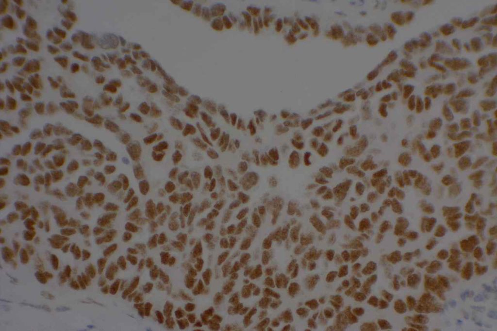
Unlike ALK, there is no known normal tissue counterpart which can be used as a control.  Therefore, known ROS-1 positive tumors or cell lines  (HCC78 cell line with the SLC34A2-ROS1 rearrangement) are generally used.  ROS-1 expression is cytoplasmic with described expression ranging from finely granular to globular cytoplasmic staining and membranous staining.  No consensuses has been established as to the minimal threshold of positivity.
 
Possible interpretation pitfalls include weak staining of type II pneumocytes and alveolar macrophages along with osteoclast-type giant cells in bone biopsies.  Like any immunostain, contextual evaluation is critical.

References
Thunnissen E, Allen TC, Adam J, Aisner DL, Beasley MB, Borczuk AC, et al. Immunohistochemistry of Pulmonary Biomarkers: A Perspective From Members of the Pulmonary Pathology Society. Arch Pathol Lab Med. 2018;142: 408–419. doi:10.5858/arpa.2017-0106-SA
 
Shaw AT, Ou S-HI, Bang Y-J, Camidge DR, Solomon BJ, Salgia R, et al. Crizotinib in ROS1-rearranged non-small-cell lung cancer. N Engl J Med. 2014;371: 1963–1971. doi:10.1056/NEJMoa1406766
 
Bubendorf L, Büttner R, Al-Dayel F, Dietel M, Elmberger G, Kerr K, et al. Testing for ROS1 in non-small cell lung cancer: a review with recommendations. Virchows Arch. 2016;469: 489–503. doi:10.1007/s00428-016-2000-3
 
Boyle TA, Masago K, Ellison KE, Yatabe Y, Hirsch FR (2015) ROS1 immunohistochemistry among major genotypes of non- small-cell lung cancer. Clin Lung Cancer 16(2):106–111. doi:10.1016/j.cllc.2014.10.003 
 
CaoB,WeiP,LiuZ,BiR,LuY,ZhangL,ZhangJ,YangY,Shen C, Du X, Zhou X (2016) Detection of lung adenocarcinoma with ROS1 rearrangement by IHC, FISH, and RT-PCR and analysis of its clinicopathologic features. Onco Targets Ther 9:131–138. doi:10.2147/OTT.S94997 
 
Sholl LM, Sun H, Butaney M, Zhang C, Lee C, Janne PA, Rodig SJ (2013) ROS1 immunohistochemistry for detection of ROS1-rearranged lung adenocarcinomas. Am J Surg Pathol 37(9):14411449. doi:10.1097/PAS.0b013e3182960fa7
 
Yoshida A, Tsuta K, Wakai S, Arai Y, Asamura H, Shibata T, Furuta, K, Kohno T, Kushima R (2014) Immunohistochemical detection of ROS1 is useful for identifying ROS1 rearrangements in lung can- cers. Mod Pathol 27(5):711720. doi:10.1038/modpathol.2013.192
 
Rogers TM, Russell PA, Wright G, Wainer Z, Pang JM, Henricksen LA, Singh S, Stanislaw S, Grille J, Roberts E, Solomon B, Fox SB (2015) Comparison of methods in the detection of ALK and ROS1 rearrangements in lung cancer. J Thorac Oncol 10(4):611618. doi:10.1097/JTO.0000000000000465
 
Rimkunas VM, Crosby KE, Li D, Hu Y, Kelly ME, Gu TL, Mack JS, Silver MR, Zhou X, Haack H (2012) Analysis of receptor tyro- sine kinase ROS1-positive tumors in non-small cell lung cancer: identification of a FIG-ROS1 fusion. Clin Cancer Res 18(16): 44494457. doi:10.1158/1078-0432.CCR-11-3351
 
Mescam-Mancini L, Lantuejoul S, Moro-Sibilot D, Rouquette I, Souquet PJ, Audigier-Valette C, Sabourin JC, Decroisette C, Sakhri L, Brambilla E, McLeer-Florin A (2014) On the relevance of a testing algorithm for the detection of ROS1-rearranged lung adenocarcinomas. Lung Cancer 83(2):168–173. doi:10.1016/j. lungcan.2013.11.019 
 
Shan L, Lian F, Guo L, Qiu T, Ling Y, Ying J, Lin D (2015) Detection of ROS1 gene rearrangement in lung adenocarcinoma: comparison of IHC, FISH and real-time RT-PCR. PLoS One 10(3): e0120422. doi:10.1371/journal.pone.0120422 

p16

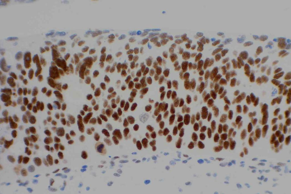
p16 is a marker primary used as a surrogate marker for high risk HPV infection.  The physiologic role of p16 when it is expressed leads to cell cycle arrest.  Normal levels are below the threshold for detection by immunohistochemical methods (IHC).  Sometimes occasional non-proliferating epithelial cells may express p16 by IHC (these cells are usually in the upper aspects of the stratified epithelium.

Continue reading p16

Vimentin

Vimentin is an intermediate filament found in mesenchymal tissue.  It is not a specific stain and there is a subset of tumors which characteristically have co-expression of cytokeratin and vimentin.  It should also be noted that poorly differentiated carcinomas of any origin may express vimentin.  
 
Some pathologists joke they use vimentin to see if IHC stains will even work in a particular piece of tissue, but vimentin can be very helpful especially when used in conjunction with a panel of stains.
Carcinomas with co-expression of vimentin
  • Renal Cell Carcinoma
  • Thyroid Carcinoma (Papillary & Anaplastic)
  • Endometrial Adenocarcinoma
  • Sarcomatoid Carcinoma
  • Mesothelioma (biphasic)
  • Metaplastic Breast Carcinoma
  • Myoepithelial Carcinoma
Sarcomas with co-expression of cytokeratin
  • Desmoplastic Small Round Cell Tumor
  • Epithelioid Sarcoma
  • Epithelioid Angiosarcoma
  • Malignant Rhabdoid Tumor
  • Synovial Sarcoma
  • Leiomyosarcoma
  • Chordoma
  • Adamantinoma
Photomicrographs
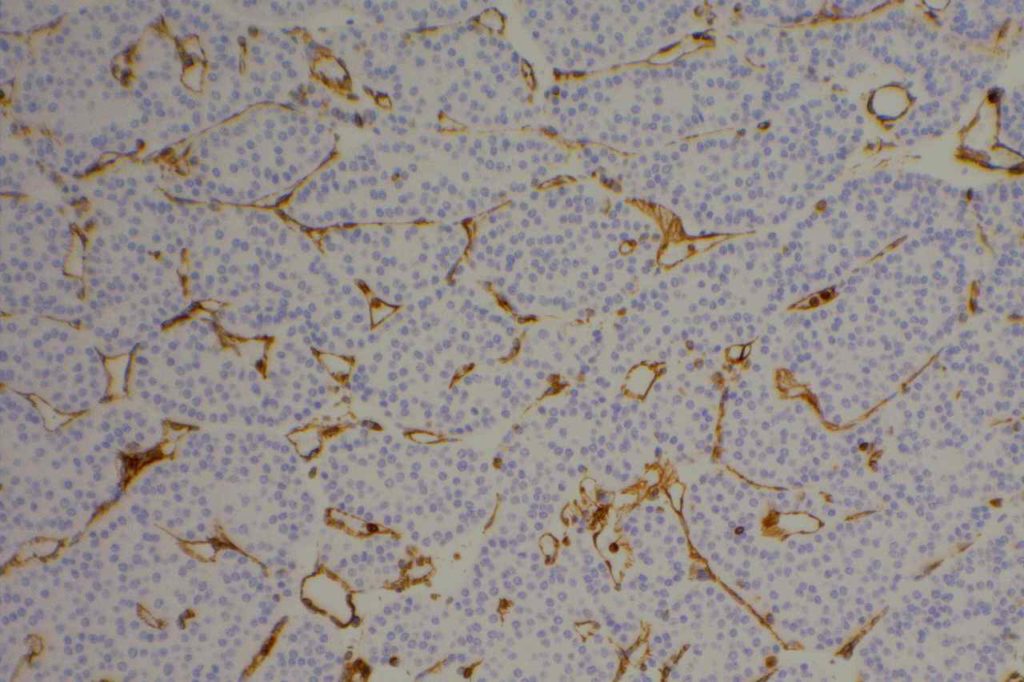
Vimentin - Renal Oncocytoma
Vimentin highlighting vessels around nest of tumor cells in a renal oncocytoma.
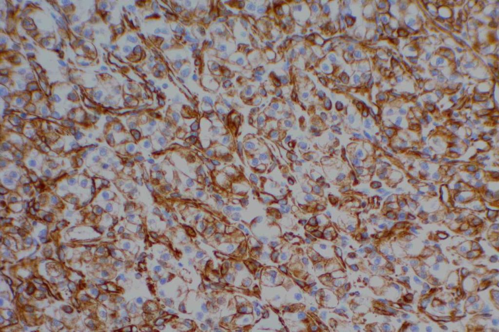
Vimentin - Renal Cell Carcinoma
Vimentin expression in renal cell carcinoma
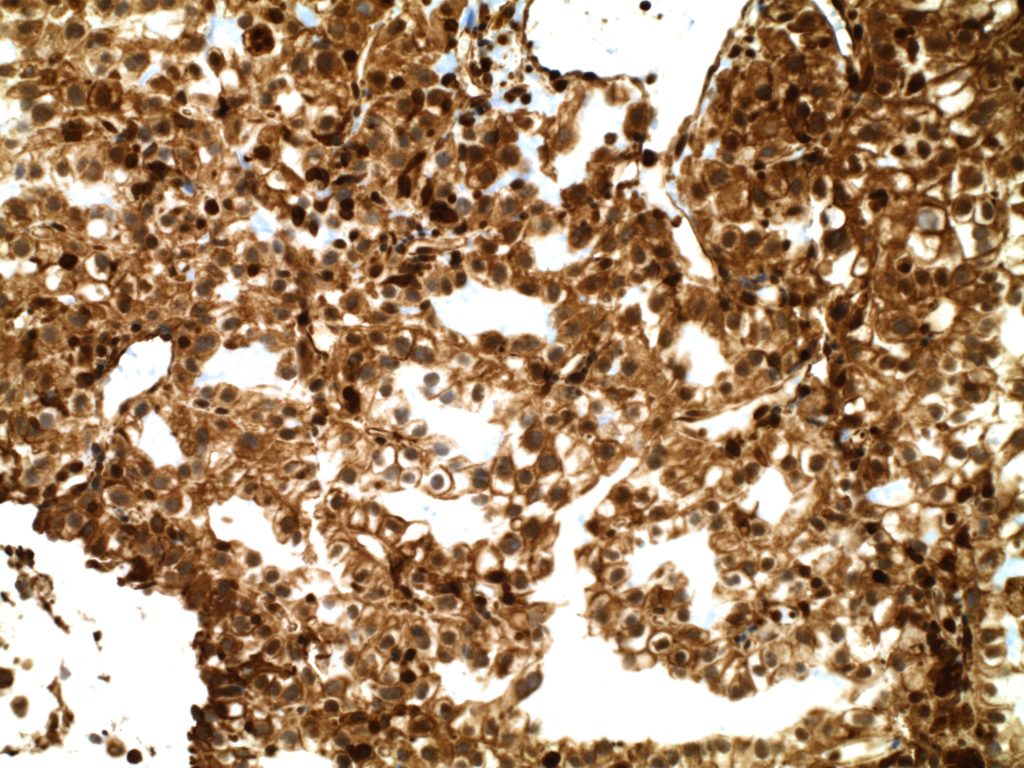
Renal Cell Carcinoma - Vimentin
Vimentin expression in a recurrent renal cell carcinoma
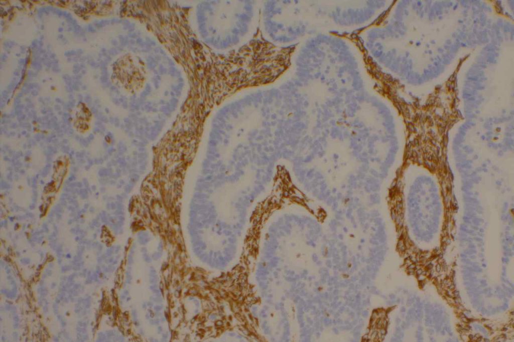
Vimentin - Colon Adenocarcinoma
Vimentin expression expressed in stromal tissue surrounding colon adenocarcinoma epithelium.
References
Kandukuri SR, Lin F, Gui L, Gong Y, Fan F, Chen L, et al. Application of Immunohistochemistry in Undifferentiated Neoplasms: A Practical Approach. Arch Pathol Lab Med. 2017;141: 1014–1032. doi:10.5858/arpa.2016-0518-RA

ZAP-70

General
ZAP-70 (zeta-associated protein-70) is a surrogate marker for the somatic mutation status of immunoglobulin heavy chain (IGHV) in CLL.  Unfortunately, attempts to utilize flow cytometry for this purpose has resulted in unreliable results.  ZAP-70 expression by IHC has been shown to have an increased risk of progression to therapy requirement (3-yr risk 83% vs. 31% for ZAP-70 negative) [Modern Pathology (2010)23,1518-1523].  ZAP-70 expression is not specific to CLL, and is not particularly useful for tumor sub-classification/prognosis outside the setting of CLL.
 
ZAP-70 expression in B-cell lymphoid neoplasms (Carreras, J, et al).
Lymphoid Disorder
No.
ZAP-70 + (%)
Lymphoblastic Lymphoma
7
28%
Chronic Lymphocytic Leukemia
52
65%
Mantle Cell Lymphoma
36
8%
     Classical
28
11%
     Blastoid
8
0%
Follicular Lymphoma
19
0%
Marginal Zone Lymphoma
23
4%
     MALT
11
0%
     Nodal
5
20%
     Splenic
7
0%
Diffuse Large B-Cell Lymphoma
45
2%
Burkitt Lymphoma
29
31%
Hodgkin Lymphoma
14
0%
Stain Interpretation
ZAP-70 is interpreted as negative or positive.  The minimum positive expression is weakly positive( 1+) staining defined as granular cytoplasmic staining with nuclear blush in a majority of tumor cells.  Strong positivity (2+) is  defined as strong expression in a majority of tumor cells.
 
ZAP-70 will also stain T-cells in the background.  Therefore, ZAP-70 should be interpreted with the accompaniment of CD3 and CD20, so that there is clear discernment between tumor and background lymphoid cells.
 
IHC on Peripheral Blood
One of the big problems to identify ZAP-70 expression in CLL is the material available for evaluation.  Most material is based on peripheral blood, and flow cytometry has been difficult to analyze reliably for ZAP-70 expression.  An alternative is to perform PERIPHERAL BLOOD MONONUCLEAR CELL (PBMC) PURIFICATION AND CELL BLOCK PREPARATION as described by Roullet, et. al. in which a cell block is prepared from peripheral blood on which IHC for ZAP-70 can be reliably performed.  Please review Roullet’s article for complete technical details.

Photomicrographs
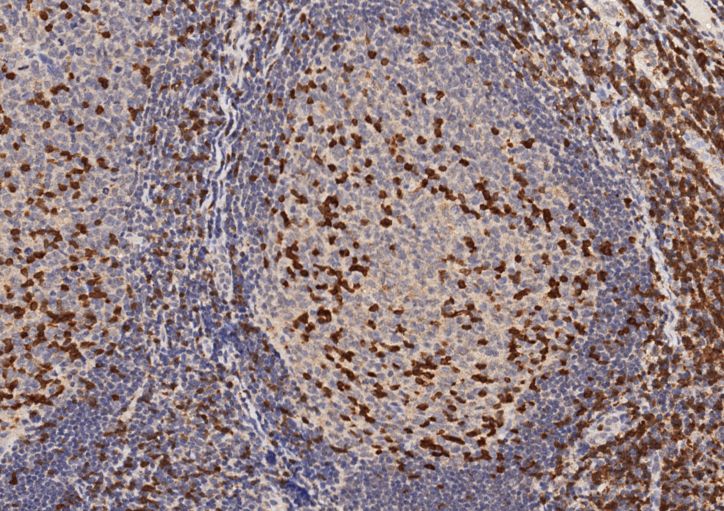
ZAP-70 - Benign Tonsil
ZAP-70 – Benign Tonsil
ZAP-70 CLL/SLL
ZAP-70 weak expression in CLL with an unmutated IgVH gene (poor prognosis). Strong staining in background T-cells.
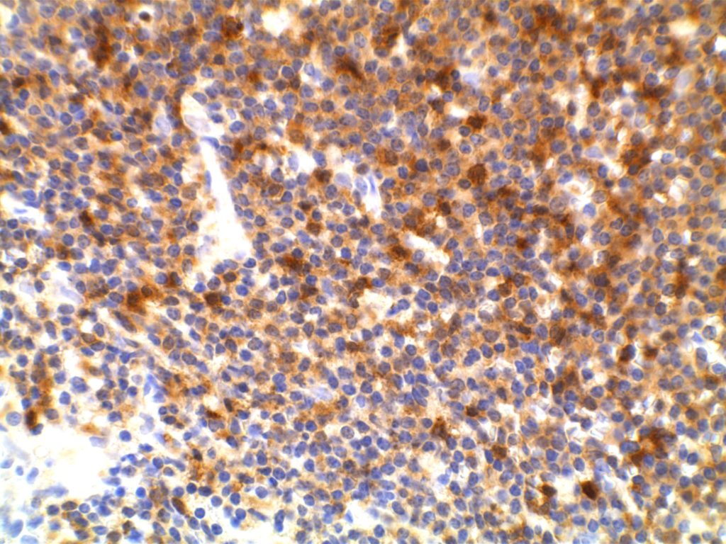
ZAP-70 CLL/SLL
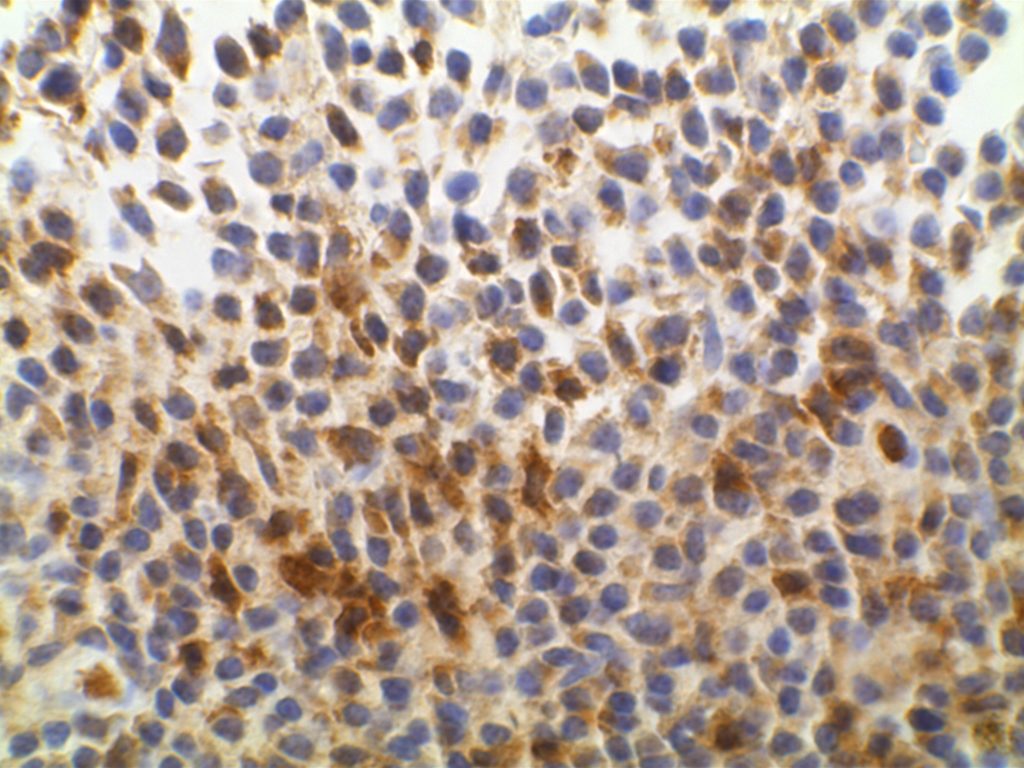
ZAP-70 weak expression in CLL with an unmutated IgVH gene (poor prognosis)

References
Admirand, J. H., Knoblock, R. J., Coombes, K. R., Tam, C., Schlette, E. J., Wierda, W. G., et al. (2010). Immunohistochemical detection of ZAP70 in chronic lymphocytic leukemia predicts immunoglobulin heavy chain gene mutation status and time to progression. Modern Pathology : an Official Journal of the United States and Canadian Academy of Pathology, Inc, 23(11), 1518–1523. doi:10.1038/modpathol.2010.131 
 
Carreras, J., Villamor, N., Colomo, L., Moreno, C., Ramón y Cajal, S., Crespo, M., et al. (2005). Immunohistochemical analysis of ZAP-70 expression in B-cell lymphoid neoplasms. The Journal of Pathology, 205(4), 507–513. doi:10.1002/path.1727
 
Roullet, M., Sargent, R., Pasha, T., Cajiao, I., Elstrom, R., Smith, T., et al. (2007). ZAP70 expression assessed by immunohistochemistry on peripheral blood: a simple prognostic assay for patients with chronic lymphocytic leukemia. Applied Immunohistochemistry & Molecular Morphology : AIMM / Official Publication of the Society for Applied Immunohistochemistry, 15(4), 471–476. doi:10.1097/01.pai.0000213152.41440.34 
 
Crespo, M., Bosch, F., Villamor, N., Bellosillo, B., Colomer, D., Rozman, M., et al. (2003). ZAP-70 expression as a surrogate for immunoglobulin-variable-region mutations in chronic lymphocytic leukemia. The New England Journal of Medicine, 348(18), 1764–1775. doi:10.1056/NEJMoa023143 

WT-1 (Wilms tumor gene product)

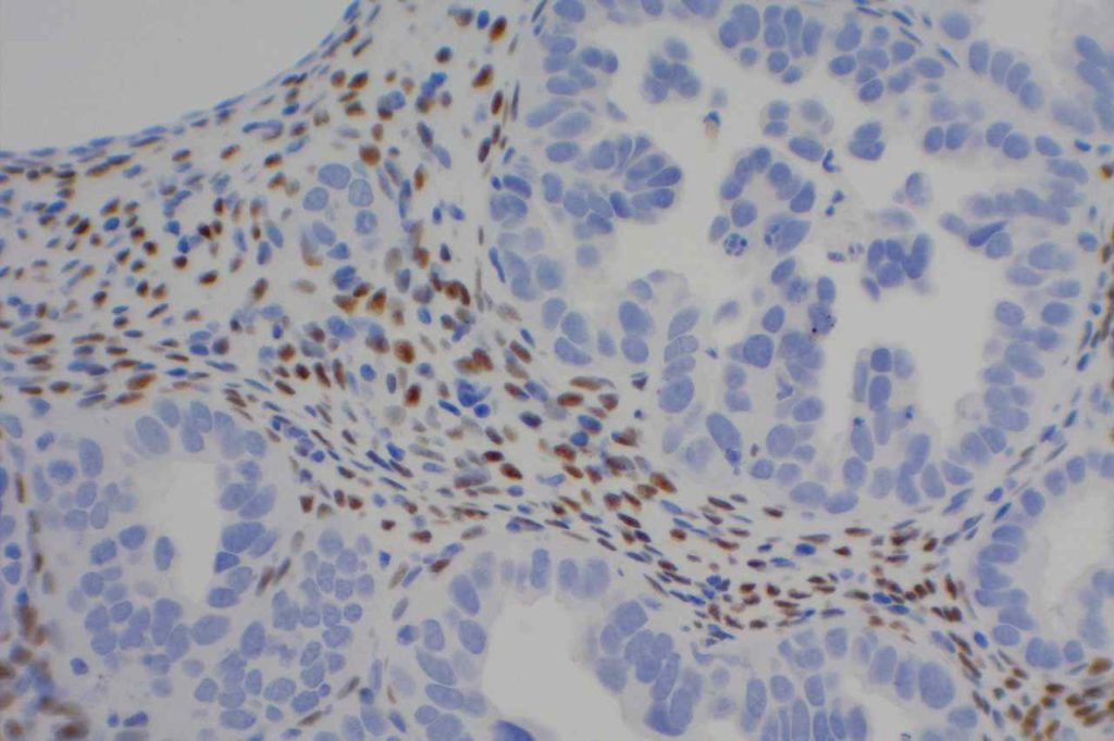
WT-1 (Wilms tumor gene product) is a marker most commonly used to identify OVARIAN SEROUS CARCINOMAS.  It is also commonly used in a panel to differentiate mesothelioma (positive) from adenocarcinoma (negative).  WT-1 has a normal expression distribution in adult tissues, which includes Sertoli cells, ovarian stromal and surface epithelium, and mesothelium.  The 6F-H2 clone has shown better sensitivity compared to other WT-1 antibodies (Ordonez).
 
Tumors / lesions expressing WT-1:
  • Mesothelioma (>75% of epithelial mesotheliomas, usually negative in sarcomatoid variants)
  • Ovarian Serous Carcinomas
  • Wilms tumor
  • Desmoplastic Small Round Cell Tumors
  • Metanephric Adenomas
 
Tumor
Expression (%)
Ovarian Serous Carcinoma
93%
Ovarian Mucinous Carcinoma
0%
Pancreatobiliary Carcinoma
0%
Breast Carcinoma
0%
Lung Carcinoma
0%
Colon Adenocarcinoma
0%
Renal Cell Carcinoma
0%
Thyroid Carcinoma
0%
Prostate Adenocarcinoma
0%
 
WT-1 expression in differentiating mesothelioma from adenocarcinoma (Marchevsky).
Tumor
Expression (%)
Epithelioid Mesothelioma
77%
Sarcomatoid Mesothelioma
13%
Adenocarcnioma
4%
Photomicrographs
WT-1 - Wilms Tumor
WT-1 – Wilms Tumor
WT-1 - Mesothelioma
WT-1 – Mesothelioma
WT-1 - Ovarian Serous Carcinoma
WT-1 – Ovarian Serous Carcinoma
WT-1 - Stromal Cells
WT-1 expression in stromal cells.

 
References:
Muir, T. E., Cheville, J. C., & Lager, D. J. (2001). Metanephric adenoma, nephrogenic rests, and Wilms’ tumor: a histologic and immunophenotypic comparison. The American Journal of Surgical Pathology, 25(10), 1290–1296.
 
Marchevsky, A. M. (2008). Application of immunohistochemistry to the diagnosis of malignant mesothelioma. Archives of Pathology & Laboratory Medicine, 132(3), 397–401.
 
Ordóñez, N. G. (2005). Immunohistochemical diagnosis of epithelioid mesothelioma: an update. Archives of Pathology & Laboratory Medicine, 129(11), 1407–1414.  
  
Hadi, AIMM Annual Meeting, “Carcinomas of Unknown Primary”, presentation, 2011.

Villin

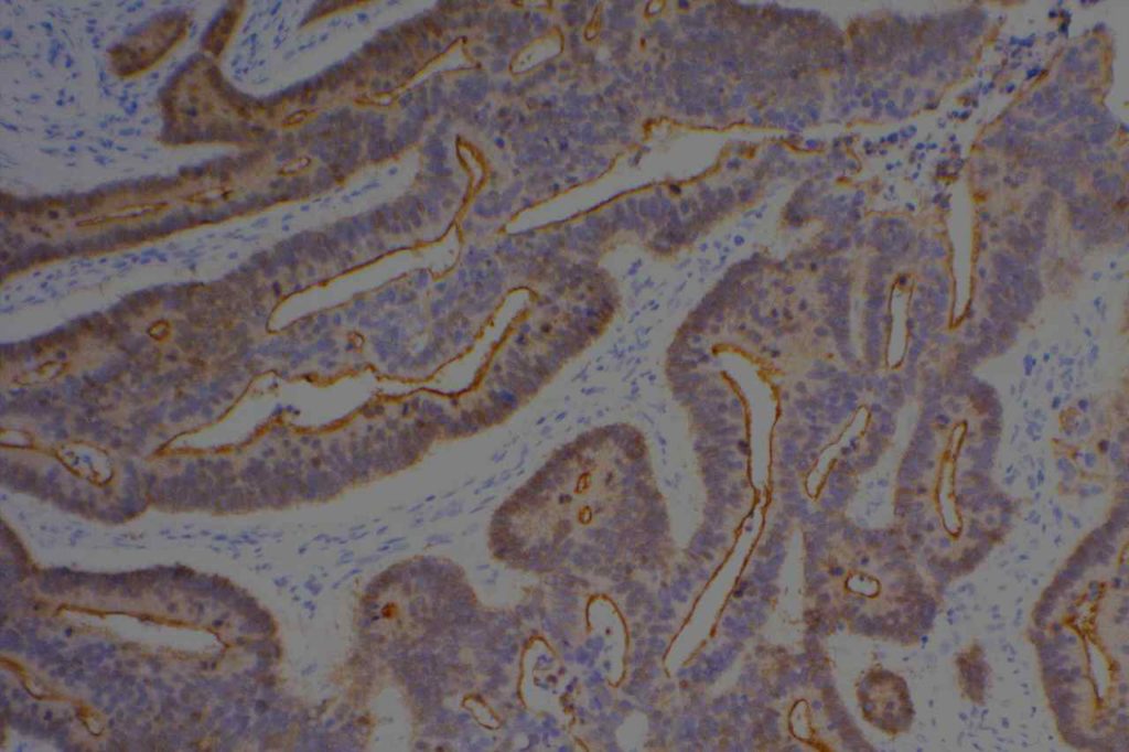
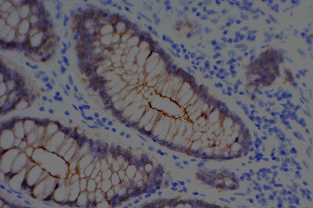
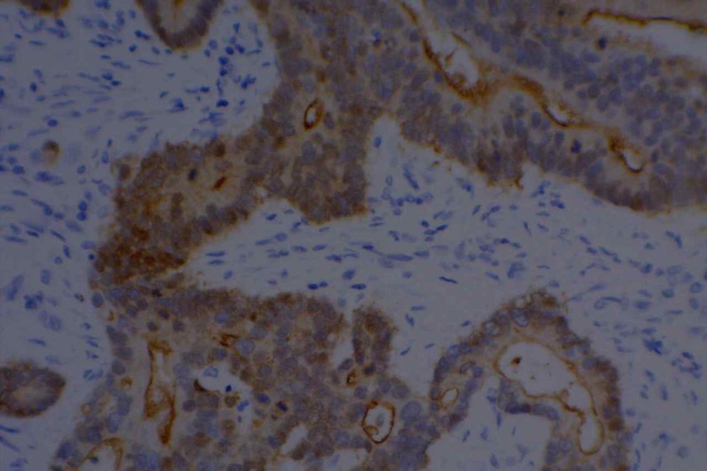
Villin is an actin-binding protein found in microvilli.  It’s expression pattern is cytoplasmic and generally restricted to epithelial cells in the GI tract.  It is a sensitive marker for colon adenocarcinomas, but not entirely specific.  It also stains renal proximal tubules and hepatic bile ducts.
 
CDX-2 and Villin expression in human tumors (2-3+ expression) (Werling, R.W., et. al.)
Tumor Type
No.
CDX2
Villin
G.I. Tract


Colon Adenocarcinoma
75
99%
82% (n=73)
Duodenal Adenoma
10
100%
100%
Gastric Adenocarcinoma
24
70%
42%
Esophageal Adenocarcinoma
9
67%
78%
Pancreatic Adenocarcinoma
22
32%
40%
Cholangiocarcinoma/GB
16
25%
60%
Hepatocellular Carcinoma
12
0%
0%
Carcinoid Tumors
12
42%
34%
Ovary


Mucinous Adenocarcinoma
14
64%
64%
Mucinous Cystadenoma
13
8%
0%
Mucinous Borderline Tumor
4
25%
0%
Non-Mucinous
36
0%
0% (n=31)
Genitourinary Tract


Urothelial Carcinoma
21
0%
0%
Adenocarcinoma
2
100%
100%
Urachal Caricinoma
1
100%
100%
Renal Cell Carcinoma
7
0%
0% (n=3)
Prostate Adenocarcinoma
27
4%
0% (n=24)
Breast Carcinoma
34
0%
0%
Lung (45 primary; 18 met)
63
0%
5%
Adenocarcinoma
11
0%
0%
Squamous Cell Carcinoma
11
0%
0%
Non-Small Cell Carcinoma NOS
33
0
9%
Mucinous Carcinoma
2
0%
0%
Small Cell Carcinoma
4
0%
0%
Mesothelioma
7
0%
0%
Head and Neck


Thyroid
36
3%
0%
Papillary Carcinoma
11
9%
0%
Follicular Adenoma/Carcinoma
25
0%
0%
Salivary Gland
12
0%
0%
Mixed Tumor
6
0%
0%
Low Grade Carcinoma
6
0%
0%
Squamous Cell Carcinoma
13
0%
0% (n=11)
Photomicrographs
Villin - Colon Adenocarcinoma
Villin – Colon Adenocarcinoma
Villin - Colon
Villin – Colon
Villin - Colon Adenocarcinoma
Villin – Colon Adenocarcinoma

References
Gastroenterol94:343-52, 1988. 
 
Hadi, AIMM Annual Meeting, “Carcinomas of Unknown Primary”, presentation, 2011.
 
Werling, R. W., Yaziji, H., Bacchi, C. E., & Gown, A. M. (2003). CDX2, a highly sensitive and specific marker of adenocarcinomas of intestinal origin: an immunohistochemical survey of 476 primary and metastatic carcinomas. The American Journal of Surgical Pathology, 27(3), 303–310.  

TTF-1

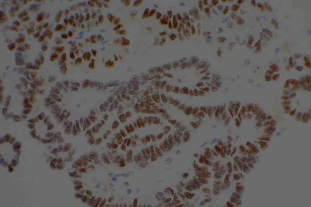
TTF-1 is a nuclear transcription factor that is expressed in thyroid and respiratory epithelium.  It is a useful marker for lung adenocarcinomas and thyroid malignancies.  In the setting of neuroendocrine carcinomas, TTF-1 expression is not specific as to the site of origin, except that Merkel cell carcinomas of the skin do not usually express TTF-1 (some have reported focal expression).  The expression pattern of TTF-1, like other transcription markers, is generally strong and diffuse.  Lower levels of positivity should bring caution to the interpretation.
 
TTF-1 use has become more important to sub-classify lung tumors on small biopsy specimens.  It is generally considered the most sensitive and specific individual marker for lung adenocarcinomas, but is often used in combination with Napsin A to maximize sensitivity and specificity for the determination of primary lung adenocarcinomas.  Current recommendations are to test non-small cell lung carcinomas (non-squamous cell carcinoma) for ALK, ROS-1 and EGFR mutations, as targeted therapies are available (this list is ever expanding and should be verified with current medical literature).

Continue reading TTF-1

Thyroglobulin

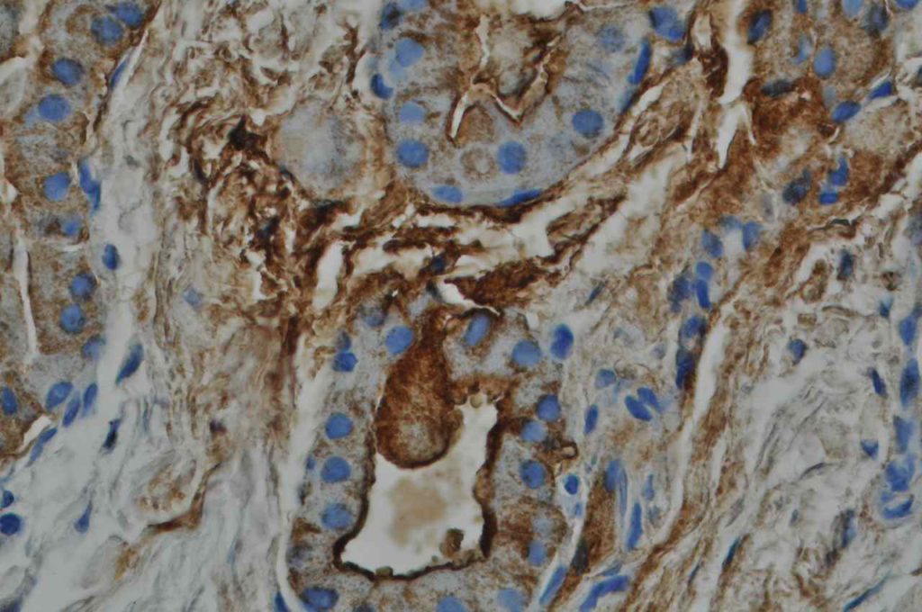
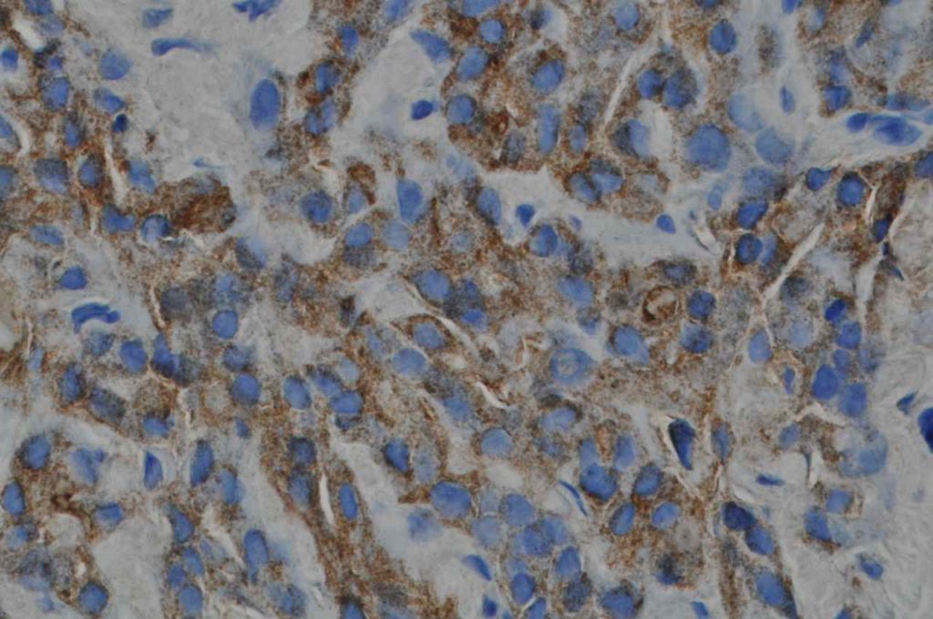
Thyroglobulin is a glycoprotein expressed by thyroid follicular cells.  It is a specific marker for thyroid derivation, and may be useful in the work-up of carcinomas of unknown primary.  It may be less sensitive in less differentiated thyroid tumors, as anapestic thyroid carcinoma is notorious for not staining with about anything.
 
Pitfalls
  • Medullary thyroid carcinoma does not express thyroglobulin.
  • Thyroglobulin can have a lot of artifactual staining, which may cause difficultly diagnosing tumors like medullary carcinoma (expected to be thyroglobulin negative).
  • Dedifferentiated thyroid tumors may not express thyroglobulin.
Photomicrographs
Thyroglobulin - Follicular Carcinoma of Thyroid
Thyroglobulin – Follicular Carcinoma of Thyroid
Thyroglobulin - Follicular Carcinoma of Thyroid
Thyroglobulin – Follicular Carcinoma of Thyroid

References:
 
Fischer, S., & Asa, S. L. (2008). Application of immunohistochemistry to thyroid neoplasms. Archives of Pathology & Laboratory Medicine, 132(3), 359–372.