
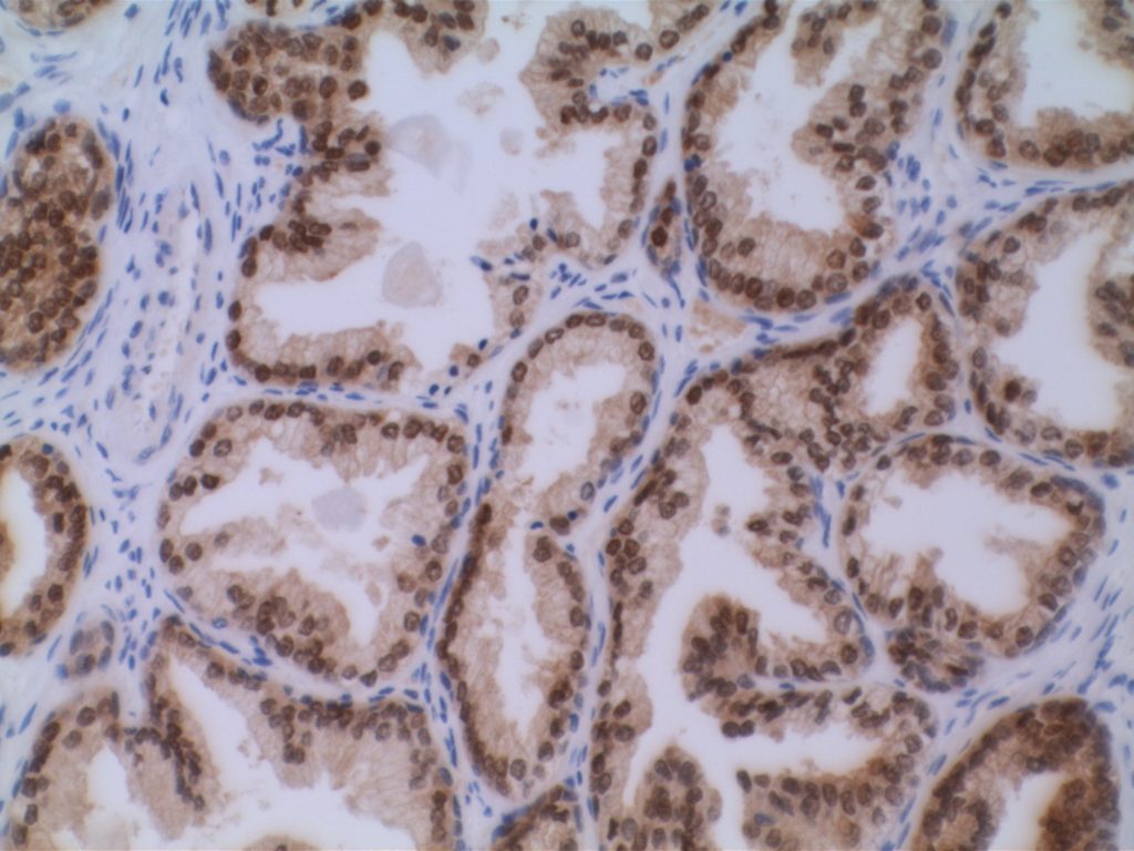
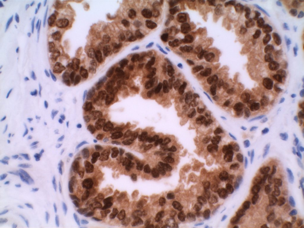
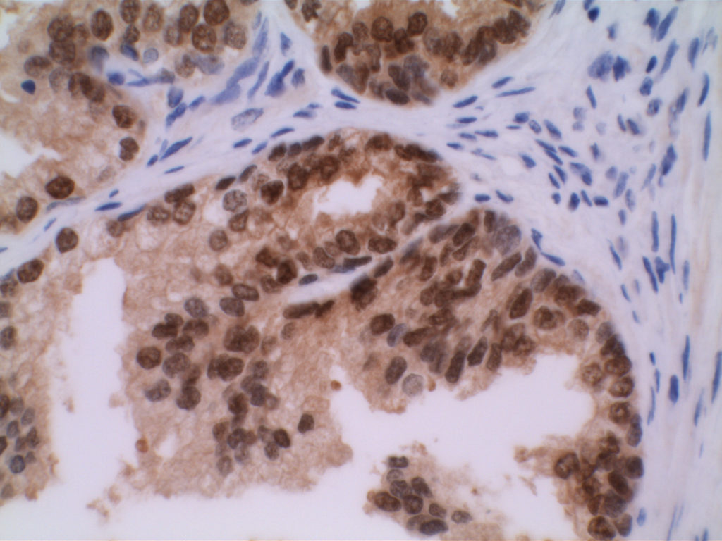
NKX3.1 is a prostate specific homeobox gene (chromosome 8p21), which as an immunohistochemisry IHC marker has shown specificity for prostate epithelium and prostate adenocarcinomas. Normal IHC expression is found in the nucleus of benign and malignant prostate glands. Unfortunately, like most other markers, it is not completely specific. Occasional mucous glands in the lung, testis, and scattered ureteral epithelial cells have shown expression (Brown, et. al.)
Two important points: (1) expression (sensitivity) decreases with hormone refractoriness and advanced stage (metastasis), and (2) expression appears independent of Gleason score (Brown, et al.). In a related article by Mohanty et. al., they showed NKX3.1 to be very sensitive (100%) and specific (100%) in the setting of poorly differentiated prostate adenocarcinomas located in the bladder trigone with the differential diagnosis of urothelial carcinoma.
NKX3.1 may be a potential prognostic marker of progression and hormone refractoriness in low stage patients, but this needs further study (Brown, et. al.). However, due to limited sensitivity in metastatic disease, this marker appears to have limited use in the workup of carcinomas of unknown primary site (CUPS).
Table 1. Expression characteristics of NKX3.1
|
|
Prostate Adenocaricnoma
Expression
|
|
Brown, et. al. (self-made clone)
|
|
|
– Overall (all grades/stages)
|
75% (N=176)
|
|
– T1 disease
|
94% (N=109)
|
|
– T3/4 disease
|
78% (N=27)
|
|
– Metastasis
|
23% (N=40)
|
|
– Hormone Refractory
|
66% (N=128)
|
|
Mohanty, et. al. (polyclonal, Biocare)
|
|
|
– Poorly differentiated prostate adenocarcinoma, bladder neck
|
100% (N=20)
|
Microscopic Images
References
Mohanty SK, Smith SC, Chang E, et al. Evaluation of contemporary prostate and urothelial lineage biomarkers in a consecutive cohort of poorly differentiated bladder neck carcinomas. Am J Clin Pathol. 2014;142(2):173–183. doi:10.1309/AJCPK1OV6IMNPFGL.
Bowen C, Bubendorf L, Voeller HJ, et al. Loss of NKX3.1 expression in human prostate cancers correlates with tumor progression. Cancer Res. 2000;60(21):6111–6115.