Category Archives: Antibody List

PIN-4

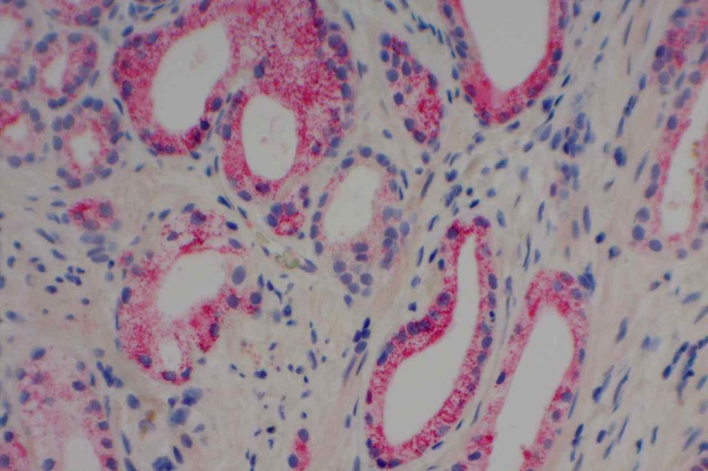
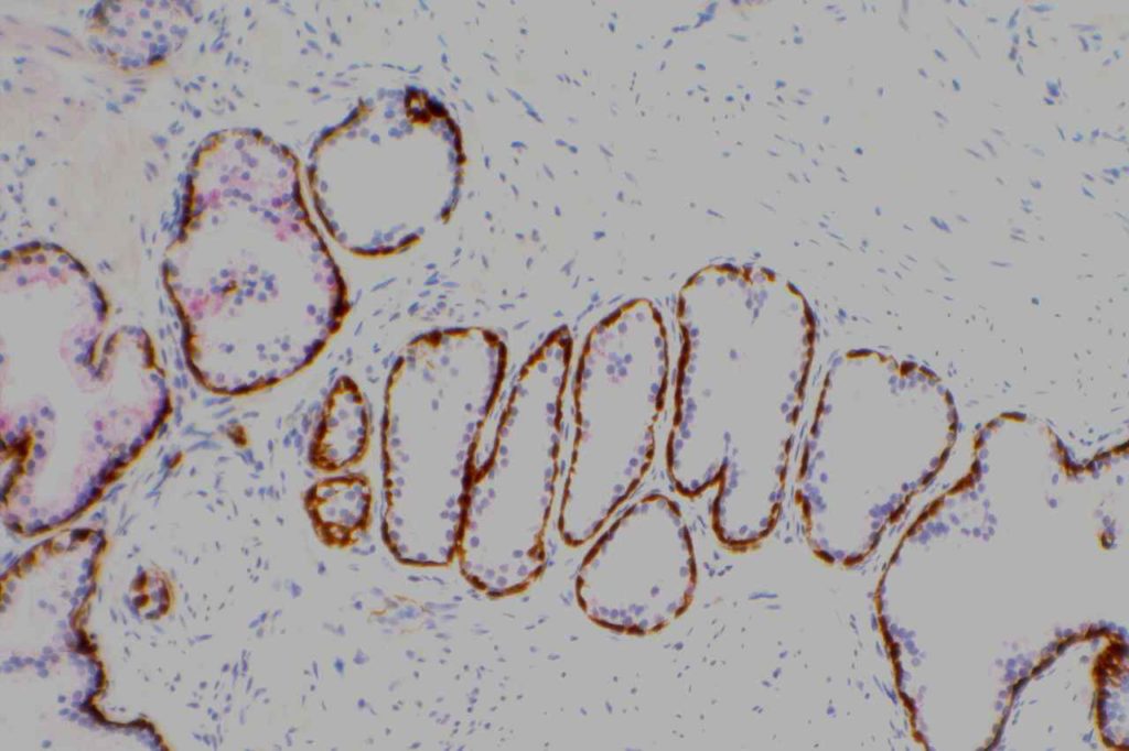
PIN-4 is a cocktail stain used in evaluation of prostate specimens, and contains antibodies CK5, CK14, p63, andP504S.  CK5 and CK14 are high molecular weight keratins, which are expressed in the basal layer of normal prostate glands.  p63 is also expressed in the nuclei of prostate basal epithelial cells.  Therefore, combining p63, CK5 and CK14 allows to increase the sensitivity in identifying a basal epithelial layer in prostate glands.  The absence of a basal epithelial layer around prostate epithelium is characteristic of an invasive prostate adenocarcinoma.
 
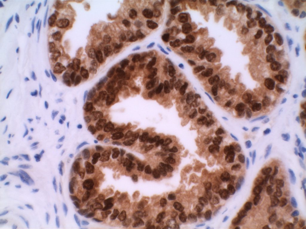
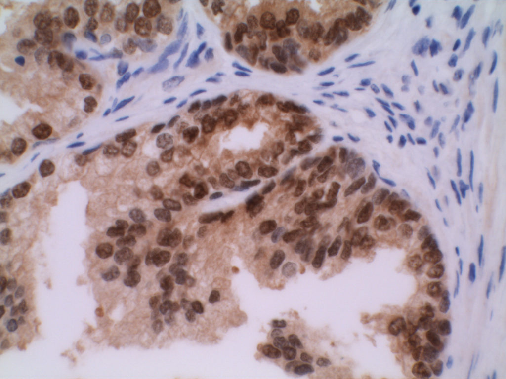
P504S is a racemase (AMACR), which is over-expressed in a significant percentage of prostate adenocarcinomas and high-grade-PIN.  Benign glands do not typically express P504S with rare exceptions.  It is also important to understand that P504S, while demonstrating good specificity for benign versus neoplastic prostate gland epithelium, is NOT specific to only neoplastic prostate epithelium.  Other (non-prostate) benign and neoplastic processes may express P504S (e.g. nephrogenic adenoma).
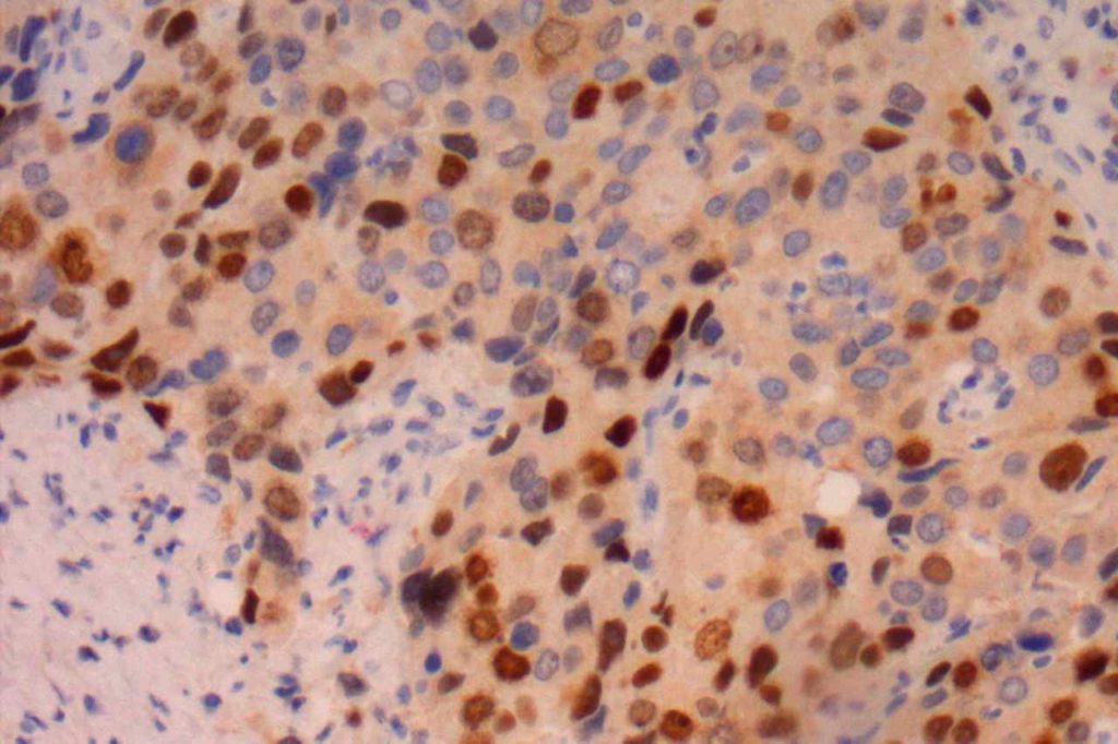
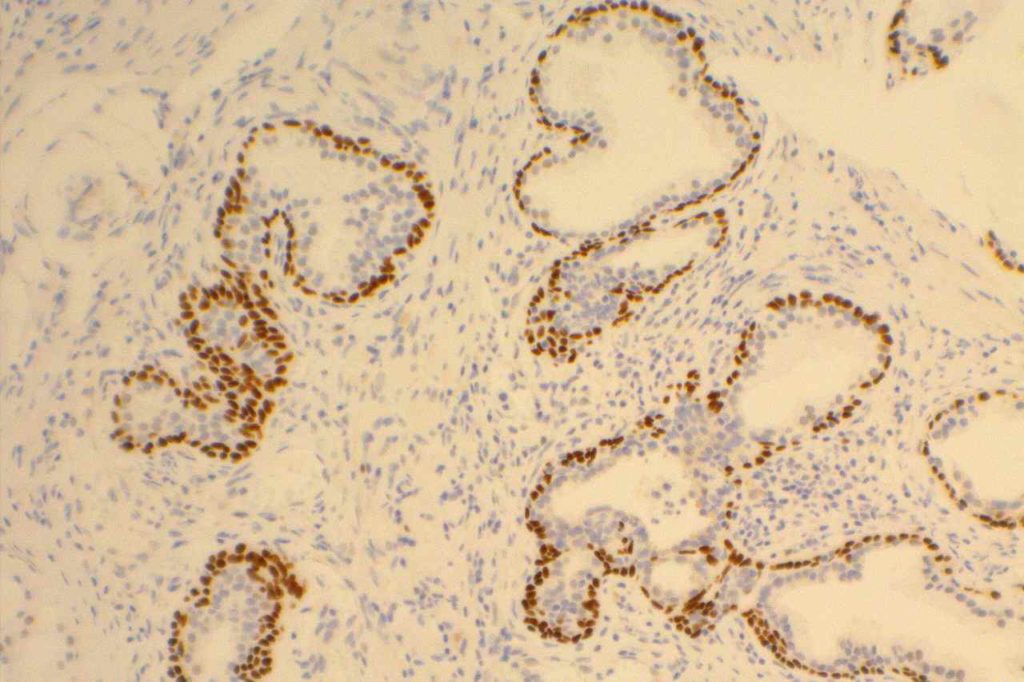
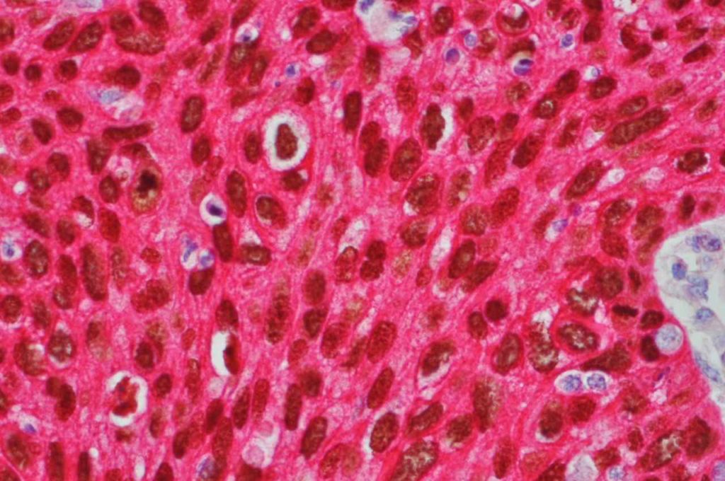
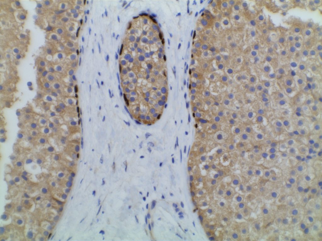
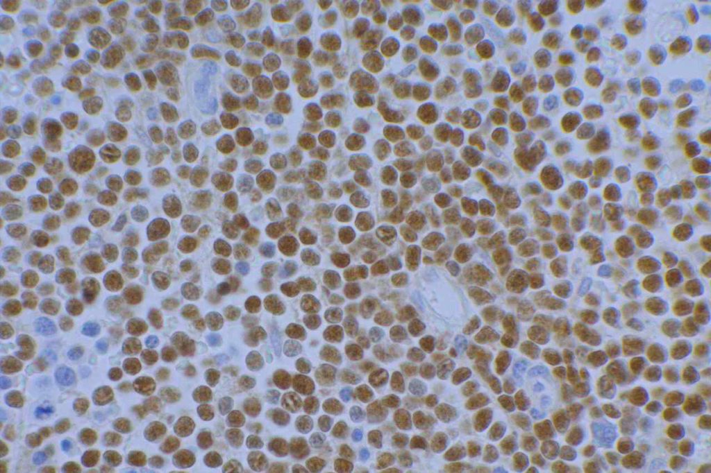
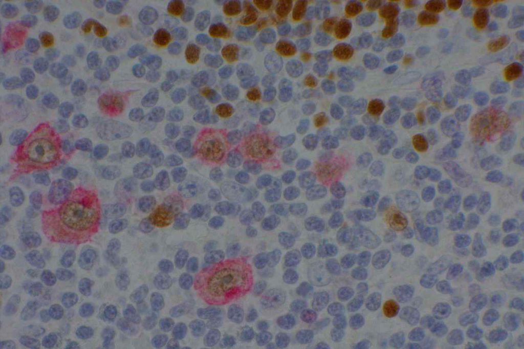
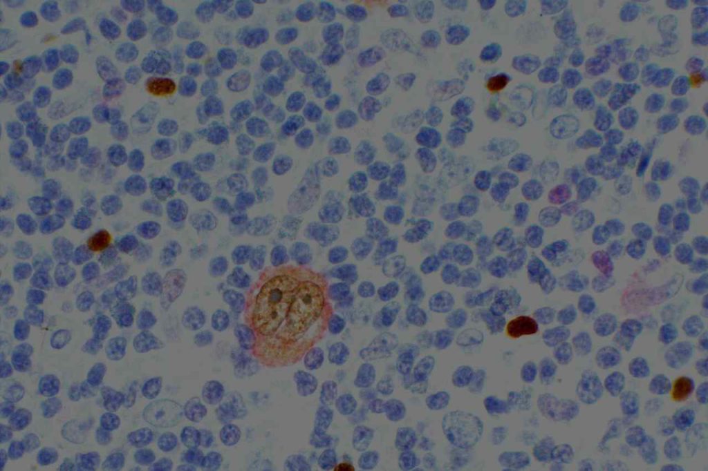
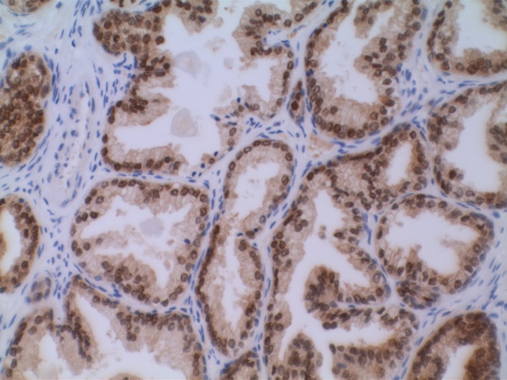
Microscopic Images
PIN-4 Prostate Adenocarcinoma
PIN-4 staining in prostate adenocarcinoma. AMACR (red) expression in neoplastic cells.
PIN-4 Benign Prostate
PIN-4 expression in benign prostate. HMWCK and p63 highlighting basal layer without significant expression of AMACR (red) in the epithelial cells.
H&E Prostate Adenocarcinoma
H&E section of prostate adenocarcinoma to compare with PIN-4 staining.
References
 Yang, X. J., Wu, C.-L., Woda, B. A., Dresser, K., Tretiakova, M., Fanger, G. R., & Jiang, Z. (2002). Expression of alpha-Methylacyl-CoA racemase (P504S) in atypical adenomatous hyperplasia of the prostate. The American Journal of Surgical Pathology, 26(7), 921–925. doi:10.1097/01.PAS.0000017328.13364.17
 
Luo, J., Zha, S., Gage, W. R., Dunn, T. A., Hicks, J. L., Bennett, C. J., et al. (2002). Alpha-methylacyl-CoA racemase: a new molecular marker for prostate cancer. Cancer Research, 62(8), 2220–2226.
 
Zhou, M., Chinnaiyan, A. M., Kleer, C. G., Lucas, P. C., & Rubin, M. A. (2002). Alpha-Methylacyl-CoA racemase: a novel tumor marker over-expressed in several human cancers and their precursor lesions. The American Journal of Surgical Pathology, 26(7), 926–931. doi:10.1097/01.PAS.0000018309.60184.52
 
Tacha, D. E., & Miller, R. T. (2004). Use of p63/P504S monoclonal antibody cocktail in immunohistochemical staining of prostate tissue. Applied Immunohistochemistry & Molecular Morphology : AIMM / Official Publication of the Society for Applied Immunohistochemistry, 12(1), 75–78. 
 
Jiang, Z., Li, C., Fischer, A., Dresser, K., & Woda, B. A. (2005). Using an AMACR (P504S)/34bE12/p63 Cocktail for the Detection of Small Focal Prostate Carcinoma in Needle Biopsy Specimens. American Journal of Clinical Pathology, 123(2), 231–236. doi:10.1309/1G1NK9DBGFNB792L 

p501s (prostein)

p501s is an antibody used to detect the expression of prostein, which is highly sensitive and specific for prostate gland epithelium. Kilos, et. al. described an 87% sensitivity (N=53) of the 10E3-G4-D3 antibody clone for prostate tumors, and negativity in 4,635 non-prostate normal and malignant tissues throughout an extensive diversity of tissue and tumor types.  Detection of expression of prostein appears to be independent of Gleason score.  This is important because PSA and PSAP have struggled to have sensitivity over 50% in high Gleason score prostate carcinomas, which can be diagnostically challenging.

Continue reading p501s (prostein)

p63

p63 is a nuclear transcription marker used in the identification of tumors subtypes and tissue structures such as:  myoepithelial cell layer (breast), basal cell layer (prostate), salivary gland tumors, squamous cell carcinoma (most sensitive marker), skin appendage tumors, and urothelial carcinoma.  As a general rule, transcription markers like p63 show a strong diffuse nuclear expression diagnostically.
Diagnostic Applications
Cell Differentiation
Comments
Myoepithelial Cells
Will stain cells in tumors of myoepithelial origin such as salivary gland tumors.
Urothelial Cells
70-100% of urothelial carcinomas will express p63
Squamous Cells
Squamous Cell Carcinomas from most locations will mark with p63 resulting in a high sensitivity.
Stromal Invasion
p63 can be very helpful to define stromal invasion, as it normally marks myoepithelial cells in breast tissue and basal cells in prostate tissue, which are lost in invasive carcinomas.  Generally recommended to be used with a cytoplasmic myoepithelial marker in breast lesions (e.g. smooth muscle myosin or calponin).
 
p63 expression in selected tumors, may have prognostic implications for Breast, Lung, and Bladder carcinomas.
GU Pathology
p63 is expressed in a high percentage of urothelial carcinomas, but is negative in renal cell carcinomas.  A small percentage of collecting duct carcinomas (14%) have been reported to express p63.  p63 may be helpful in differentiation of renal urothelial carcinoma and poorly differentiated renal cell carcinoma (RCC).
 
Arch Pathol Lab Med-Vol.135, January, 2011
Tumor
p63 Expression (%)
Urothelial Carcinoma
70-100%
Renal Cell Carcinoma
0%
Sarcomatoid Urothelial Carcinoma
50%
Collecting Duct Carcinoma
14%
Placental Sit Trophoblastic Tumor
p63 is expressed in intermediate trophoblasts associated with placental site nodules, but is negative in placental site trophoblastic tumors (epithelioid trohphoblastic tumors also express p63) [Mittal, et. al.].
Undifferentiated Carcinomas
CK5/6 co-expression with p63 is a sensitive (77%) and specific (96%) marker for squamous cell carcinoma.   p63 appears to be very specific for squamous and urothelial origin.  Only rare other non-squamous cell carcinomas show >50% expression of p63. [Kaufman, O., et. al.]
Small Cell Carcinoma
Small cell carcinomas express p63 in approximately 77% of cases (n=14). [Au, NH, et. al.]  This could be a potential pitfall if one is considering a poorly differentiated squamous cell carcinoma and not a poorly differentiated neuroendocrine carcinoma. 
Microscopic Images
p63 - Lung Adenocarcinoma
Variable subset nuclear expression of p63 in a lung adenocarcinoma, which is characteristic in a subset of cases. SCC expression should be diffuse and consistent.
p63 - Benign Prostate
p63 highlighting the basal layer of benign prostate gland epithelium.
p63-CK5 (double stain) lung squamous cell carcinoma
Co-expression of CK5 (red) and p63 (DAB) in a lung squamous cell carcinoma. Note the diffuse consistent expression of p63.
PIN-4 Benign Prostate
PIN-4 expression in benign prostate. HMWCK and p63 highlighting basal layer without significant expression of AMACR (red) in the epithelial cells.
p63 - LCIS
p63 highlighting nuclear expression in the myoepithelial cell layer surrounding LCIS. Best used in combination with another cytoplasmic myoepithelial marker (e.g. smooth muscle myosin, calponin, etc.).

References
Hadi, AIMM Annual Meeting, “The Thirty Most Important Antibodies”, presentation, 2011.
 
Truong, LD, et. al. “Immunohistochemical Diagnosis of Renal Neoplasms.” Archives of Pathology and Laboratory Medicine. 2011;135:92-109.
 
Mittal, K, et. al. “Aplplication of Immunohistochemistry to Gynecologic Pathology.”  Archives of Pathology and Laboratory Medicine. Vol. 132, March 2008.
 
Kaufmann, O., et. al. “Value of p63 and CK 5/6 as IHC Markers for the Differential Dx. of Poorly Differentiated and Undifferentiated Carcinomas.” American Journal of Clinical Pathology. 2001;116:823-830
 
Au NHC, Gown AM, Cheang M, et al. P63 expression in lung carcinoma: a tissue microarray study of 408 cases. Appl Immunohistochem Mol Morphol. 2004;12(3):240–247.
 
Zhao L, Yang X, Khan A, Kandil D. Diagnostic role of immunohistochemistry in the evaluation of breast pathology specimens. Arch Pathol Lab Med. 2014;138: 16–24. doi:10.5858/arpa.2012-0440-RA

p40

p40 is a member of the p53 family, and is an isoform of p63.  It is also known as delta-Np63.  p63 can have different isoforms, and p40 reacts with truncated dominant isoforms of the p63 gene. (Brown, et. al.; Bishop, et. al.; and Ross, et. al.)  p40 performs in a similar fashion to p63, but with so-called better specificity. (Bishop)
 
p40 appears very sensitive for lung squamous cell carcinomas (>90%) and urothelial carcinomas (>88%) from multiple studies. (Gaily, et. al. and Bishop, et. al.)  Rarely (<5%) breast, biliary tract/pancreas, endometrium, and prostate showed reactivity, and evaluation of multiple cases (40-100 each) of renal cell carcinoma, thyroid, hempatocellular carcinoma, colon adenocarcinoma, stomach adenocarcinoma, and ovarian epithelium failed to show any reactivity (negative). (Brown, et. al.)
 
General Comments
Ross, et. al. claims superiority of p40 over p63 because only 3% of lung adenocarcinomas showed any positivity with p40 compared to 31% with p63.  To the inexperienced pathologist using p63 in lung cancer subcategorization, partial positivity with p63 may lead to comments like “focal squamous differentiation” or “cannot exclude a component of squamous cell carcinoma”.  These comments should be avoided, if possible.  Often oncologists will not give the patient Avastin, which is an important therapy option, if there is any evidence of squamous differentiation due to the risk of pulmonary hemorrhage.
 
Where some pathologists find variable subset nuclear expression with p63 in lung adenocarcinomas, others find such expression pattern as re-inforcing support for the diagnosis of lung adenocarcinoma.  It is important to remember a basic tenant (general rule) in interpreting nuclear transcription markers like p40 and p63; expression should be consistent and diffuse for the tumor cells of interest.  If expression does not meet this threshold, then one may be wise to consider additional markers in a panel to confirm differentiation.   Pathologists must NOT think of stains as positive and negative, but in the context of the pattern of expression, and what that means.

 
References:
Brown AF, Sirohi D, Fukuoka J, Cagle PT, Policarpio-Nicolas M, et al. (2013) Tissue-preserving antibody cocktails to differentiate primary squamous cell carcinoma, adenocarcinoma, and small cell carcinoma of lung. Arch Pathol Lab Med 137: 1274–1281. doi:10.5858/arpa.2012-0635-OA.
 
Bishop JA, Teruya-Feldstein J, Westra WH, Pelosi G, Travis WD, et al. (2012) p40 (ΔNp63) is superior to p63 for the diagnosis of pulmonary squamous cell carcinoma. Mod Pathol 25: 405–415. doi:10.1038/modpathol.2011.173.
 
Rossi G, Pelosi G, Barbareschi M, Graziano P, Cavazza A, et al. (2013) Subtyping Non-Small Cell Lung Cancer: Relevant Issues and Operative Recommendations for the Best Pathology Practice. International Journal of Surgical Pathology 21: 326–336. doi:10.1177/1066896913489346.
 
Gailey MP, Bellizzi AM (2013) Immunohistochemistry for the novel markers glypican 3, PAX8, and p40 (ΔNp63) in squamous cell and urothelial carcinoma. Am J Clin Pathol 140: 872–880. doi:10.1309/AJCP4NSKW5TLGTDS.

PAX-8

PAX-8 along with PAX-2 are transcription factors (nuclear expression) important in the development of Mullein system, kidney, and thyroid.  They both are expressed in normal kidney and most renal neoplasms.  PAX-8 expression may also be seen in carcinomas of the lower urinary tract and nephrogenic adenomas.  In the study by Ozacan et. al., PAX-8 had better performance characteristics than PAX-2.
 
PAX-8 is not expressed in cancers from rectum, pituitary, adrenal, colon, prostate, breast, lung, stomach, liver, soft tissue, melanoma, and lymphoma.  The rabbit monoclonal PAX-8 antibody shows staining in lymph nodes, pancreas, and stomach/colon neuroendocrine cells.  The mouse PAX-8 antibody clone BC12 does not appear to have expression in lymph nodes, pancreas and stomach/colon neuroendocrine cells. (Tacha, D., et. al.)  In an experience by Ortiz-Rey, et al., they found PAX-8 to be helpful in differentiated seminal vesicle epithelium from prostate epithelium in needle core biopsies.  Sometimes this can be difficult to differentiate from carcinoma, and could be helpful in a panel to differentiate epithelial types.
 
Ozcan, A., et. al.
Tumor Type
No.
PAX-2
PAX-8
Renal Cell Carcinoma


     Clear Cell
95
95%
97%
     Papillary
38
76%
100%
     Chromophobe
25
56%
88%
     Collecting Duct
7
43%
71%
     Acquired Cystic Kidney
     Disease-related RCC
8
75%
100%
     Oncocytoma
13
54%
85%
 
Galley, M.P., et. al.
Tumor Type
PAX-8 (%)
Uterine Cervix SCC
3%
Urothelial Carcinoma
10%
 
Normal Tissue Expression for PAX-8  (Chan, J.K.C.)
Tissue
Expression Pattern
Thyroid
Follicular epithelium
Kidney
Tubular epithelium and Bowman capsule lining cells
Female Genital Tract
Ovary – inclusion cysts, rete ovarii, not surface mesothelium
Fallopian Tube – epithelium
Uterus – epithelium
Endocervix – epithelium (weak to moderate staining)
Male Genital Tract
Epithelium from tete testis to ejaculatory duct
Thymus
Thymic epithelium (weak staining)
Pancreas
Islet cells (rabbit polyclonal ab, mouse clone BC12 does not appear to cross react)
Lymphocytes
Subset of lymphoid cells, thought to be due to cross-reactivity with PAX-5 (rabbit polyclonal ab, mouse clone BC12 does not appear to cross react)
 
References:
Shen, SS. “Role of Immunohistochemistry in Diagnosing Renal Neoplams: When Is It Really Useful?”Arch Pathol Lab Med, Vol. 136, April 2012.  pp. 410-417. 
 
Al-Ghawi, H., Asojo, O. A., Truong, L. D., Ro, J. Y., Ayala, A. G., & Zhai, Q. J. (2010). Application of Immunohistochemistry to the Diagnosis of Kidney Tumors. Pathology Case Reviews, 15(1), 25–34. doi:10.1097/PCR.0b013e3181d51c70 
 
Ozcan, A., la Roza, de, G., Ro, J. Y., Shen, S. S., & Truong, L. D. (2012). PAX2 and PAX8 expression in primary and metastatic renal tumors: a comprehensive comparison. Archives of Pathology & Laboratory Medicine, 136(12), 1541–1551. doi:10.5858/arpa.2012-0072-OA 
 
Chan, J. K. C. (2013). Newly Available Antibodies With Practical Applications in Surgical Pathology. International Journal of Surgical Pathology, 21(6), 553–572. doi:10.1177/1066896913507601 
 
Gailey, M. P., & Bellizzi, A. M. (2013). Immunohistochemistry for the novel markers glypican 3, PAX8, and p40 (ΔNp63) in squamous cell and urothelial carcinoma. American Journal of Clinical Pathology, 140(6), 872–880. doi:10.1309/AJCP4NSKW5TLGTDS 
 
Tacha, D., Qi, W., Zhou, D., Bremer, R., & Cheng, L. (2013). PAX8 mouse monoclonal antibody [BC12] recognizes a restricted epitope and is highly sensitive in renal cell and ovarian cancers but does not cross-react with b cells and tumors of pancreatic origin. Applied Immunohistochemistry & Molecular Morphology : AIMM / Official Publication of the Society for Applied Immunohistochemistry, 21(1), 59–63. doi:10.1097/PAI.0b013e318257cc1c 
 
Ortiz-Rey – PAX8 AIMM editorial

PAX-5

PAX-5 (B-cell specific activator protein: BSAP) is a nuclear transcription marker that is normally found onPre-B and mature B-lymphocytes (not plasma cells).  Expression is seen in Pre-B ALL, B-cell lymphomas and classical Hodgkin lymphoma (>90%, dimmer expression compared to background B-cells), but not plasmacytomas.  It should be noted that some cases of small cell carcinoma (73%), Merkel cell carcinoma (93%), and alveolar rhabdomyosarcoma may show PAX-5 expression.
 
PAX-5 is particularly useful in evaluating cases of B-cell lymphomas, which have been treated with rituximab.  At some point during treatment, approximately 20% of these cases treated with rituximab therapy may lose expression of CD20 (flow cytometry appears to be more sensitive in detecting down regulation of CD20 with rituximab therapy).  PAX-5 is helpful in identifying such B-cell with loss of CD20 expression.  This is important clinically, because such cases may not respond to additional rituximab therapy.  It is important to note that there may be down regulation of other B-cell markers with rituximab therapy.  In a relatively small set of cases by Chu, PG, et al, PAX-5 expression was present in 21 of 24 cases of CD20 negative mature B-cell neoplasms treated with rituximab.  Therefore, a panel of B-cell markers may be needed in cases treated with rituximab.
PAX-5 Expression

Microscopic Images
PAX-5 - B-Cell ALL
PAX-5 expression in B-cell acute lymphoblastic lymphoma.
CD30-PAX5 double stain in classical Hodgkin lymphoma
CD30-PAX-5 double stain in CHL. Note the dim PAX-5 expression in the Hodgkin cells, which are also highlighted with CD30 (red).
CD30-PAX5 double stain in classical Hodgkin lymphoma
CD30-PAX-5 double stain in CHL. Note the dim PAX-5 expression in the Hodgkin cells, which are also highlighted with CD30 (red).

 References
Hadi Yaziji, AIMM Annual Meeting, “New Antibodies in Diagnostic Immunohistochemistry”, presentation, 2010.
 
AJSP 2005;29:687
 
Bone Marrow IHC.  Torlakovic, EE, et. al. American Society for Clinical Pathology Pathology Press © 2009.  pp. 221.
 
Chu, P. G., Loera, S., Huang, Q., & Weiss, L. M. (2006). Lineage Determination of CD20- B-Cell Neoplasms: An Immunohistochemical Study. American Journal of Clinical Pathology, 126(4), 534–544. doi:10.1309/3WG32YRAMQ7RB9D4 

PAX-2

PAX-2 is nuclear marker that is expressed in early renal organogenesis.  It appears to be a sensitive marker of renal cell carcinoma.   However, caution should be applied in the setting of papillary neoplasms (particularly uterine and ovarian serous papillary carcinomas).
 
PAX-8 along with PAX-2 are transcription factors (nuclear expression) important in the development of mullein, kidney, and other organs.  They both are expressed in normal kidney and most renal neoplasms.  However, PAX-2 is typically negative in thyroid neoplasms.
 
Ozcan, A., et. al. (PAX-2 and PA-8 expression in various renal tumors)
Tumor Type
No.
PAX-2
PAX-8
Renal Cell Carcinoma


     Clear Cell
95
95%
97%
     Papillary
38
76%
100%
     Chromophobe
25
56%
88%
     Collecting Duct
7
43%
71%
     Acquired Cystic Kidney
     Disease-related RCC
8
75%
100%
     Oncocytoma
13
54%
85%
 
Sharma, S.G., et. al.  (PAX-2 and RCCma expression in various papillary tumors)
Tumor Type
No.
PAX-2
RCCma
Papillary RCC
24
67%
96%
Ovarian Papillary Serous Carcinoma
10
40%
80%
Uterine Papillary Serous Carcinoma
9
56%
44%
Papillary Thyroid Carcinoma
9
0%
100%
Papillary Urothelial Carcinoma
10
0%
10%
Intraductal Papillary Mucinous Tumor of the Pancreas
2
0%
50%
Chroroid Plexus Papilloma
1
0%
100%
Pituitary Adenoma with Papillary Features
1
0%
100%
Lung Adenocarcinoma with Papillary Features
2
0%
50%
 
References:
Hadi, AIMM Annual Meeting, “Carcinomas of Unknown Primary”, presentation, 2011.
 
Mazal PR et al.  Mod Pathol 2005;18:535-540.
 
Shen, SS. “Role of Immunohistochemistry in Diagnosing Renal Neoplams: When Is It Really Useful?”Arch Pathol Lab Med, Vol. 136, April 2012.  pp. 410-417. 
 
Ozcan, A., la Roza, de, G., Ro, J. Y., Shen, S. S., & Truong, L. D. (2012). PAX2 and PAX8 expression in primary and metastatic renal tumors: a comprehensive comparison. Archives of Pathology & Laboratory Medicine, 136(12), 1541–1551. doi:10.5858/arpa.2012-0072-OA  
 
Sharma, S. G., Gokden, M., McKenney, J. K., Phan, D. C., Cox, R. M., Kelly, T., & Gokden, N. (2010). The utility of PAX-2 and renal cell carcinoma marker immunohistochemistry in distinguishing papillary renal cell carcinoma from nonrenal cell neoplasms with papillary features. Applied Immunohistochemistry & Molecular Morphology : AIMM / Official Publication of the Society for Applied Immunohistochemistry, 18(6), 494–498. doi:10.1097/PAI.0b013e3181e78ff8 

NKX3.1

NKX3.1 is a prostate specific homeobox gene (chromosome 8p21), which as an immunohistochemisry IHC marker has shown specificity for prostate epithelium and prostate adenocarcinomas.  Normal IHC expression is found in the nucleus of benign and malignant prostate glands.  Unfortunately, like most other markers, it is not completely specific.  Occasional mucous glands in the lung, testis, and scattered ureteral epithelial cells have shown expression (Brown, et. al.)
 
Two important points: (1) expression (sensitivity) decreases with hormone refractoriness and advanced stage (metastasis), and (2) expression appears independent of Gleason score (Brown, et al.).  In a related article by Mohanty et. al., they showed NKX3.1 to be very sensitive (100%) and specific (100%) in the setting of poorly differentiated prostate adenocarcinomas located in the bladder trigone with the differential diagnosis of urothelial carcinoma.
 
NKX3.1 may be a potential prognostic marker of progression and hormone refractoriness in low stage patients, but this needs further study (Brown, et. al.).  However, due to limited sensitivity in metastatic disease, this marker appears to have limited use in the workup of carcinomas of unknown primary site (CUPS).
 
Table 1.  Expression characteristics of NKX3.1
 
Prostate Adenocaricnoma
Expression
Brown, et. al. (self-made clone)
 
–  Overall (all grades/stages)
75% (N=176)
–  T1 disease
94% (N=109)
–  T3/4 disease
78% (N=27)
–  Metastasis
23% (N=40)
–  Hormone Refractory
66% (N=128)
Mohanty, et. al. (polyclonal, Biocare)
 
–  Poorly differentiated prostate adenocarcinoma, bladder neck
100% (N=20)
Microscopic Images
NKX3.1 - Prostate
NKX3.1 expression in prostate gland tissue.
NKX3.1 - Prostate
NKX3.1 expression in prostate gland tissue.
NKX3.1
NKX3.1 expression in prostate gland tissue.

References
 
Mohanty SK, Smith SC, Chang E, et al. Evaluation of contemporary prostate and urothelial lineage biomarkers in a consecutive cohort of poorly differentiated bladder neck carcinomas. Am J Clin Pathol. 2014;142(2):173–183. doi:10.1309/AJCPK1OV6IMNPFGL.
 
 Bowen C, Bubendorf L, Voeller HJ, et al. Loss of NKX3.1 expression in human prostate cancers correlates with tumor progression. Cancer Res. 2000;60(21):6111–6115. 

Napsin A

Napsin A is an aspartic proteinase, and is normally found in lung pneumocytes and alveolar macrophages, pancreatic acini and ducts, and renal tubules.  Neoplastic tissue which stains with Napsin A includes:  lung adencarcinoma, renal cell carcinoma (clear cell and papillary), thyroid (papillary) carcinoma, and ovarian clear cell carcinoma.
 
Napsin A is primary used in combination with TTF-1 in identifying lung adencarcinomas (TTF-1 appears to be a little more sensitive in poorly differentiated tumors).  While approximately 97% of Napsin A expressing lung adenocarcinomas express TTF-1, up to 13% of TTF-1 negative cases express Napsin A.  The more recent description of Napkin A expression in 100% of ovarian clear cell carcinomas by Kandalaft, et al. further emphasizes the importance of using an appropriate panel of IHC stains for a given differential diagnosis to obtain optimal sensitivity and specificity.
 
In the setting of ovarian tumors (Kandalaft, et al.), Napsin A was 100% sensitive for ovarian clear cell carcinoma.  Endometrioid carcinomas of the ovary had focal Napsin A expression in 10% of cases, but no cases of high grade serous carcinoma or serous borderline tumor demonstrated expression.

Continue reading Napsin A