Category Archives: Antibody List

GATA-3

GATA-3 is a member of a subfamily of zinc finger transcription proteins, which has been found to be highly expressed in breast (>90%) (especially lunimal A breast carcinomas) and urothelial carcinomas (>80%).  Current evidence suggests that this marker has a better balance of sensitivity and specificity for breast carcinoma than GCDFP-15 and mamaglobin (MGB).  Especially, in the setting of ER-negative tumors.  GATA-3 use should be considered as part of IHC panels in the setting of carcinoma of unknown primary site. (Lin, et al)  Mccluskey, et al. found high GATA-3 expression to be associated with favorable survival and relapse free course in advanced cases.
 
GATA-3 may also have a role in the evaluation of possible primary bladder tumors.  As with most IHC markers, there are very few “silver bullets,” and interpretation should take into consideration the clinical-radiologic context along with the known performance characteristics of the antibody.  
 
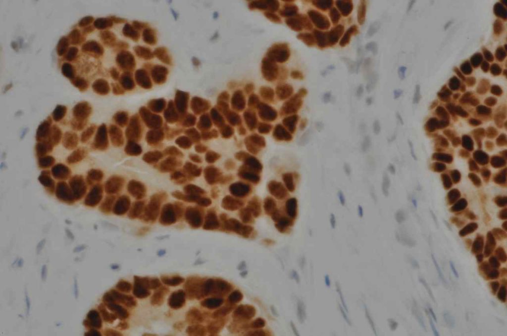
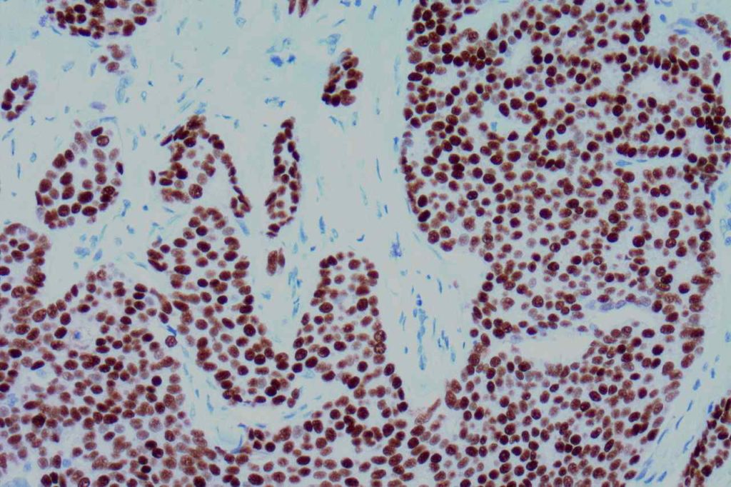
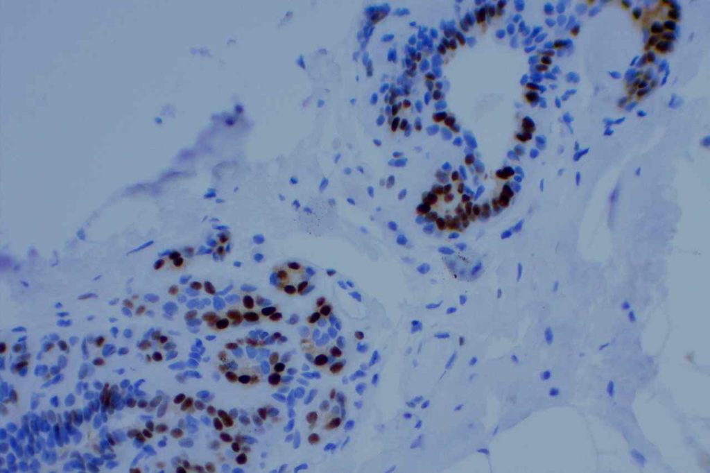
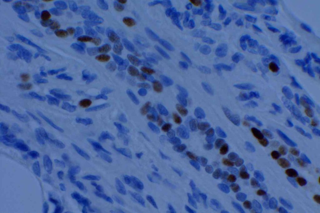
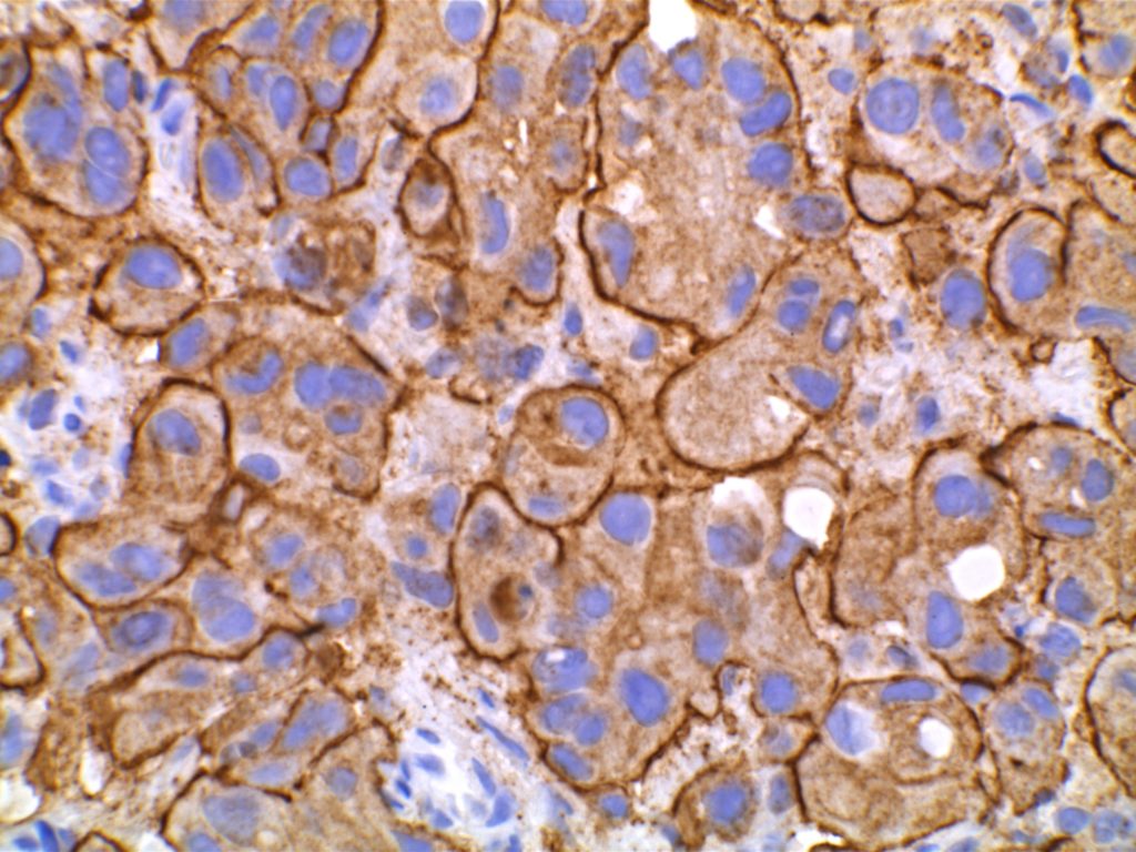
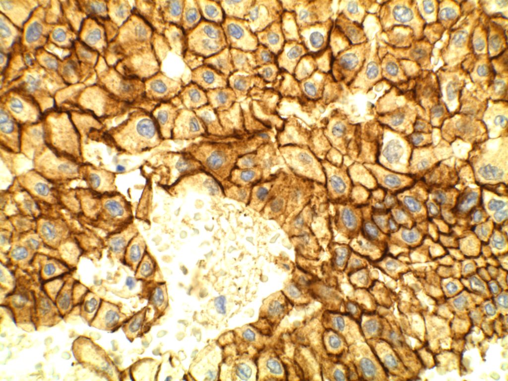
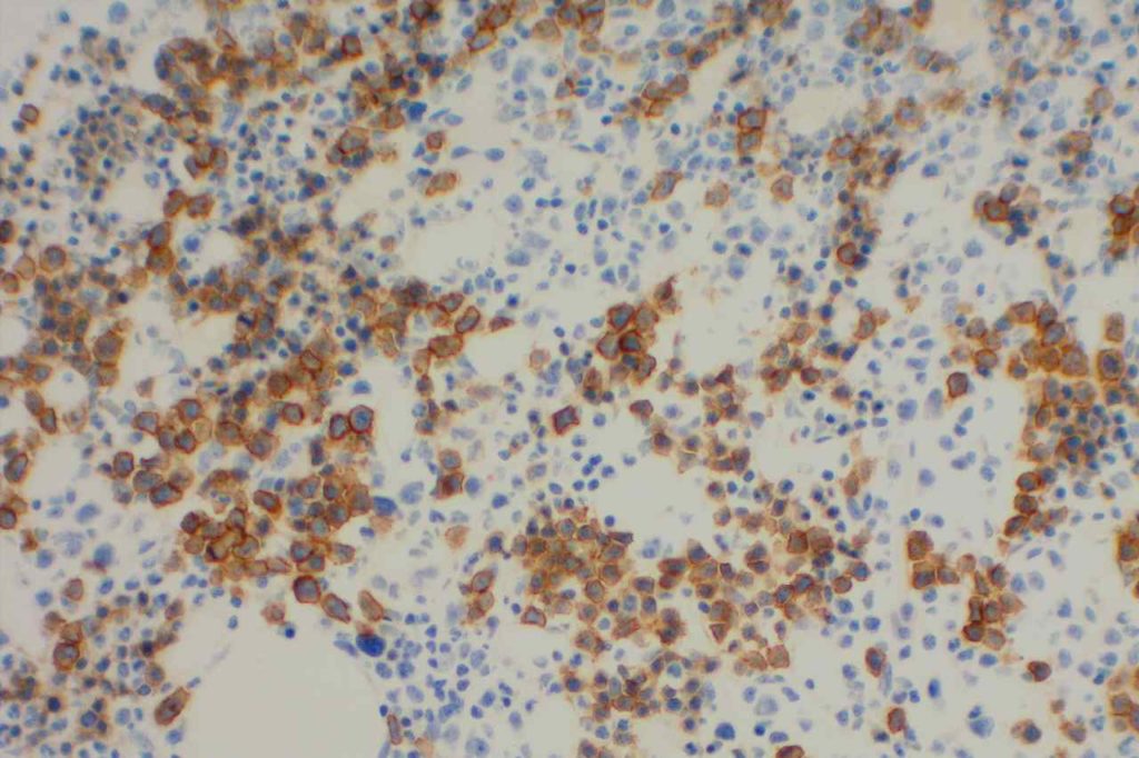
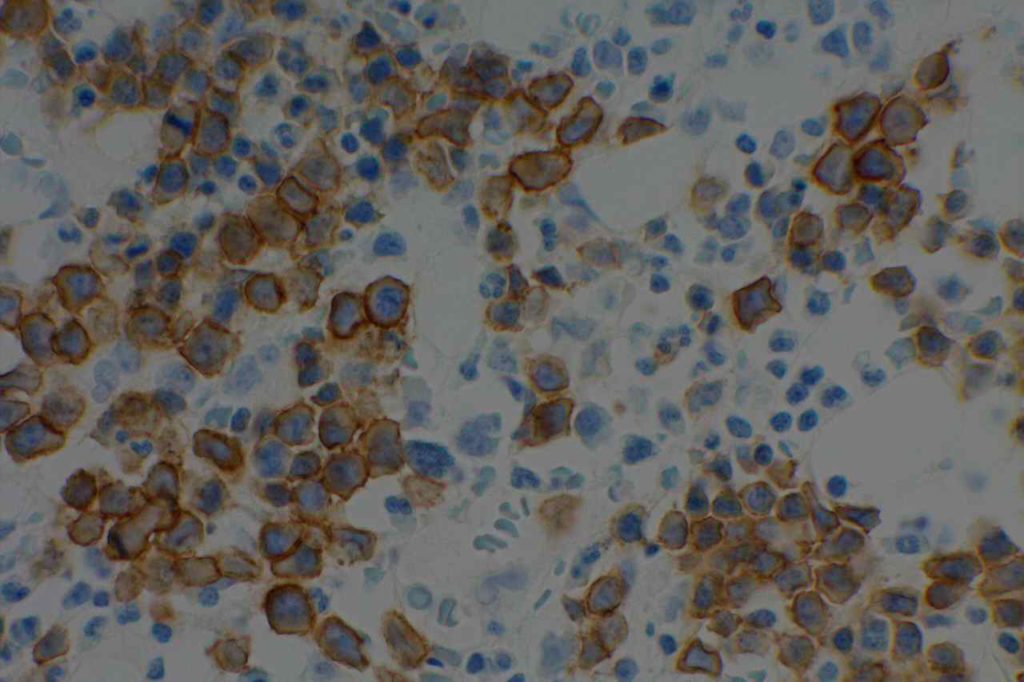
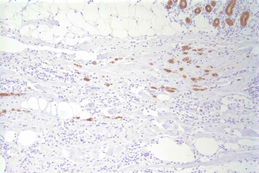
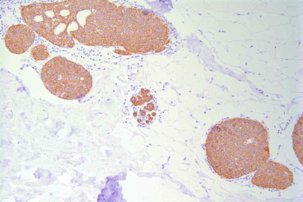
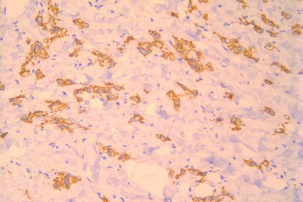
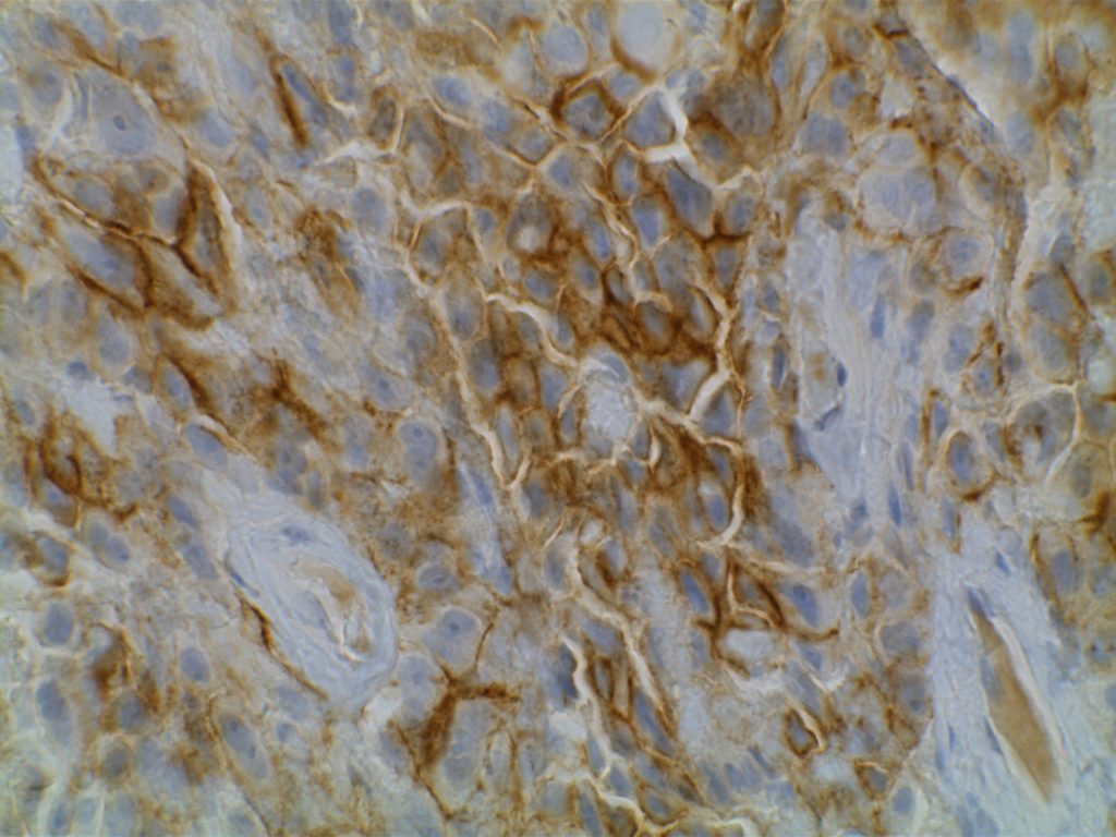
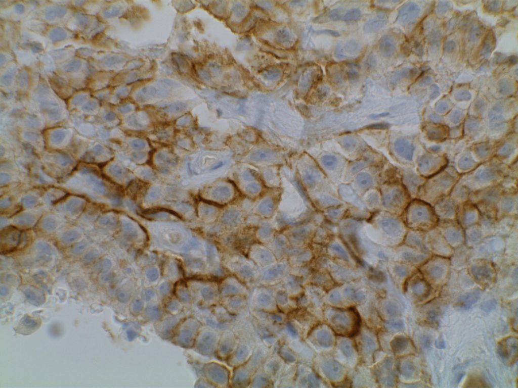
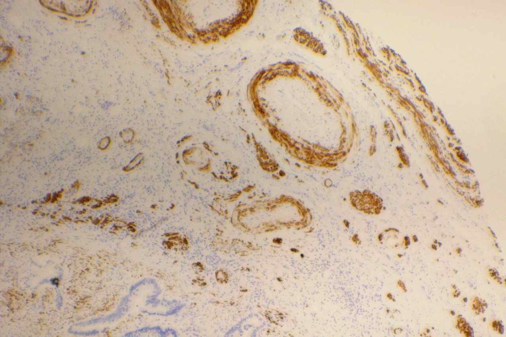
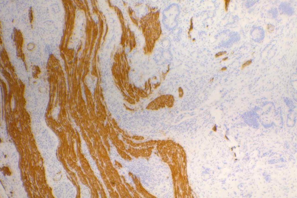
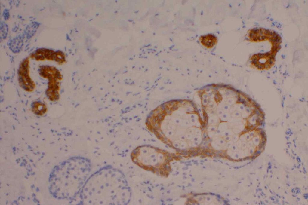
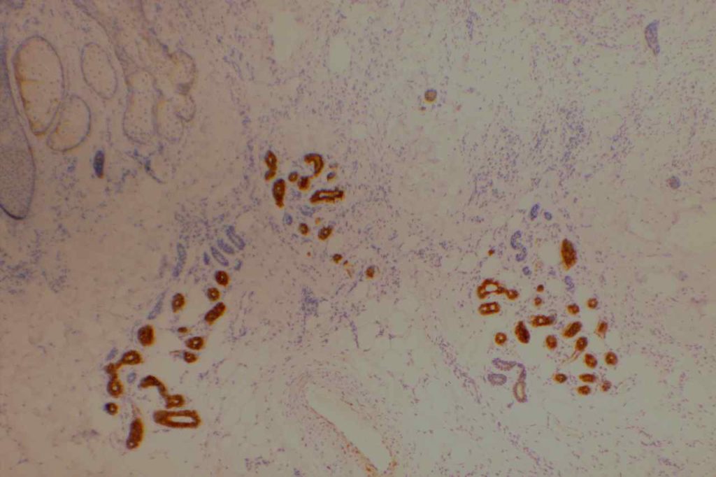
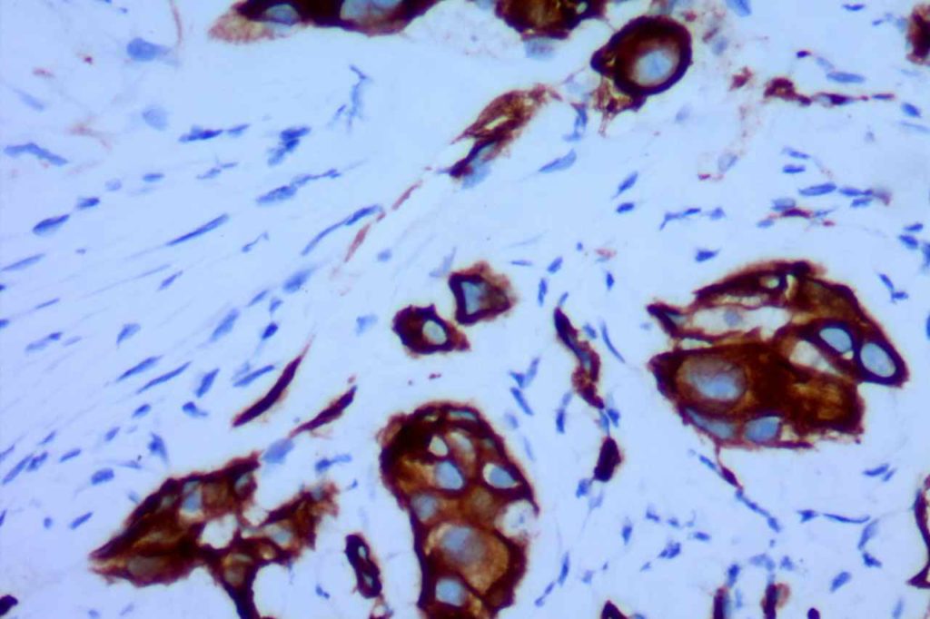
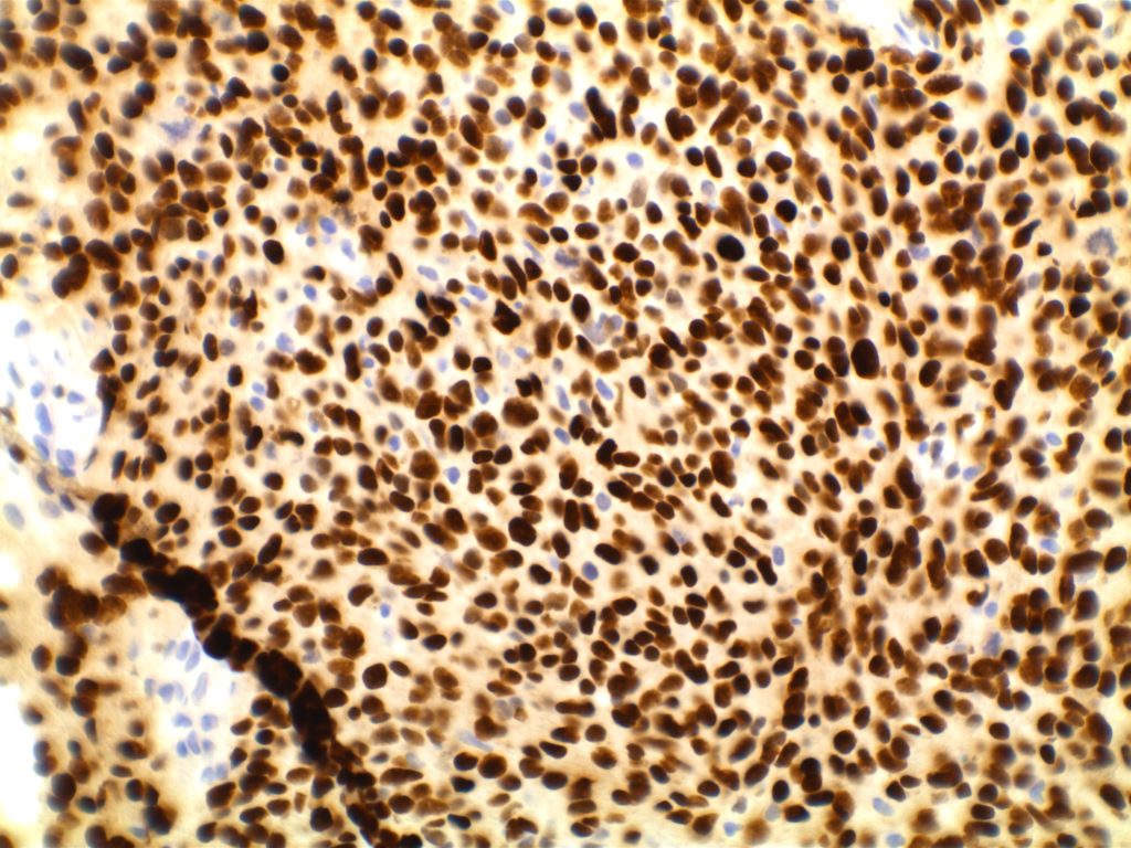
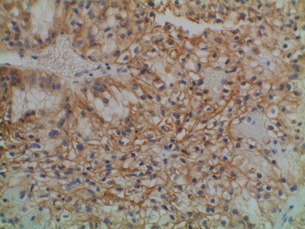
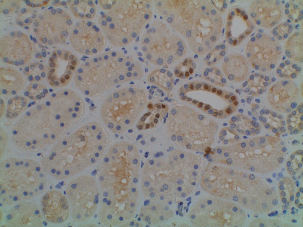
The expression pattern is nuclear, which is typically moderate to strong and diffuse.  Some variability of the sensitivity of GATA-3 in breast carcinomas has been noted in the literature, and confirmation of one’s assay compared to the medical literature is recommended during the validation process.  The following tables show data from multiple papers in the pathology literature.
 
Liu, et al (Biocare Medical, Concord, CA)
Tumor
GATA3
GCDFP15
MGB
Breast Carcinoma
94%
35-55%
65-70%
ER-negative breast ca.
69%
15%
35%
Urothelial Carcinoma
86%


Miettinen, et al (clone L50-823, dilution of 1:500; Biocare Medical, Concord, CA)
Tumor Type
%
N
Adrenocortical Carcinoma
11%
N=27
Basal Cell Ca., Skin
98%
N=62
Benign Skin Adnexal Tumors
100%
N=24
Breast, Ductal Ca., Primary
92%
N=179
Breast, Ductal Ca., Metastatic
96%
N=51
Breast, Lobular Carcinoma
100%
N=38
Malignant Mesothelioma
58%
N=64
Germ Cell Tumor, Choriocarcinoma
100%
N=11
Germ Cell Tumor, Endodermal Sinus Tumor
100%
N=6
Pancreas, Adenocarcinoma
37%
N=62
Renal Cell Carcinoma, Chromophobe
51%
N=35
Renal Oncocytoma
17%
N=35
Salivary Gland, Adenoid Cystic Carcinoma
29%
N=17
Salivary Gland, Ductal Carcinoma
43%
N=14
SCC – Skin
81%
N=31
SCC – Cervix
33%
N=21
SCC – Larynx
16%
N=36
SCC – Lung
12%
N=74
Urothelial Carcinoma – Low Grade
100%
N=22
Urothelial Carcinoma – High Grade
84%
N=32
 
No GATA-3 expression found in the following:  Seminoma (n=76), Pure Embryonal Carcinoma (n=5), Lung Small Cell Carcinoma (n=30), Lung Carcinoid (n=11), Small Intestine Carcinoid (n=18), Merkel Cell Carcinoma (n=4), Ovary Non-Serous Carcinomas (n=25), Pancreatic Neuroendocrine Tumor, Rectal Adenocarcinoma (n=27), and Thymoma (n=41). (Miettinen, et al)
 
GATA-3 expression in 0-5% of tumors was found in the following: Stomach Adenocarcinoma (n=133), Thyroid Papillary and Follicular Carcinomas (n=75), Renal Cell Carcinoma NOT Chromophobe (n=154), Prostate Adenocarcinoma (n=95), Hepatocellular Carcinoma (n=47), and Colon Adenocarcinoma (n=142).  (Miettinen, et al)
 
GATA-3 expression in 6-10% of tumors was found in the following:  Anaplastic Thyroid Carcinoma (n=11), Ovarian Serous Carcinoma (n=73), Lung Adenocarcinoma (n=71), Cholangiocarcinoma (n=57), and Endometrial Adenocarcinoma (n=89).  (Miettinen, et al)
 
Liu, et al. (GATA-3 [HG3-31]:sc-268; Santa Cruz Biotech, Santa Cruz, CA)
Tumor
%
N
Seminoma
0%
N=30
Embryonal Carcinoma
0%
N=24
Yolk Sac Tumor
0%
N=12
Lung Neuroendocrine Carcinoma
0%
N=61
Lung Adenocarcinoma
0%
N=61
Lung SCC
0%
N=49
Papillary Thyroid Carcinoma
0%
N=47
Follicular Thyroid Carcinoma
0%
N=37
Medullary Thyroid Carcinoma
0%
N=10
Clear Cell RCC
0%
N=82
Papillary RCC
0%
N=20
Colon Adenocarcinoma
0%
N=43
Esophageal Adenocarcinoma
0%
N=30
Gastric Adenocarcinoma
0%
N=21
Pancreatic Adenocarcinoma
0%
N=50
Urothelial Carcinoma
86%
N=72
Prostatic Adenocarcinoma
0%
N=136
Cholangiocarcinoma
0%
N=11
Breast Ductal Carcinoma
91%
N=99
Breast Lobular Carcinoma
100%
N=48
Endocervical Adenocarcinoma
0%
N=17
Endometrial Carcinoma
2%
N=96
Ovarian Serous Carcinoma
0%
N=56
Hepatocellular Carcinoma
0%
N=18
Pancreatic Endocrine Neoplasm
0%
N=15
Skin Melanoma
0%
N=100
 
Clark, et al.
Tumor
%
N
Breast
 
 
–  HR+/Her2=
99%
N=131
–  HR+/Her2+
100%
N=18
–  HR=/Her2+
100%
N=7
–  Triple Negative
73%
N=30
Endocervix
18%
N=34
Vulva/Cervix SCC
60%
N=10
Endometrium
7%
N=55
Ovary
10%
N=50
Bladder
95%
N=22
Liver (cholangiocarcinoma)
3%
N=62
Pancreas
10%
N=30
Stomach
2%
N=62

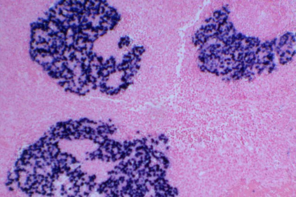
Parathyroid vs. Thyroid

GATA-3 has been shown to be expressed in >95% of parathyroid samples (n=25 combined) consisting of hyperplastic glands and adenomas, whereas no thyroid tissues showed GATA-3 expression.  

Interestingly, IHC for PTH only stained between 1/3 and 2/3rds of parathyroid samples.  Therefore, GATA-3 combined with TTF-1 may be very helpful in differentiating between thyroid and parathyroid lesions in small samples (e.g. thyroid FNAs). (Takada, et al.)
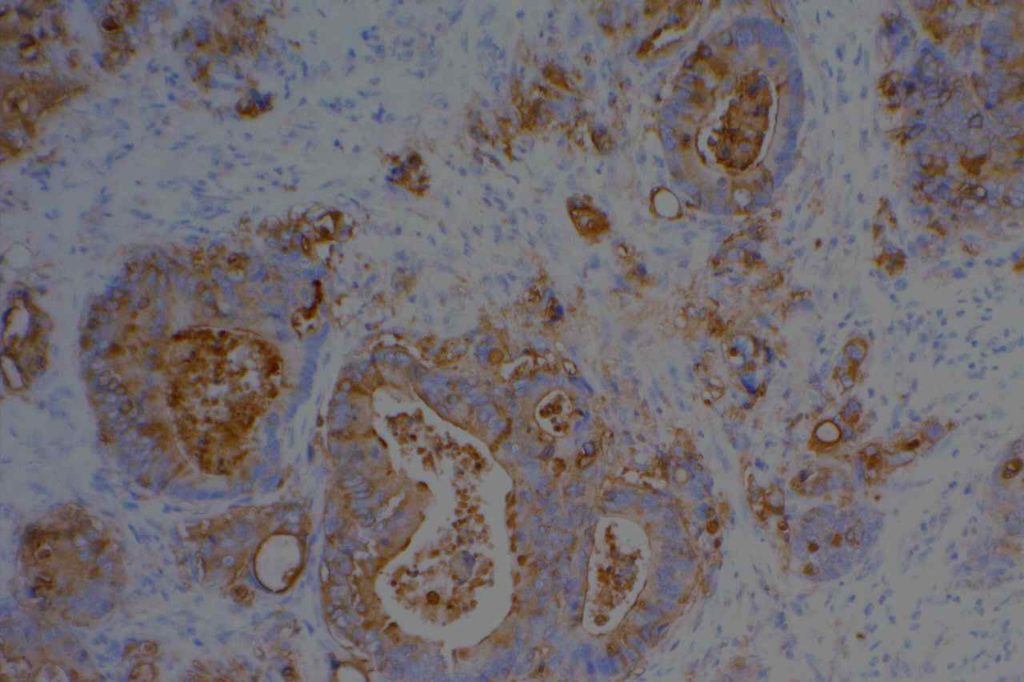
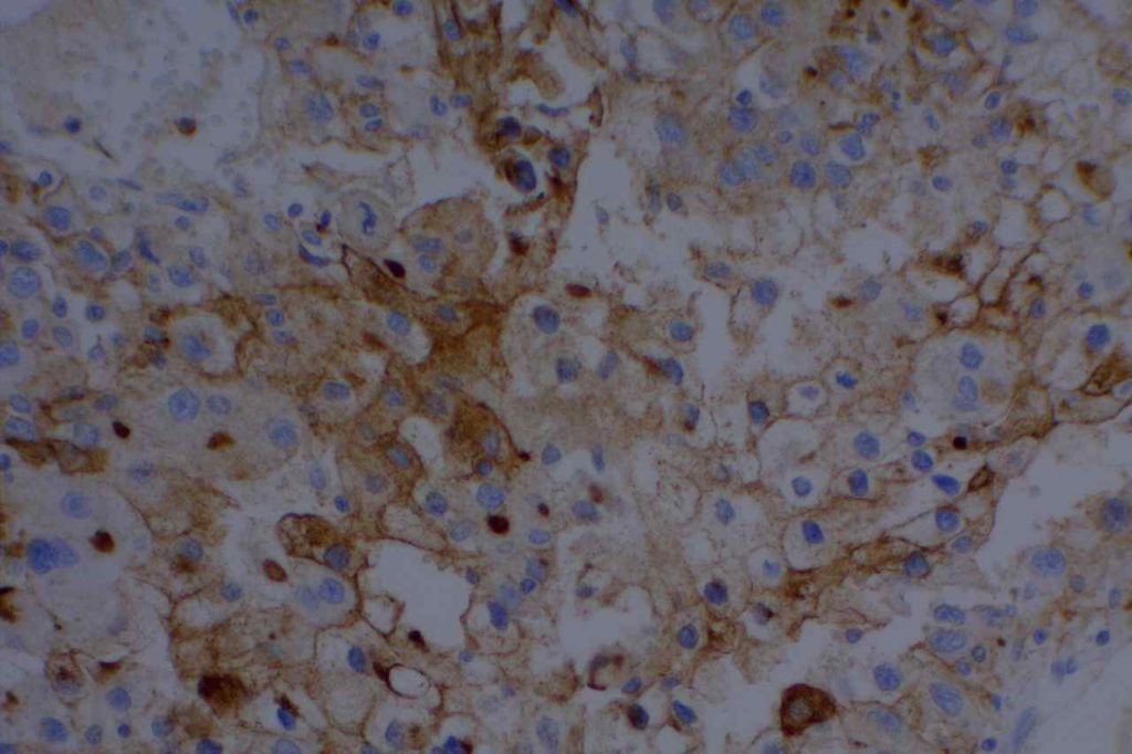
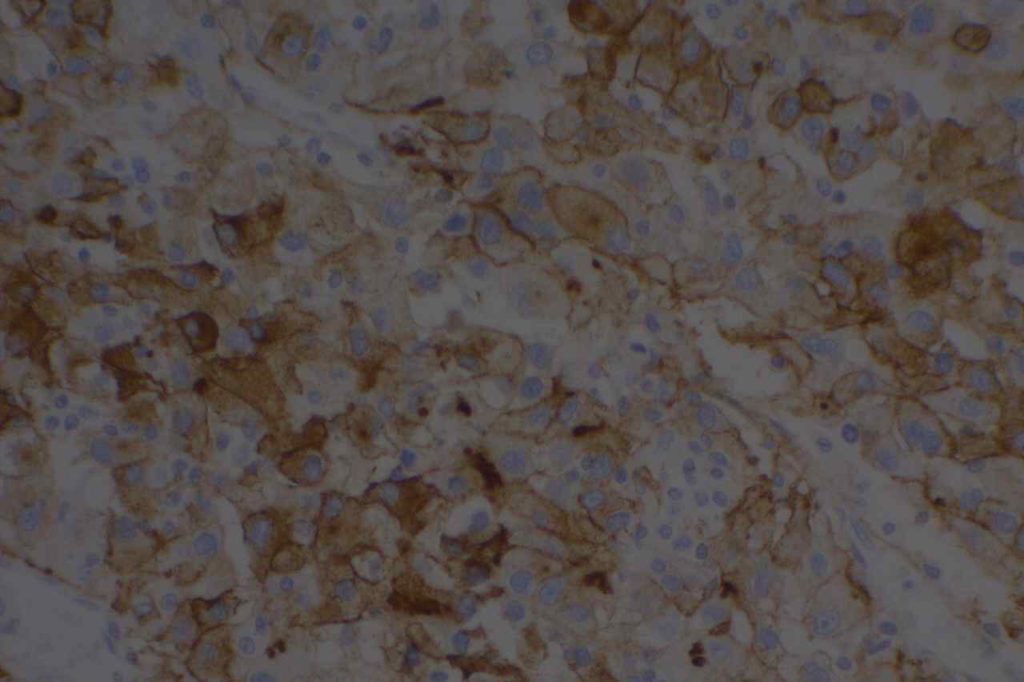
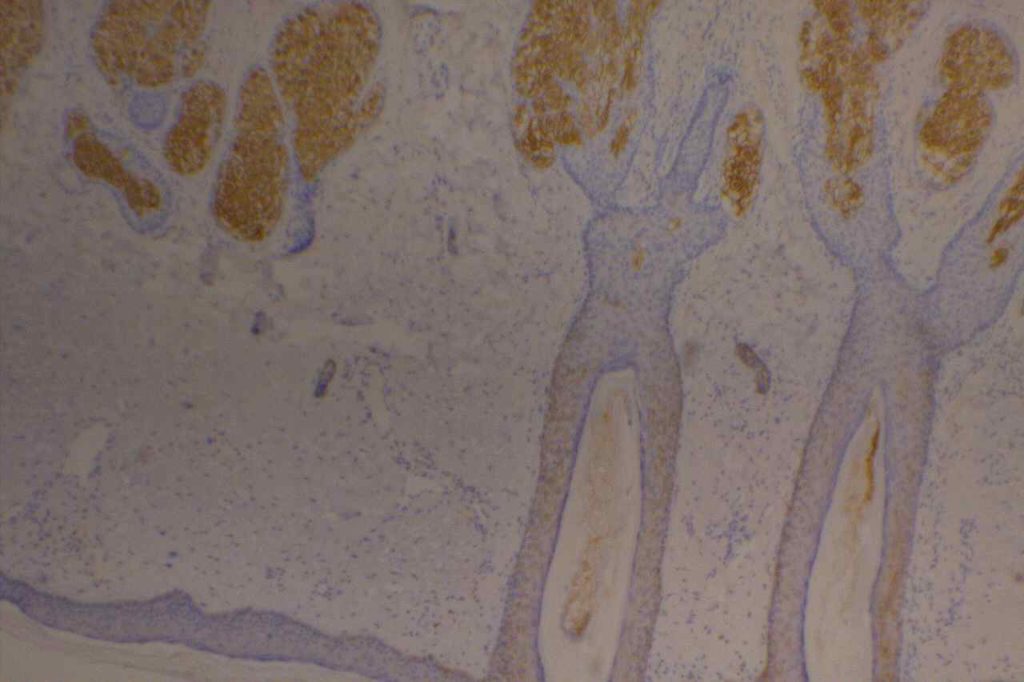
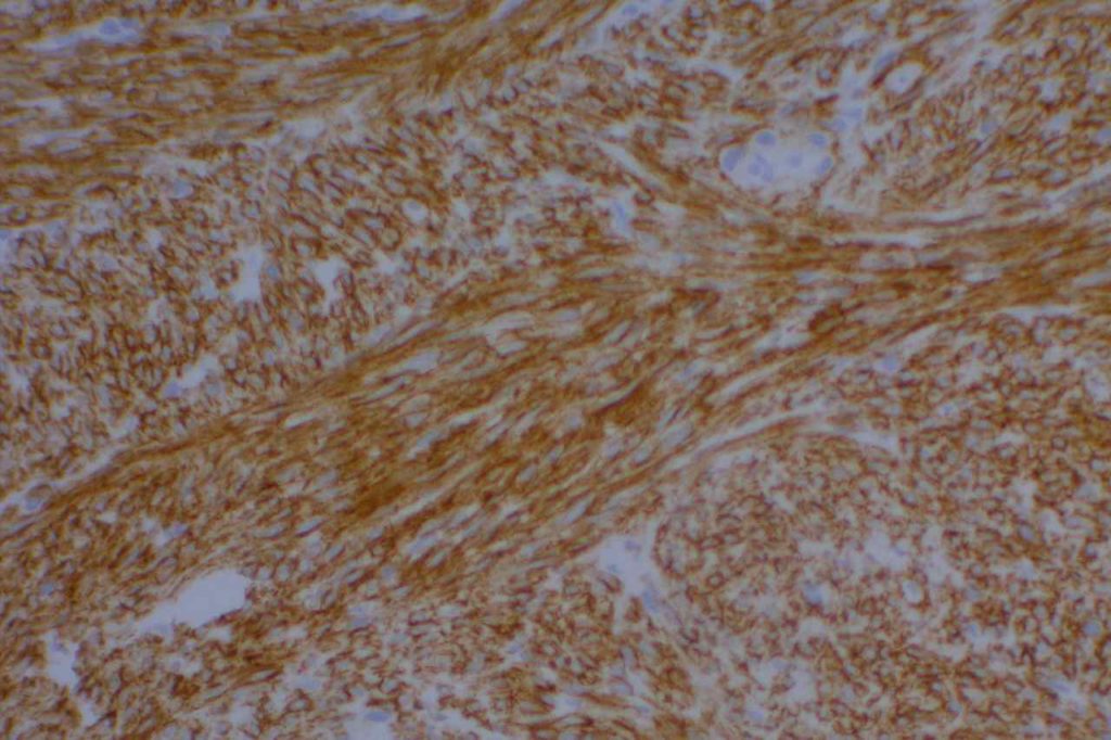
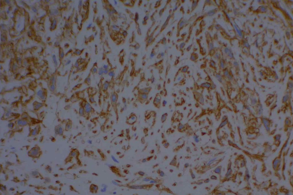
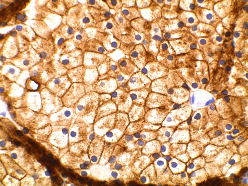
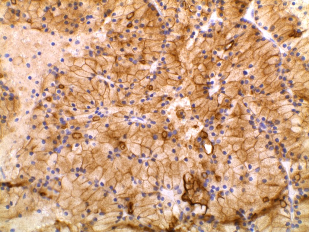
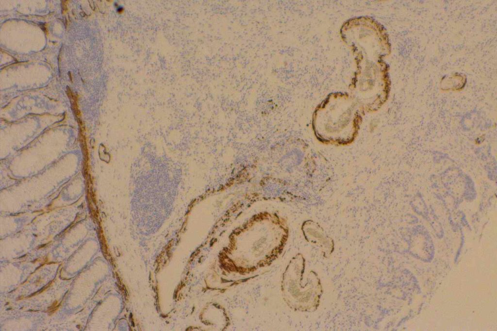
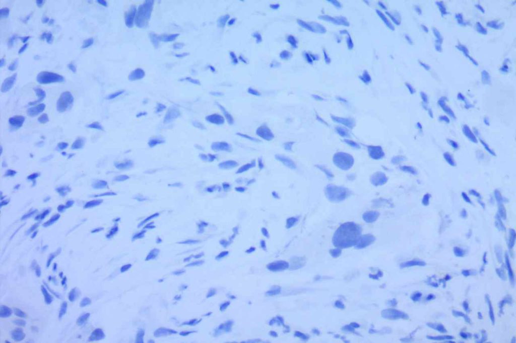
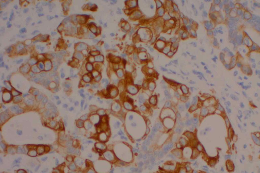
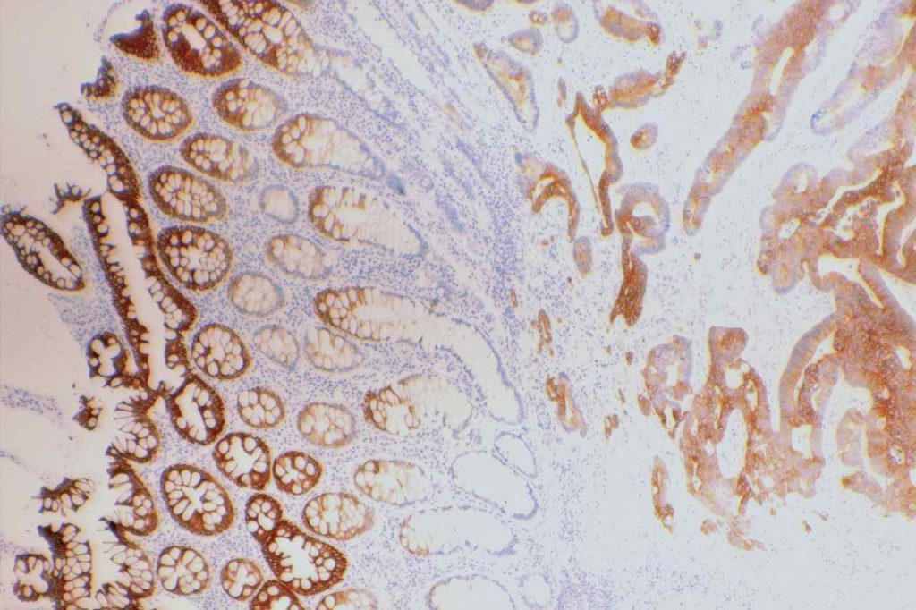
Photomicrographs
GATA-3 - Breast
GATA-3 – Breast
GATA-3 Renal Cell Carcinoma
GATA-3 expression in a clear cell renal cell carcinoma
GATA-3 Benign Kidney
GATA-3 expression in benign renal parenchyma.

References
Liu, H., Shi, J., Wilkerson, M. L., & Lin, F. (2012). Immunohistochemical evaluation of GATA3 expression in tumors and normal tissues: a useful immunomarker for breast and urothelial carcinomas. American Journal of Clinical Pathology, 138(1), 57–64. doi:10.1309/AJCP5UAFMSA9ZQBZ
 
Miettinen, M., McCue, P. A., Sarlomo-Rikala, M., Rys, J., Czapiewski, P., Wazny, K., et al. (2014). GATA3: A Multispecific But Potentially Useful Marker in Surgical Pathology: A Systematic Analysis of 2500 Epithelial and Nonepithelial Tumors. The American Journal of Surgical Pathology, 38(1), 13–22. doi:10.1097/PAS.0b013e3182a0218f
 
Ellis, C. L., Chang, A. G., Cimino-Mathews, A., Argani, P., Youssef, R. F., Kapur, P., et al. (2013). GATA-3 immunohistochemistry in the differential diagnosis of adenocarcinoma of the urinary bladder. The American Journal of Surgical Pathology, 37(11), 1756–1760. doi:10.1097/PAS.0b013e31829cdba7
 
Zhao, L., Antic, T., Witten, D., Paner, G. P., Taxy, J. B., Husain, A., et al. (2013). Is GATA3 expression maintained in regional metastases?: a study of paired primary and metastatic urothelial carcinomas. The American Journal of Surgical Pathology, 37(12), 1876–1881. doi:10.1097/PAS.0b013e31829e2525
 
Clark, B. Z., Beriwal, S., Dabbs, D. J., & Bhargava, R. (2014). Semiquantitative GATA-3 Immunoreactivity in Breast, Bladder, Gynecologic Tract, and Other Cytokeratin 7-Positive Carcinomas. American Journal of Clinical Pathology, 142(1), 64–71. doi:10.1309/AJCP8H2VBDSCIOBF
 
McCleskey BC, Penedo TL, Zhang K, Hameed O, Siegal GP, Wei S. GATA3 Expression in Advanced Breast Cancer: Prognostic Value and Organ-Specific Relapse. Am J Clin Pathol. 2015;144: 756–763.
doi:10.1309/AJCP5MMR1FJVVTPK
 
Takada, N., Hirokawa, M., Suzuki, A., Higuchi, M., Kuma, S., Miyauchi, A. (2016). Diagnostic value of GATA-3 in cytological identification of parathyroid tissues Endocrine Journal 63(7), 621-626. https://dx.doi.org/10.1507/endocrj.ej15-0700

Estrogen Receptor (ER)

Summary (Breast Carcinoma)
  • Nuclear Marker
  • Stain is reported as PERCENT STAINING OF TUMOR CELLS and STAIN INTENSITY (1+, 2+, 3+)
  • 1% or greater nuclear expression in tumor cells is considered positive, and therefore eligible to receive hormonal therapy.
  • CAP-ASCO recommendations are for <1 hr. from time of excision/biopsy to having a cut edge of tumor in 10% neutral bufferedormalin fixative.  Fixation window of 6-72 hrs.  These times should be noted in the pathology report (time of excision, time in gross room, and time in fixative).
  • Negative staining results in biopsy material without an internal control should be repeated on the excisional specimen using blocks with both tumor and benign breast parenchyma.

General
Estrogen Receptor (ER) is a nuclear marker, which is most commonly used to identify breast carcinomas that may be responsive hormonal therapy (e.g. Tamoxifen).  It also conveys prognostic information (ER+ has a more favorable prognosis).  In normal breast duct epithelium ER will show variable patchy expression, but does not normally stain myoepithelial cells.
 
Interpretation
Interpretation of ER expression in breast carcinoma is based on percentage positive (+), and stain intensity (1+, 2+, 3+).  Benign breast duct epithelium in the background serves as an internal barometer for stain intensity.  Breast tumors are considered positive if 1% or more of tumor cells express ER.  An important side note:  if a case is negative for ER expression, and there is NO INTERNAL CONTROL (i.e. benign duct epithelium), which is positive, then repeat testing on the excision specimen is recommended.  Block selection for testing should include benign background tissue, which can serve as an internal control.
 
In a breast case, if PR is positive and ER is negative, then the ER assay may not be working or the slides have been switched.  PR expression should, practically, only occur in the setting of ER expression.
 
Marker Specificity
Sometimes ER is utilized as a marker to resolve the primary origin of an adenocarinoma.  ER is relatively specific for breast origin, but far from perfect.  Any “hormonally driven organ” (i.e. ovary, breast, uterus) may commonly show ER expression.  Less commonly, practically any organ may show ER expression.
 
ER is often used as part of a panel to differentiate an endocervical adenocarcinoma (ER negative) from an endometrial adenocarcinoma (ER positive).
 
Common expression patterns in carcinoma (Dennis, JL, et al)
Tumor
Expression (%)
Breast
30-60%
Colon
<5%
Lung
<10%
Ovary
10-50%
Pancreas
0%
Stomach
<5%
Prostate
~10%
Microscopic Images 
ER - Breast
ER expression in an invasive ductal carcinoma of the breast.
ER - Breast
ER expression in an invasive breast cancer.
ER
ER expression in benign ductal structures of the breast.
ER
ER expression in benign ductal structures of the breast.
References
Yaziji, H., Taylor, C. R., Goldstein, N. S., Dabbs, D. J., Hammond, E. H., Hewlett, B., et al. (2008). Consensus recommendations on estrogen receptor testing in breast cancer by immunohistochemistry. Applied Immunohistochemistry & Molecular Morphology : AIMM / Official Publication of the Society for Applied Immunohistochemistry, 16(6), 513–520. doi:10.1097/PAI.0b013e31818a9d3a 
 
Hammond, M. E. H., Hayes, D. F., Dowsett, M., Allred, D. C., Hagerty, K. L., Badve, S., et al. (2010). American Society of Clinical Oncology/College of American Pathologists guideline recommendations for immunohistochemical testing of estrogen and progesterone receptors in breast cancer (unabridged version). Archives of Pathology & Laboratory Medicine, 134(7), e48–72. 
 
Dennis, J. L., Hvidsten, T. R., Wit, E. C., Komorowski, J., Bell, A. K., Downie, I., et al. (2005). Markers of adenocarcinoma characteristic of the site of origin: development of a diagnostic algorithm. Clinical Cancer Research : an Official Journal of the American Association for Cancer Research, 11(10), 3766–3772. doi:10.1158/1078-0432.CCR-04-2236  

EMA (MUC-1)

Epithelial Membrane Antigen (EMA), originally described in 1979, targets a complex membrane glycoprotein originally isolated from milk fat globules.  It is also known as MUC-1.  EMA is helpful in identifying epithelial differentiation, but is not entirely specific.  In general EMA expression is similar to cytokeratin expression, although hepatocellular carcinomas, adrenocortical neoplasms, and germ cell neoplasms do not express EMA.  Some non-epithelial processes, such s ALCL, plasmacytomas, epithelioid sarcoma, synovial sarcoma, meningioma, and T-cell lymphomas may show at least focal positivity. (Wick, M.R.)
 
In a recent study by Minato, H. et al, EMA was found to be expressed in 79% (n=34) of cases of mesothelioma compared to 13% (n=40) of cases of reactive mesothelial cells.
Photomicrographs
EMA - Colon Adenocarcinoma
EMA expression in colon adenocarcinoma.
EMA - Renal Cell Carcinoma
EMA expression in conventional clear cell renal cell carcinoma.
EMA - Renal Cell Carcinoma
EMA expression in renal cell carcinoma.
EMA - Skin
EMA expression in skin.
EMA - Micropapillary Breast Carcinoma
Characteristic EMA expression in micropapillary brest carcinoma.
References
Wick, M. R. (2008). Immunohistochemical approaches to the diagnosis of undifferentiated malignant tumors. Annals of Diagnostic Pathology, 12(1), 72–84. doi:10.1016/j.anndiagpath.2007.10.003 
 
Minato, H., Kurose, N., Fukushima, M., Nojima, T., Usuda, K., Sagawa, M., et al. (2014). Comparative Immunohistochemical Analysis of IMP3, GLUT1, EMA, CD146, and Desmin for Distinguishing Malignant Mesothelioma From Reactive Mesothelial Cells. American Journal of Clinical Pathology, 141(1), 85–93. doi:10.1309/AJCP5KNL7QTELLYI 

E-Cadherin

E-cadherin is an intercellular adhesion marker, and has a membraneous expression pattern.  Its most common use is to help differentiate between DCIS (positive) and LCIS (negative).  It is less helpful in differentiating invasive ductal from invasive lobular, because invasive ductal carcinoma will not infrequently lose E-cadherin expression.  
 
If E-cadherin is positive in an invasive breast carcinoma, that would support ductal origin, but negative staining is not helpful / diagnostic between  a ductal or lobular process.  Practically, if one is having difficulty differentiating an invasive lobular vs. invasive ductal, it is possible that E-cadherin would be negative as the cells become dis-cohesive and lose the intercellular adhesion complexes.
 
Up to 15% of lobular lesions may exhibit some membraneous E-cadherin expression.  In such equivocal cases, p120 catenin or Beta-catenin may be helpful adjunct stains to help resolve discordance between morphology and E-cadherin expression.  As is the case with immunohistochemistry in general, a well devised panel of antibodies for a given differential diagnosis is often the best practice, when results or the diagnosis is not straight forward.
DCIS vs. LCIS
IHC Stain
DCIS
LCIS
E-Cadherin
+
=
34BetaE12
=
+
Brattauer, 2002.  S. Schnitt, UAMS Lecture, 2002.
Hematopathology
E-cadherin is a sensitive and specific marker for immature erythroid cells.  It may be useful (similar to CD71) in identifying immature erythroid precursors, which may be difficult to differentiate from myeloblasts in certain circumstances.  CD34 and CD117 (blast markers) may be combined with CD71 and E-cadherin (erythroid markers) to achieve this goal.
Other
E-cadheirn expression has also been studied in numerous other organ tissues individually (e.g. chromophobe renal cell carcinoma) and as part of panels.  Use of this stain in close conjunction with current medical literature is recommended.
Photomicrographs
Chomophobe Renal Cell Carcinoma - E-Cadherin
E-Cadherin expression in a chromophobe RCC.
E-Cadherin - Erythroid Precursors
E-Cadherin expression in bone marrow erythroid precursors.
E
E-Cadherin expression in bone marrow erythroid precursors.
E-Cadherin - Invasive Ductal Carcinoma
E-Cadherin expression in an invasive ductal carcinoma of the breast.
E-Cadherin - Breast DCIS
E-Cadherin expression in breast DCIS.
E
E-Cadherin expression in an invasive ductal carcinoma of breast origin.
References
Modern Pathology 24, 375-383 (March 2011)
 
Dabbs, D. J., Schnitt, S. J., Geyer, F. C., Weigelt, B., Baehner, F. L., Decker, T., et al. (2013). Lobular neoplasia of the breast revisited with emphasis on the role of E-cadherin immunohistochemistry. The American Journal of Surgical Pathology, 37(7), e1–11. doi:10.1097/PAS.0b013e3182918a2b 
 
Klein, G. E-Cadherin Is Functionally Involved in the Maturationof the Erythroid Lineage. The Journal of Cell Biology, October 1, 1995. pp. 1–7. 
 
Ohgami, R. S., Chisholm, K. M., Ma, L., & Arber, D. A. (2014). E-Cadherin Is a Specific Marker for Erythroid Differentiation and Has Utility, in Combination With CD117 and CD34, for Enumerating Myeloblasts in Hematopoietic Neoplasms. American Journal of Clinical Pathology, 141(5), 656–664. doi:10.1309/AJCP8M4QQTAZPGRP

EBV (EBER)

Detection of EBV virus may be performed as stains in tissue sections in one of two ways:  (1) EBV IHC or (2) EBER (EBV-encoded RNA) ISH.  EBV IHC antibody reacts with the BNLF1 gene product that forms the latent membrane protein (LMP).  This marker has limited sensitivity in the 30% range.
 
EBER expression is localized to the nucleus, while the IHC LMP stains the surface membrane.
Pitfalls
EBER expression can identify lymphocytes latently infected with EBV.  Therefore, in CHL for example, the tumor cells must show expression for the case to be considered EBV-related.  On the other hand IHC stains for LMP rarely mark latently infected cells in the background, but may show false positivity in poorly fixed tissue, cells in the nervous system, and some uninfected hematopoietic elements (eosinophils and plasma cells).  False negative results are more common with IHC LMP in decalcified tissues.
 
Inter-observer agreement is greater for the interpretation of EBER compared to LMP.
EBV Expression Profile
  • DLBCL – EBV is identified in cases of EBV+ DLBCL of the elderly.
  • Hodgkin Lymphoma – Approximately 40% of Hodgkin lymphoma cases express EBV in the Hodgkin cells. (Mixed cellularity HL is ~70%+)
  • Burkitt Lymphoma – Endemic form
  • Nasopharyngeal Carcinoma
  • Infectious Mononucleosis
Photomicrographs
EBV (EBER)
EBV (EBER)
References
American Journal of Clinical Pathology. 2002;117(2)

DOG-1

General
DOG-1 is a calcium-activated chloride channel protein (a.k.a. anoctamin-1 or TMEM16a), and has been found to be ubiquitously expressed in GI stromal tumors irrespective of KIT or PDGFRA mutation status.  This antibody appears to stain a significant subset of CD117 (c-kit) negative cases (36-46%).  Approximately 50% of DOG-1 negative GISTs will express CD117.  In the setting of a GIST differential diagnosis, DOG-1 appears to have specificity comparable to CD117.  There appears to be expression on a small subset of synovial sarcomas.  Normal staining by DOG-1 can be found in interstitial cells of Cajal.  Membraneous and apical staining / reactivity may be seen with DOG-1 in non-GIST tumors / cells.
 
In addition to normal expression in interstitial cells of Cajal in the GI tract, luminal expression can be seen in the gastric mucosa, pancreatic centroacinar cells, intrahepatic bile duct epithelium, bladder, and gallbladder epithelium.  Acinic cells in the salivary gland express DOG-1 (cytoplasmic), and may be helpful in confirming the diagnosis of an acinic cell carcinoma.
 
Clone:  K9 (no joke)
Titer: 1:400
Photomicrographs
Do
DOG-1 expression in a GIST tumor.
DOG-1 - GIST
DOG-1 expression in a GIST tumor.
DOG-1 - Acinic Cell Carcinoma (Parotid)
DOG-1 – Acinic Cell Carcinoma (Parotid)
DOG-1 - Acinic Cell Carcinoma (Parotid)
DOG-1 – Acinic Cell Carcinoma (Parotid)
References
Hadi Yaziji, AIMM Annual Meeting, “New Antibodies in Diagnostic Immunohistochemistry”, presentation, 2010.
 
West RB, et al. American Journal of Pathology.  Vol. 165, No. 1, July, 2004.
 
Chan, J. K. C. (2013). Newly Available Antibodies With Practical Applications in Surgical Pathology. International Journal of Surgical Pathology, 21(6), 553–572. doi:10.1177/1066896913507601 
 
Liegl-Atzwanger, B., Fletcher, J. A., & Fletcher, C. D. M. (2010). Gastrointestinal stromal tumors. Virchows Archiv : an International Journal of Pathology. doi:10.1007/s00428-010-0891-y
 
Patil, D. T., & Rubin, B. P. (2011). Gastrointestinal stromal tumor: advances in diagnosis and management. Archives of Pathology & Laboratory Medicine, 135(10), 1298–1310. doi:10.5858/arpa.2011-0022-RA
 
Chan JKC. Newly Available Antibodies With Practical Applications in Surgical Pathology. International Journal of Surgical Pathology. 2013;21: 553–572. doi:10.1177/1066896913507601 

D2-40

D2-40 is a marker for a sialoglycoprotein found on lymphatic endothelium.  This marker is most commonly used to identify lymphatic invasion, and can help differentiate between lymphatic or vascular invasion with the additional support of a general endothelial marker (e.g. CD31 or CD34), which mark both vascular and lymphatics.  It is not recommended to use D2-40 without CD31 and/or CD34 when evaluating for lymphatic invasion.  Other cell types (including myoepithelial cells in the breast) can also express D2-40, which may result in misidentification of lymphatic invasion.
 
In addition expression is also found in germ cell neoplasia and fetal testicular monocytes.  (Mohammad and Ellis)
 
D2-40 will also be positive in mesotheliomas, but negative in adenocarcinomas.  Chu et al. showed 96% of epithelioid mesotheliomas and 65% of ovarian serous carcinomas reacted with D2-40 with membraneous staining.  Other carcinomas generally did not express D2-40 in their study.  There is no evidence that D2-40 is helpful with spindle-cell/sarcomatoid mesotheliomas.
Kaposi Sarcoma
D2-40 is expressed in approximately 2/3rds of cases of Kaposi’s Sarcoma.  It stains in a similar pattern as CD31 and CD34.
Germ Cell Tumors
Seminomas show expression for D2-40, along with a subset of embryonic carcinomas.  Yolk sac tumors are not know to express D2-40. (Bai, et al.)
Photomicrographs
D2-40 - Mesothelioma
D2-40 – Mesothelioma
D2-40 - Mesothelioma
D2-40 – Mesothelioma

References
Bai S, Wei S, Pasha TL, Yao Y, Tomaszewski JE, Bing Z. Immunohistochemical Studies of Metastatic Germ-Cell Tumors in Retroperitoneal Dissection Specimens: A Sensitive and Specific Panel. International Journal of Surgical Pathology. 2013;21: 342–351. doi:10.1177/1066896912471849
 
Rosado FGN, Itani DM, Coffin CM, Cates JM. Utility of immunohistochemical staining with FLI1, D2-40, CD31, and CD34 in the diagnosis of acquired immunodeficiency syndrome-related and non-acquired immunodeficiency syndrome-related Kaposi sarcoma. Arch Pathol Lab Med. 2012;136: 301–304. doi:10.5858/arpa.2011-0213-OA
 
Ordóñez NG. Immunohistochemical diagnosis of epithelioid mesothelioma: an update. Arch Pathol Lab Med. 2005;129: 1407–1414.
 
Wick MR. Immunohistochemical approaches to the diagnosis of undifferentiated malignant tumors. Annals of Diagnostic Pathology. 2008;12: 72–84. doi:10.1016/j.anndiagpath.2007.10.003
 
Marchevsky AM. Application of immunohistochemistry to the diagnosis of malignant mesothelioma. Arch Pathol Lab Med. 2008;132: 397–401.
 
Mohammed RAA, Martin SG, Gill MS, Green AR, Paish EC, Ellis IO. Improved methods of detection of lymphovascular invasion demonstrate that it is the predominant method of vascular invasion in breast cancer and has important clinical consequences. Am J Surg Pathol. 2007;31: 1825–1833. doi:10.1097/PAS.0b013e31806841f6


Desmin

Desmin is a muscle marker for intermediate filaments, which are present in smooth and striated muscle.  It shows variable myofibroblastic expression.  It is most commonly used to identify muscle differentiation in neoplastic processes (e.g. leiomyoma, rhabdomyosarcoma, etc.).
 
It is interesting that reactive mesothelial cells are often positive for desmin (>50%), compared to mesothelioma (<10%) or carcinoma (<5%).
Photomicrographs
Desmin - Colon Vessels
Desmin highlighting smooth muscle in the vessels of colon tissue.
Desmin - Colon muscularis propria
Desmin highlighting muscular propria of colon tissue.
Desmin - Colon
Desmin highlighting smooth muscle in the vessels submucosal interface of colon tissue.
References
 
Davidson, B., Nielsen, S., Christensen, J., Asschenfeldt, P., Berner, A., Risberg, B., Johansen, P.  (2001).  The role of desmin and N-cadherin in effusion cytology: a comparative study using established markers of mesothelial and epithelial cells.  American Journal of Surgical Pathology, Nov;25(11):1405-12.
 
Minato, H., Kurose, N., Fukushima, M., Nojima, T., Usuda, K., Sagawa, M., et al. (2014). Comparative Immunohistochemical Analysis of IMP3, GLUT1, EMA, CD146, and Desmin for Distinguishing Malignant Mesothelioma From Reactive Mesothelial Cells. American Journal of Clinical Pathology, 141(1), 85–93. doi:10.1309/AJCP5KNL7QTELLYI 

CK20

CK20 is an intermediate filament with a selective expression pattern in different carcinomas, which when combined with CD7 is useful in the work up of carcinomas of unknown primary origin.
 
Common expression patterns in carcinoma (Dennis, JL, et al).
Tumor
(%)
Breast
<10%
Colon
>80%
Lung
<10%
Ovary
<10%
Pancreas
35-50%
Stomach
30-50%
Prostate
<10%
 
Moll, RT, et al.  Cytokeratin expression in various tumors.
Tumor
CK8/CK18
CK19
CK7
CK20
CK5
Hepatocellular Ca.
+
+/-
+/-
+/-
=
Colorectal ACA
+
+
+/-
+
=
Stomach ACA
+
+
+/-
+/-
=
Pancreas Ductal ACA
+
+
+
+/-
+/-
Lung ACA
+
+
+
=
=
Breast Inv. Ductal
+
+
+
=
+/-
Endometrium ACA
+
+
+
=
+/-
Ovary ACA 
+
+
+
=
=
RCC, Clear Cell Type
+
+/-
=
=
=
RCC, Papillary Type
+
+
+
=
=
RCC, Chromophobe
+
+/-
+
=
=
Mesothelioma
+
+
+/-
=
+
Lung, Small Cell Ca.
+
+/-
=
=
=
Merkel Cell Ca.
+
+
=
+
=
Urothelial Carcinoma
+
+
+
+/-
+/-
Squamous Cell Ca.
+/-
+/-
=
=
+
Key:  “+/-“, focal staining in some cases. “=“, negative, “+”, positive.
Photomicrographs
CK20 - Lung Adenocarcinoma
CK20 expression (or lack thereof) in lung adneocarcinoma (CK7+/CK20-).
CK20 - Colon Adenocarcinoma
CK20 expression in colon adenocarcinoma.
CK20 - Colon Adenocarcinoma
CK20 expression in underlying colon adenocarcinoma, and normal expression in overlying benign mucosa (increased expression as it becomes more superficial).
References
Hadi, AIMM Annual Meeting, “The Thirty Most Important Antibodies”, presentation, 2011.
 
Dennis, J. L., Hvidsten, T. R., Wit, E. C., Komorowski, J., Bell, A. K., Downie, I., et al. (2005). Markers of adenocarcinoma characteristic of the site of origin: development of a diagnostic algorithm. Clinical Cancer Research : an Official Journal of the American Association for Cancer Research, 11(10), 3766–3772. doi:10.1158/1078-0432.CCR-04-2236  
 
Moll, R., Divo, M., & Langbein, L. (2008). The human keratins: biology and pathology. Histochemistry and Cell Biology, 129(6), 705–733. doi:10.1007/s00418-008-0435-6

CK7

CK7 is an intermediate filament with a selective expression pattern in different carcinomas, which when combined with CD20 is useful in the work up of carcinomas of unknown primary origin.
 
Common expression patterns in carcinoma (Dennis, JL, et al).
Tumor
Expression %
Breast
>80%
Colon
<10%
Lung
>90%
Ovary
>80%
Pancreas
>80%
Stomach
35-70%
Prostate
~10%
 
Moll, RT, et al.  Cytokeratin expression in various tumors.
Tumor
CK8/CK18
CK19
CK7
CK20
CK5
Hepatocellular Ca.
+
+/-
+/-
+/-
=
Colorectal ACA
+
+
+/-
+
=
Stomach ACA
+
+
+/-
+/-
=
Pancreas Ductal ACA
+
+
+
+/-
+/-
Lung ACA
+
+
+
=
=
Breast Inv. Ductal
+
+
+
=
+/-
Endometrium ACA
+
+
+
=
+/-
Ovary ACA 
+
+
+
=
=
RCC, Clear Cell Type
+
+/-
=
=
=
RCC, Papillary Type
+
+
+
=
=
RCC, Chromophobe
+
+/-
+
=
=
Mesothelioma
+
+
+/-
=
+
Lung, Small Cell Ca.
+
+/-
=
=
=
Merkel Cell Ca.
+
+
=
+
=
Urothelial Carcinoma
+
+
+
+/-
+/-
Squamous Cell Ca.
+/-
+/-
=
=
+
Key:  “+/-“, focal staining in some cases. “=“, negative, “+”, positive.
Photomicrographs
CK7 - Benign Skin
CK7 expression in skin highlighting duct epithelium of eccrine/adnexal structures. CK7 staining pattern is the inverse of CK5 expression.
CK7 - Benign Skin
CK7 expression in eccrine/adnexal structures from a skin section. CK7 with stain in an “inverse” pattern with epithelium compared to CK5
CK7 - Lung Adenocarcinoma
CK7 expression in lung adenocarcinoma
References
Hadi, AIMM Annual Meeting, “The Thirty Most Important Antibodies”, presentation, 2011.
 
Dennis, J. L., Hvidsten, T. R., Wit, E. C., Komorowski, J., Bell, A. K., Downie, I., et al. (2005). Markers of adenocarcinoma characteristic of the site of origin: development of a diagnostic algorithm. Clinical Cancer Research : an Official Journal of the American Association for Cancer Research, 11(10), 3766–3772. doi:10.1158/1078-0432.CCR-04-2236  
 
Moll, R., Divo, M., & Langbein, L. (2008). The human keratins: biology and pathology. Histochemistry and Cell Biology, 129(6), 705–733. doi:10.1007/s00418-008-0435-6