Category Archives: Antibody List

CK5

CK5 is a high molecular weight cytokeratin that stains stratified squamous epithelium (e.g. skin, tongue mucosa, etc.), basal cells of the prostate gland, myoepithelial cells in breast tissue, and mesothelial cells/mesothelioma.  Therefore CK5 is an excellent marker, which can be used in a variety of diagnostic applications.
 
CK5 is useful in the diagnosis of squamous cell carcinoma, urothelial carcinoma, mesothelioma, thymic tumors, and skin appendage tumors.  It is also helpful in identifying the myoepithelial layer in breast tissue, and the basal layer in prostate tissue.
 
The combined expression of p63 and CK5 in poorly differentiated tumors is highly suggestive of squamous origin (Kaufmann, O., et al)
Lung
CK5 will stain approximately 70-80% of squamous cell carcinomas with a cytoplasmic staining pattern.  p63 is considered slightly more sensitive, but may give varying positivity in cases of lung adenocarcinoma that can be confusing.  Therefore, CK5 and p63 are often used as part of a panel to diagnose squamous cell carcinoma.  Since p63 is a nuclear marker, these two stains can be performed as a double stain on a single slide with one or two different chromogens.
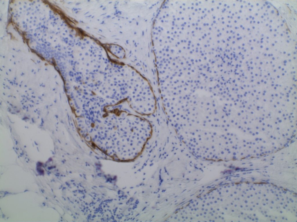
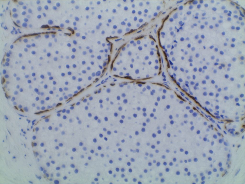
Prostate
CK5 is an excellent marker for the basal layer, and stains in a similar pattern to 34βE12.  Again, CK5 can be combined with p63, which also stains the basal layer of prostate glands.
 
Bladder
Urothelial carcinoma will typically express p63 diffusely with variable reports of CK5 or CK5/6 expression in urothelial carcinoma.
Breast
CK5 will stain the myoepithelial layer of breast tissue, but other markers such as smooth muscle myosin and calponin are typically used more commonly.  Basal-like breast carcinomas usually express CK5, especially with the monoclonal CK5 antibody compared to CK5/6 (Bhargava, R, et al).  Metaplastic/sarcomatoid breast carcinomas have also been reported to express CK5.
Skin
CK5 will stain the stratified squamous epithelium.  If there are cells in the squamous epithelium, which may be Paget cells or melanocytes, CK5 can be used along with CK7 or Melan A/HMB-45 to differentiate between odd appearing keratinocytes, Paget’s disease, or melanocytes.
CK5 vs. CK5/6
There is occasionally discussion as to which is a better marker.  Some feel CK5/6 is a better marker since it has more than one cytokeratin in it’s cocktail.  However, there is a very strong consensus among pathologists who have used both that CK5 performs significantly better than CK5/6, and is therefore, the preferred antibody.  
 
Common expression patterns in carcinoma (Chu, PG, et al).
Tumor
Expression %
Differentiation
Skin: BCC & SCC
100%
Squamous
Squamous Salivary Gland Tumors
100%
Myoepithelial
Mesothelioma
76%
Mesothelial
Urothelial Ca. 
62%
Transitional
Endometrial Ca.
50%
Squamous
Ovarian Ca. (Serous)
75%
Serous Epithelium
 
Note:  other references note a much higher + expression pattern in meotheliomas.  This may be dependent upon the mix of epithelial and sarcomatous variants.
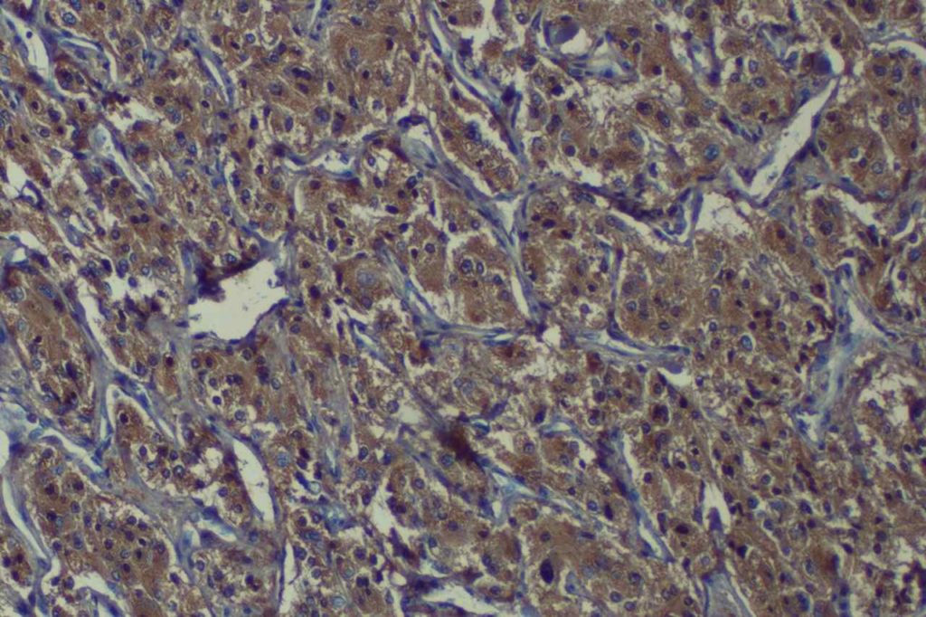
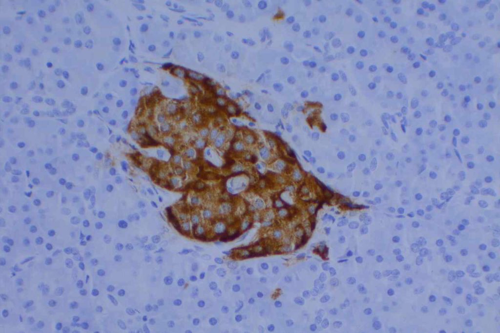
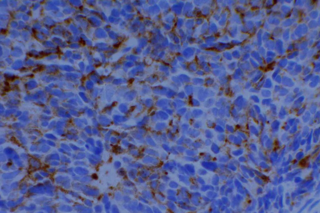
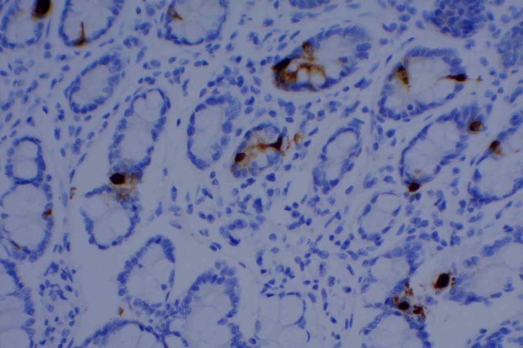
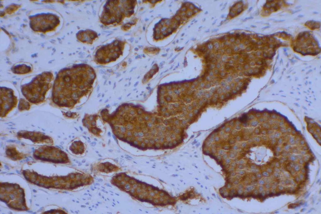
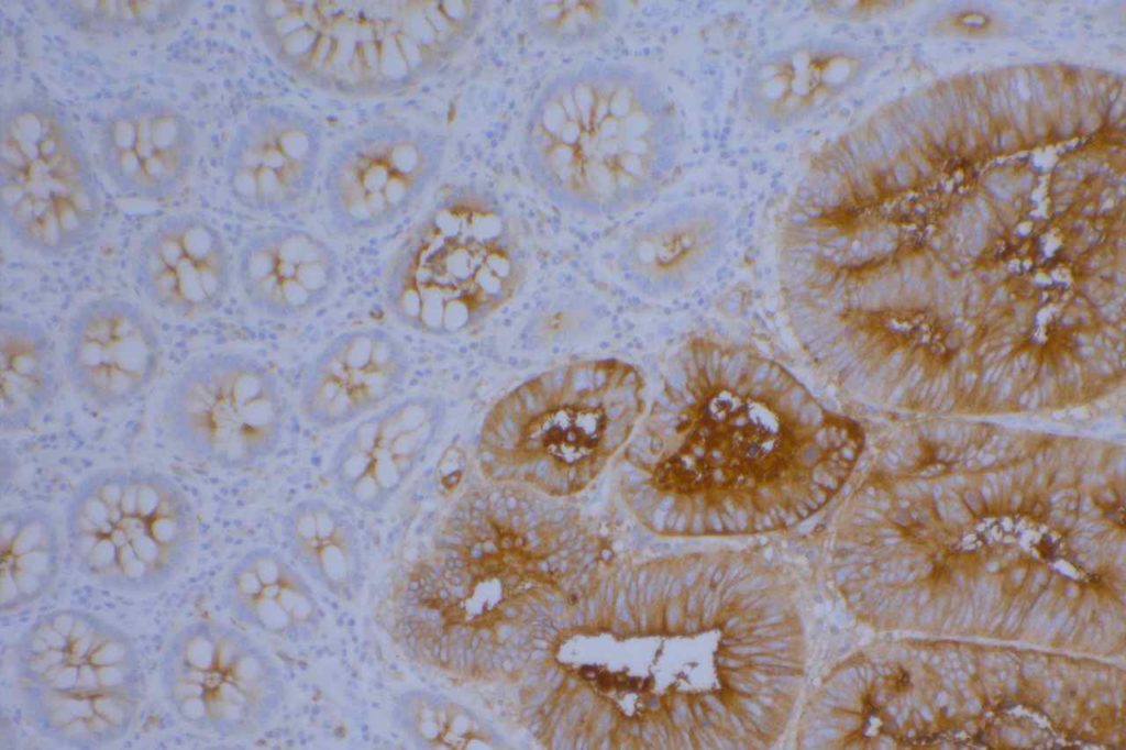
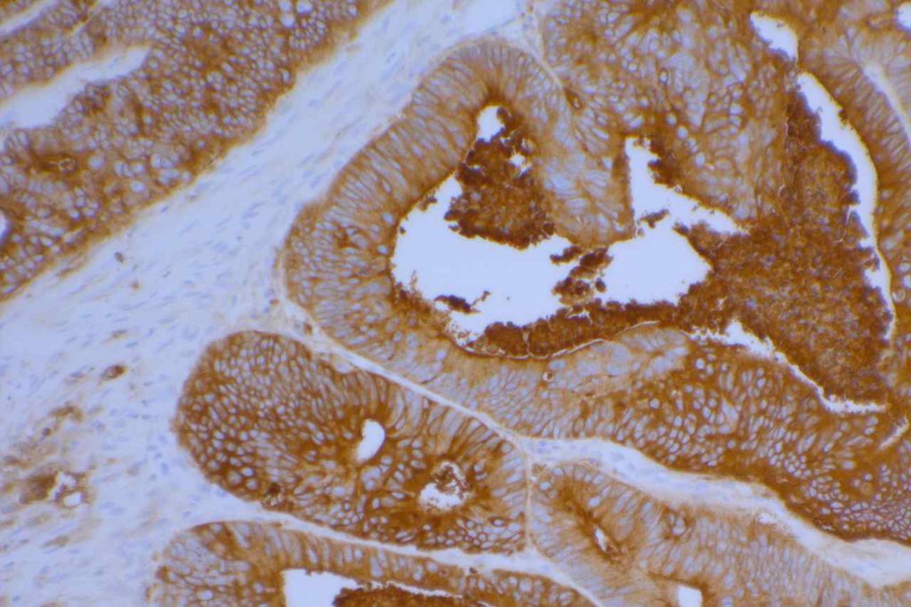
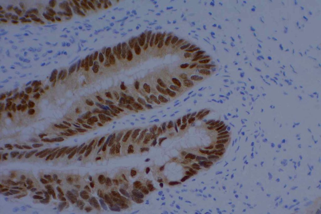
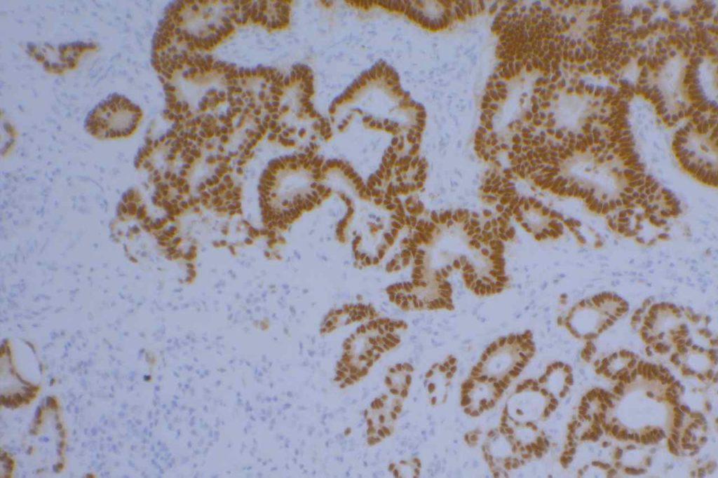
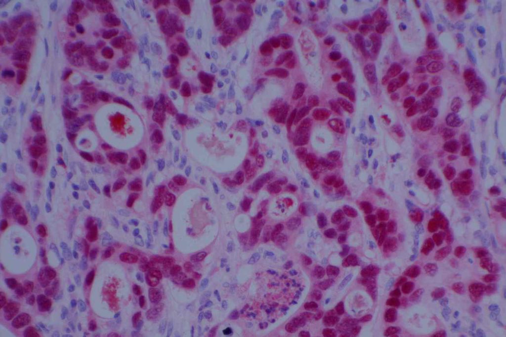
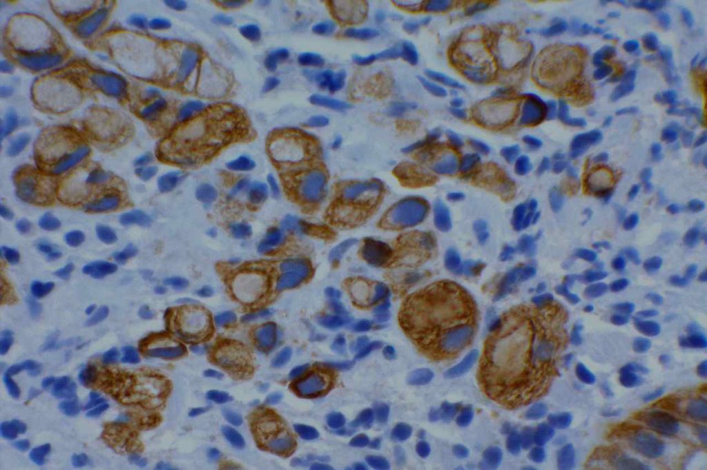
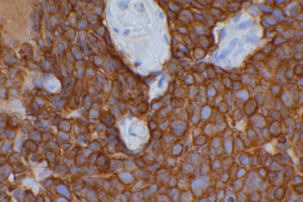
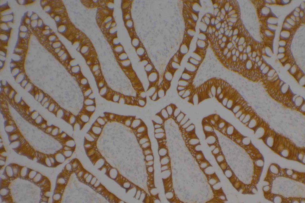
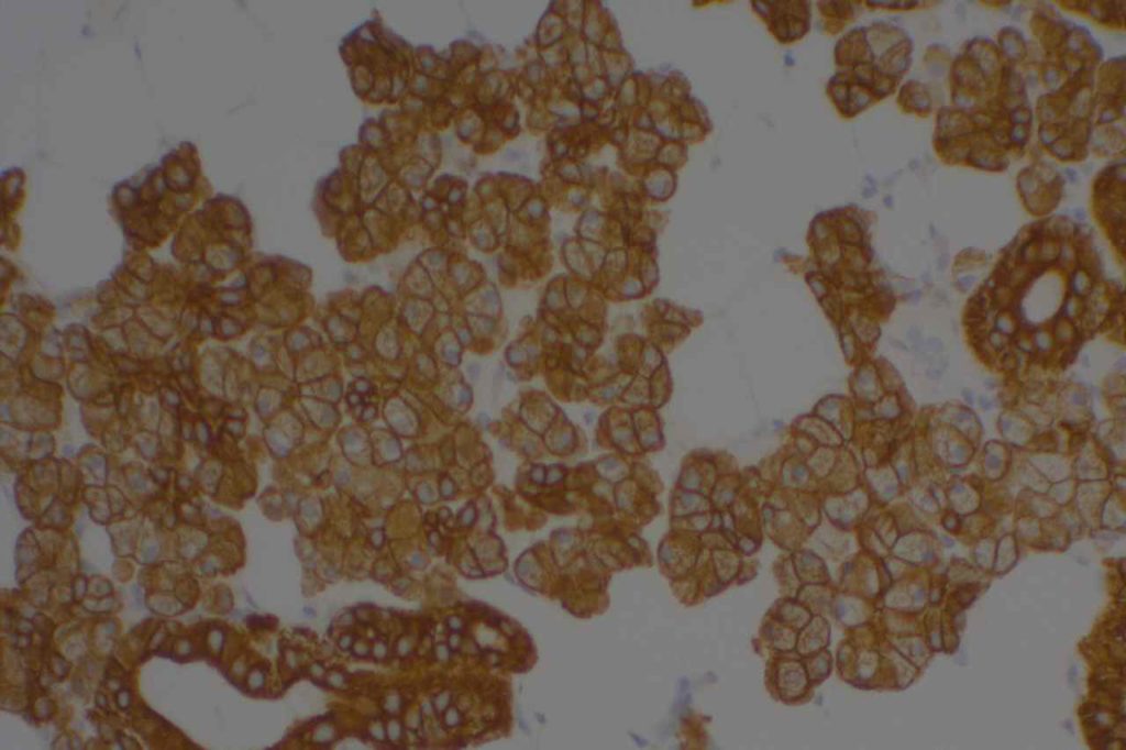
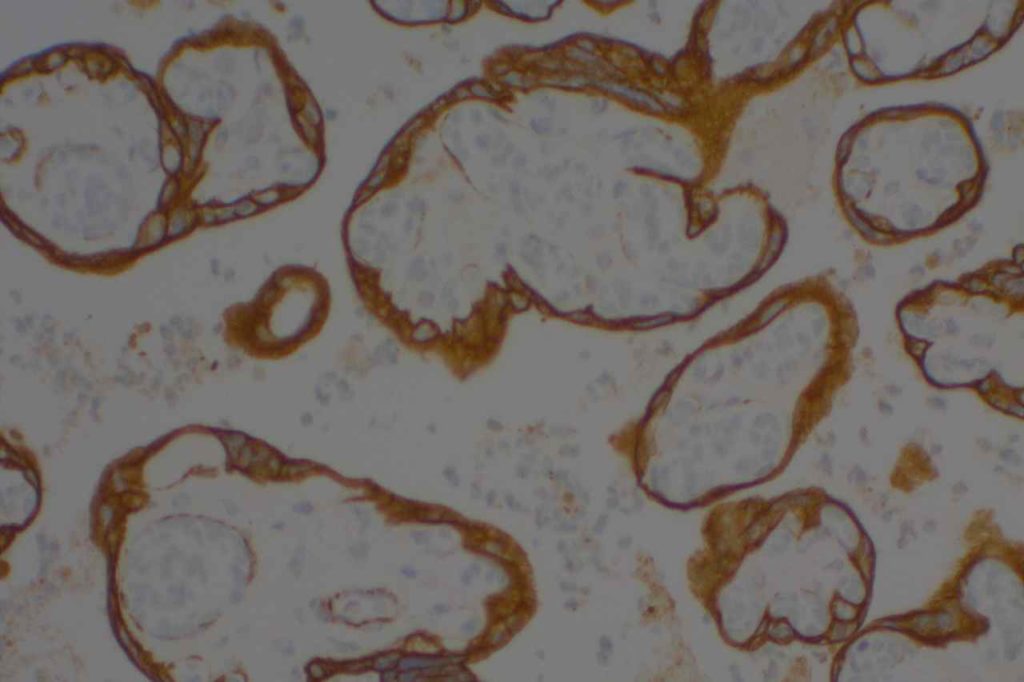
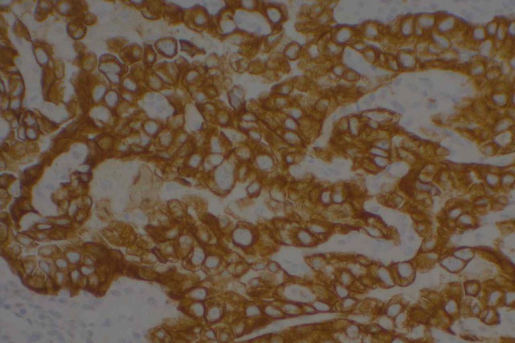
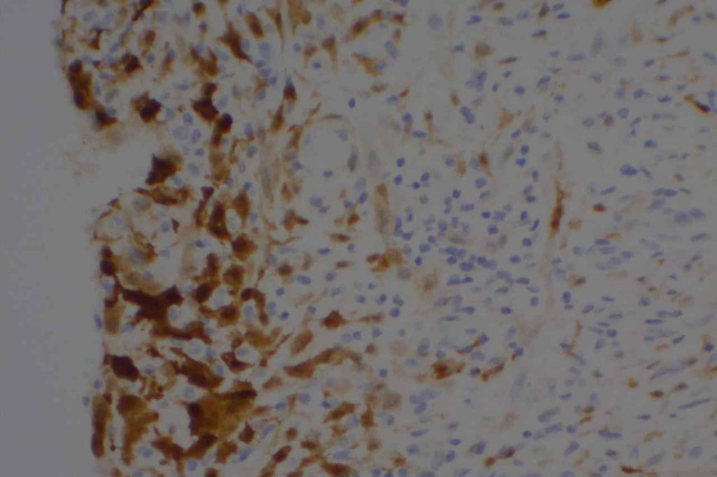
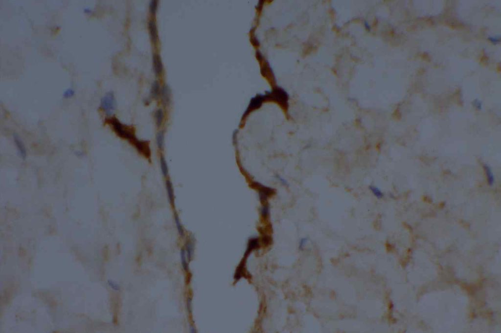
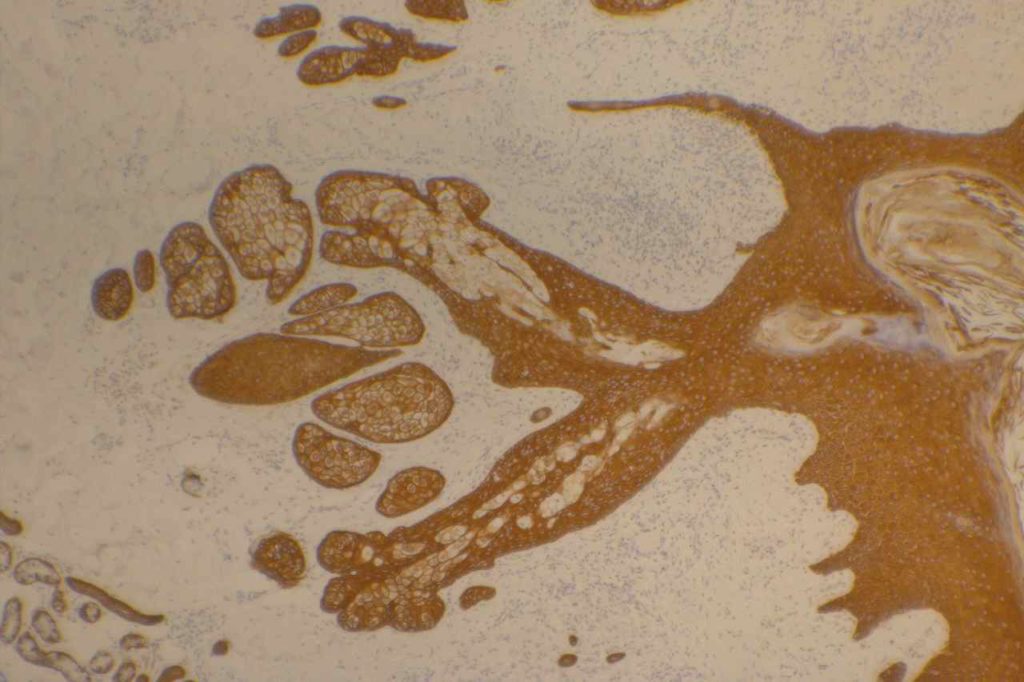
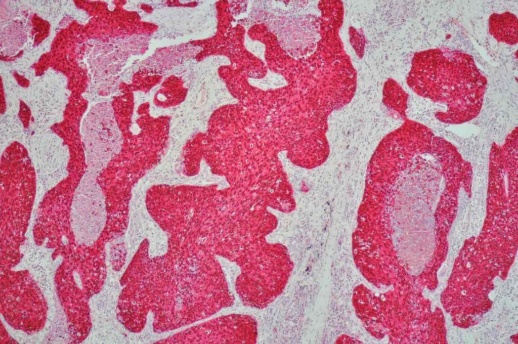
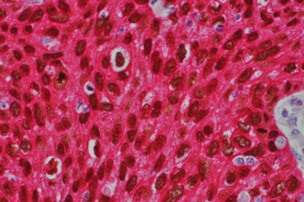
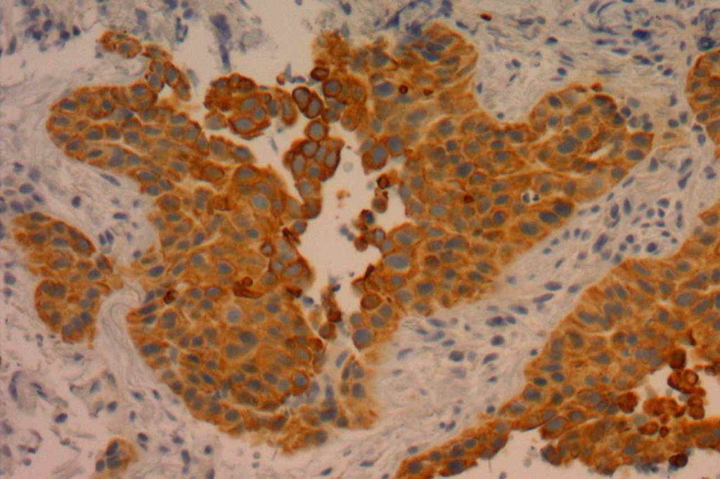
Photomicrographs
CK5 - Benign Skin
CK5 is a high molecular weight cytokeratin expression by stratified epthelium (epidermis) and myoepithlial/basal layer cells.
p63-CK5 (double stain) lung squamous cell carcinoma
p63/CK5 (double stain) with p63 nuclear expression (DAB) and CK5 cytoplasmic expression (red)
p63-CK5 (double stain) lung squamous cell carcinoma
Lung squamous cell carcinoma with p63 nuclear expression (DAB) and cytoplasmic CK5 expression (red).
CK5/6 - Lung Squamous Cell Carcinoma
CK5/6 expression in a lung squamous cell carcinoma. CK5/6 tends to have more varied expression compared to the monoclonal CK5 antibody.
Reference 
Fichtenbaum EJ, Marsh WL, Zynger DL. CK5, CK5/6, and double-stains CK7/CK5 and p53/CK5 discriminate in situ vs invasive urothelial cancer in the prostate. Am J Clin Pathol. 2012;138: 190–197. doi:10.1309/AJCP5ZC4GQVNWTYR
 
Bhargava, R., Beriwal, S., Mcmanus, K., & Dabbs, D. J. (2008). CK5 is more sensitive than CK5/6 in identifying the “basal-like” phenotype of breast carcinoma. American Journal of Clinical Pathology, 130(5), 724–730. doi:10.1309/AJCP3KFF1LTYWQIY 
 
Mukhopadhyay, S., & Katzenstein, A.-L. A. (2011). Subclassification of non-small cell lung carcinomas lacking morphologic differentiation on biopsy specimens: Utility of an immunohistochemical panel containing TTF-1, napsin A, p63, and CK5/6. The American Journal of Surgical Pathology, 35(1), 15–25. doi:10.1097/PAS.0b013e3182036d05
 
Kaufmann, O., Fietze, E., Mengs, J., & Dietel, M. (2001). Value of p63 and cytokeratin 5/6 as immunohistochemical markers for the differential diagnosis of poorly differentiated and undifferentiated carcinomas. American Journal of Clinical Pathology, 116(6), 823–830. doi:10.1309/21TW-2NDG-JRK4-PFJX  
 
Chu, P. G., & Weiss, L. M. (2002). Expression of cytokeratin 5/6 in epithelial neoplasms: an immunohistochemical study of 509 cases. Modern Pathology : an Official Journal of the United States and Canadian Academy of Pathology, Inc, 15(1), 6–10. doi:10.1038/modpathol.3880483 
 
Sutton LM, Han JS, Molberg KH, Sarode VR, Cao D, Rakheja D, et al. Intratumoral expression level of epidermal growth factor receptor and cytokeratin 5/6 is significantly associated with nodal and distant metastases in patients with basal-like triple-negative breast carcinoma. Am J Clin Pathol. 2010;134: 782–787. doi:10.1309/AJCPRMD3ARUO5WPN
 
Hadi, AIMM Annual Meeting, “The Thirty Most Important Antibodies”, presentation, 2011.
Mod Path 15:6-10, 2002.
 
Moll R, Divo M, Langbein L. The human keratins: biology and pathology. Histochem Cell Biol. 2008;129: 705–733. doi:10.1007/s00418-008-0435-6

Cyclin D1 (bcl-1)

Cyclin D1 (bcd-1) is most commonly used diagnostically for confirming the diagnosis of mantle cell lymphoma (positivity should be diffuse, but intensity may be variable).   It has also been described to be expressed in invasive breast carcinoma.  While it is generally considered a specific marker (given the differential diagnosis usually with CLL/SLL), and can also be expressed in plasma cell neoplasms (40%) and hairy cell leukemia (25%).  One can also often see focal nuclear expression in vascular endothelial cells and other stromal cells, which should not be interpreted as focal positivity.
 
Given the small but variable expression of CD23 between CLL/SLL and Mantle cell lymphoma, including CyclinD1 in the standard IHC panel for this differential is recommended.
Pitfalls
Recently cyclin D1 expression in proliferation centers of cases of CLL/SLL have been described, which should not be confused with diffuse expression encountered in cases of mantle cell lymphoma. 
Cyclin D1 Expression
There are several clones available, but the most sensitive marker is the rabbit monoclonal antibody from Biocare Medical.  Other clones are not recommended.
Microscopic Images
Cyclin D1 (bcl-1) - Mantle Cell Lymphoma
Cyclin D1 (bcl-1) expression mantle cell lymphoma.

References
Gradowski, J. F., Sargent, R. L., Craig, F. E., Cieply, K., Fuhrer, K., Sherer, C., & Swerdlow, S. H. (2012). Chronic Lymphocytic Leukemia/Small Lymphocytic Lymphoma With Cyclin D1 Positive Proliferation Centers Do Not Have CCND1 Translocations or Gains and Lack SOX11 Expression. American Journal of Clinical Pathology, 138(1), 132–139. doi:10.1309/AJCPIVKZRMPF93ET
 
Bone Marrow IHC.  Torlakovic, EE, et. al. American Society for Clinical Pathology Pathology Press © 2009.  pp. 221.

Chromogranin A

Chromogranin A is a very specific marker for neuroendocrine cell differentiation (reacts with cytoplasmic neurosecretory granules).  Unfortunately, it is not highly sensitive.  It is often used as part of a panel to identify neuroendocrine neoplasms (e.g. synaptophysin, CD56).  It is more often positive in well-differentiated lesions, and stains the neurosecretory granules in the cell cytoplasm.
 
Chromogranin A is expressed in approximately 30% of small cell carcinomas and neuroblastomas.
Photomicrographs
Chromogranin A - Pheochromocytoma
Pheochromocytoma stained with chromogranin A (strong expression).
Chromogranin A - Pancreas Islet Cells
Pancreatic islet cells stained with chromogranin A (strong expression).
Chromogranin A - Merkel Cell Carcinoma
Perinuclear dot-like expression of chromogranin A in Merkel cell carcinoma.
Chromogranin A - Benign Colon
Scattered neuroendocrine cells present in benign colon.
Chromogranin A - Carcinoid
Chromogranin A expression in a carcinoid tumor (strong diffuse)
Reference
Wick, MR. “Immunohistochemical approaches to the diagnosis of undifferentiated malignant tumor.”Annals of Diagnostic Pathology12(2008):72-84.

CEA

CEA (carcinoembryonic antigen), CD66e, comes as either a polyclonal (pCEA) or monoclonal (mCEA) stain.  CEA is generally used as an epithelial marker with strong expression in adencarcinomas.  CEA is strongly expressed in colorectal adenocarcinomas, but it is not a specific marker as many other epithelial tumors may show expression.  CEA is most commonly used as part of a panel to discriminate between metastatic carcinoma (positive) and mesothelioma (negative).
Photomicrographs
CEA - Benign Colon and Adenocarcinoma
CEA – Benign Colon and Adenocarcinoma
CEA - Colon Adenocarcinoma
CEA – Colon Adenocarcinoma
References
Wick, MR. “Immunohistochemical approaches to the diagnosis of undifferentiated malignant tumor.”Annals of Diagnostic Pathology12(2008):72-84.

CDX-2

CDX-2 is a nuclear transcription marker that regulates differentiation and proliferation of intestinal epithelial cells, and also serves as tumor suppressor gene.  CDX-2 is normally expressed in small and large intestine epithelium.  It is a sensitive marker for colorectal adenocarcinomas (especially with strong diffuse expression).  It can also mark non-colorectal GI adenocarcinomas, columnar cell variant of papillary thyroid carcinoma (and other variants of papillary thyroid carcinoma, classic and modular variant),  ovarian mutinous carcinomas, bladder adenocarcinomas, goblet cells in Barrett’s esophagus, along with other tumors (data in tables below). Practically speaking, if a particular tumor can have any type of intestinal differentiation, then it most likely can have CDX-2 expression.
Pitfalls
It is important to note that antibody sensitivity can have significant inter laboratory variation due to both clone selection and antibody optimization (Borrisholt, et. al.).  Each laboratory should have a good understanding of the performance characteristics of their CDX-2 clone and optimization.
 
IHC Survey of Primary and Metastatic Caricnomas (Werling, R. W., et. al.)
Tumor
Expression (%)
Gastric
  Adencarcinoma
70% (N=24)
Pancreatic
  Carcinoma
32% (N=22)
Cholangiocarcinoma
25% (N=16)
Carcinoid Tumor
42% (N=12)
Ovarian Carcinoma
  (Mucinous)
64% (N=14)
Bladder
  Adenocarcinoma
100% (N=2)
 
Common expression patterns in carcinoma (Dennis, J. L, et. al.)
Tumor
Expression (%)
Breast
0%
Colon
>90%
Lung
<5%
Pancreas
0-32%
Stomach
20-70%
Prostate
<5%
 
(Enriquez, M.L., et. al.)
Tumor
Expression (%)
Columnar Cell Variant
  Papillary Thyroid Carcnioma
55% (N=11)
 
CDX-2 and Villin expression in human tumors (2-3+ expression) (Werling, R.W., et. al.)
Tumor Type
No.
CDX2
Villin
G.I. Tract


Colon Adenocarcinoma
75
99%
82% (n=73)
Duodenal Adenoma
10
100%
100%
Gastric Adenocarcinoma
24
70%
42%
Esophageal Adenocarcinoma
9
67%
78%
Pancreatic Adenocarcinoma
22
32%
40%
Cholangiocarcinoma/GB
16
25%
60%
Hepatocellular Carcinoma
12
0%
0%
Carcinoid Tumors
12
42%
34%
Ovary


Mucinous Adenocarcinoma
14
64%
64%
Mucinous Cystadenoma
13
8%
0%
Mucinous Borderline Tumor
4
25%
0%
Non-Mucinous
36
0%
0% (n=31)
Genitourinary Tract


Urothelial Carcinoma
21
0%
0%
Adenocarcinoma
2
100%
100%
Urachal Caricinoma
1
100%
100%
Renal Cell Carcinoma
7
0%
0% (n=3)
Prostate Adenocarcinoma
27
4%
0% (n=24)
Breast Carcinoma
34
0%
0%
Lung (45 primary; 18 met)
63
0%
5%
Adenocarcinoma
11
0%
0%
Squamous Cell Carcinoma
11
0%
0%
Non-Small Cell Carcinoma NOS
33
0
9%
Mucinous Carcinoma
2
0%
0%
Small Cell Carcinoma
4
0%
0%
Mesothelioma
7
0%
0%
Head and Neck


Thyroid
36
3%
0%
Papillary Carcinoma
11
9%
0%
Follicular Adenoma/Carcinoma
25
0%
0%
Salivary Gland
12
0%
0%
Mixed Tumor
6
0%
0%
Low Grade Carcinoma
6
0%
0%
Squamous Cell Carcinoma
13
0%
0% (n=11)
Photomicrograph
CDX-2 Benign Colon
CDX-2 expression in benign colon epithelium.
CDX-2 Colon Adenocarcinoma
CDX-2 staining in colon adenocarcinoma.
CDX-2 Colon Adenocarcinoma
Colon adenocarcinoma stained with CDX-2 using a red alkaline phosphatase staining kit. Brown (DAB) staining tends to be easier to interpret with nuclear markers (red is good with cytoplasmic markers).
References
Werling, R. W., Yaziji, H., Bacchi, C. E., & Gown, A. M. (2003). CDX2, a highly sensitive and specific marker of adenocarcinomas of intestinal origin: an immunohistochemical survey of 476 primary and metastatic carcinomas. The American Journal of Surgical Pathology, 27(3), 303–310.  Link: http://www.ncbi.nlm.nih.gov/pubmed/?term=American+Journal+of+Surgical+Pathology%2C+27(3)%2C+303%E2%80%93310. 
 
Dennis, J. L., Hvidsten, T. R., Wit, E. C., Komorowski, J., Bell, A. K., Downie, I., et al. (2005). Markers of adenocarcinoma characteristic of the site of origin: development of a diagnostic algorithm. Clinical Cancer Research : an Official Journal of the American Association for Cancer Research, 11(10), 3766–3772. doi:10.1158/1078-0432.CCR-04-2236  Link:  http://www.ncbi.nlm.nih.gov/pubmed/?term=Markers+of+adenocarcinoma+characteristic+of+the+site+of+origin%3A+development+of+a+diagnostic+algorithm 
 
Enriquez, M. L., Baloch, Z. W., Montone, K. T., Zhang, P. J., & LiVolsi, V. A. (2012). CDX2 expression in columnar cell variant of papillary thyroid carcinoma. American Journal of Clinical Pathology, 137(5), 722–726. doi:10.1309/AJCPXE3PUBWVZCGZ Link:  http://www.ncbi.nlm.nih.gov/pubmed/22523209
 
Cameselle-Teijeiro, J., Alberte-Lista, L., Peteiro-Gonzalez, D., Abdulkader-Nallib, I., Reyes-Santias, R., Soares, P., et al. (2012). CDX2 Expression in Some Variants of Papillary Thyroid Carcinoma. American Journal of Clinical Pathology, 138(6), 907–910. doi:10.1309/AJCP1BGCA6MFCNKH  Link:  http://www.ncbi.nlm.nih.gov/pubmed/23161723 
 
Borrisholt, M., Nielsen, S., & Vyberg, M. (2013). Demonstration of CDX2 is highly antibody dependant. Applied Immunohistochemistry & Molecular Morphology : AIMM / Official Publication of the Society for Applied Immunohistochemistry, 21(1), 64–72. doi:10.1097/PAI.0b013e318257f8aa Link: http://www.ncbi.nlm.nih.gov/pubmed/?term=Demonstration+of+CDX2+is+highly+antibody+dependant 
 
Werling, R. W., Yaziji, H., Bacchi, C. E., & Gown, A. M. (2003). CDX2, a highly sensitive and specific marker of adenocarcinomas of intestinal origin: an immunohistochemical survey of 476 primary and metastatic carcinomas. The American Journal of Surgical Pathology, 27(3), 303–310. Link: http://www.ncbi.nlm.nih.gov/pubmed/?term=CDX2%2C+a+highly+sensitive+and+specific+marker+of+adenocarcinomas+of+intestinal+origin%3A+an+immunohistochemical+survey+of+476+primary+and+metastatic+carcinomas 

Calponin

Calponin is an actin filament and is expressed in smooth muscle.  It is often used individually or combined with other IHC markers to identify the myoepithelial layer in breast epithelium.  It is important to remember, like other smooth muscle markers, that myofibroblasts may also express calponin.  Therefore, care should be taken not to mistake myofibroblast positivity for myoepithelial positivity. 
 
It is also expressed in cases of collagenous spherulosis (positive), but not adenoid cystic carcinoma of the breast.  Calponin is not a specific stain, and has been identified in numbers pathologic disease processes.  It is recommended to refer to specific medical literature for the IHC performance based on the differential diagnosis.
 
Positive Expression:
  • Atypical fibroxanthoma (30%) [Virchows Arch 200;437:58]
  • Benign Fibrous Histiocytoma (65%)
  • Collagenous Spherulosis[Mod Path 2006;19:1351]
  • DFSP (40%)
  • Fibromatosis [Am J Dermatopathol 2006;28:105]
  • Fibrosarcoma (60%)
  • Glomus Tumor [AJSP 2002;26:301]
  • Leiomyoma
  • Leiomyosarcoma
  • MFH of bone (47%) [J Clin Pathol 2002;55:853]
  • MPNST (40%)
  • Myoepithelioma-skin
  • Solitary fibrous tumor (70%)
  • Synovial Sarcoma [Histopathology 2003;42:588]
  • Myofibroblastic lesions
Photomicorgraphs
Calponin - Pleomorphic Lobular Carcinoma of Breast
Calponin – Pleomorphic Lobular Carcinoma of Breast
Calponin - Pleomorphic Lobular Carcinoma of Breast
Calponin – Pleomorphic Lobular Carcinoma of Breast
Calponin - Pleomorphic Lobular Carcinoma of Breast
Calponin – Pleomorphic Lobular Carcinoma of Breast
References
Zhao L, Yang X, Khan A, Kandil D. Diagnostic role of immunohistochemistry in the evaluation of breast pathology specimens. Arch Pathol Lab Med. 2014;138: 16–24. doi:10.5858/arpa.2012-0440-RA
 
Dewar R, Fadare O, Gilmore H, Gown AM. Best practices in diagnostic immunohistochemistry: myoepithelial markers in breast pathology. Arch Pathol Lab Med. 2011;135: 422–429. doi:10.1043/2010-0336-CP.1
 
Werling RW, Hwang H, Yaziji H, Gown AM. Immunohistochemical distinction of invasive from noninvasive breast lesions: a comparative study of p63 versus calponin and smooth muscle myosin heavy chain. Am J Surg Pathol. 2003;27: 82–90. 
 
Liu H. Application of immunohistochemistry in breast pathology: a review and update. Arch Pathol Lab Med. 2014;138: 1629–1642. doi:10.5858/arpa.2014-0094-RA

CA-125

Common expression patterns in carcinoma [Clin Cancer Res 2005;11(10) May 15, 2005]
Tumor
Expression (%)
Breast
10-25%
Colon
0-15%
Lung
20-40%
Ovary
60-95%
Pancreas
50%
Stomach
~10%
Prostate
<5%
References
Dennis, J. L., Hvidsten, T. R., Wit, E. C., Komorowski, J., Bell, A. K., Downie, I., et al. (2005). Markers of adenocarcinoma characteristic of the site of origin: development of a diagnostic algorithm. Clinical Cancer Research : an Official Journal of the American Association for Cancer Research, 11(10), 3766–3772. doi:10.1158/1078-0432.CCR-04-2236 

CAM5.2

CAM5.2 is an antibody clone manufactured by Becton Dickinson (BD), which shows reactivity to CK8 primarily and CK7 to a less extent.  It does not demonstrate reactivity to CK18 or CK19.  Sometimes CAM5.2 has been used interchangeably with CK8/18 in the medical literature, which is not correct (Hsu, JD, et al).  It is an excellent screening marker for poorly differentiated carcinomas, micro-metastasis of carcinoma in lymph nodes, and for characteristic peri-nuclear dot-like reactivity in Merkel cell carcinoma.
 
AE1/AE3 is generally the most common screening cytokeratin marker used, which has both high and low molecular weight cytokeratins.  Rarely AE1/AE3 may not stain a poorly differentiated carcinoma, which will mark with CAM5.2.
 
CAM5.2 may occasionally stain myofibroblasts and smooth muscle cells, which may express cytokeratins in certain situations (Moll, RT, et al).
 
Moll, RT, et al.  Cytokeratin expression in various tumors.
Tumor
CK8/CK18
CK19
CK7
CK20
CK5
Hepatocellular Ca.
+
+/-
+/-
+/-
=
Colorectal ACA
+
+
+/-
+
=
Stomach ACA
+
+
+/-
+/-
=
Pancreas Ductal ACA
+
+
+
+/-
+/-
Lung ACA
+
+
+
=
=
Breast Inv. Ductal
+
+
+
=
+/-
Endometrium ACA
+
+
+
=
+/-
Ovary ACA 
+
+
+
=
=
RCC, Clear Cell Type
+
+/-
=
=
=
RCC, Papillary Type
+
+
+
=
=
RCC, Chromophobe
+
+/-
+
=
=
Mesothelioma
+
+
+/-
=
+
Lung, Small Cell Ca.
+
+/-
=
=
=
Merkel Cell Ca.
+
+
=
+
=
Urothelial Carcinoma
+
+
+
+/-
+/-
Squamous Cell Ca.
+/-
+/-
=
=
+
Key:  “+/-“, focal staining in some cases. “=“, negative, “+”, positive.
Microscopic Images
CAM5.2 - Colon Adenocarcinoma
Poorly differentiated sigmoid colon adenocarcinoma with CAM5.2 expression.
CAM5.2 - Lung Small Cell Carcinoma
Small cell carcinoma of the lung stained with CAM5.2.
CAM5.2 - Small Intestine
Normal small bowel epithelium stained with CAM5.2.
CAM5.2 - Salivary Gland
Salivary gland epithelium stained with CAM5.2.
CAM5.2 - Placenta
CAM5.2 expression in placental tissue.
CAM5.2 - Lung Adenocarcinoma
Lung adenocarcinoma stained with CAM5.2.

References
Tamas, EF, Epstein, JI. (2007).  Detection of residual tumor cells in bladder biopsy specimens:  pitfalls in the interpretation of cytokeratin stains.  American Journal of Surgical Pathology, Mar;31(3):390-7.
 
Moll, R., Divo, M., & Langbein, L. (2008). The human keratins: biology and pathology. Histochemistry and Cell Biology, 129(6), 705–733. doi:10.1007/s00418-008-0435-6 
 
Hsu, J.-D., Yao, C.-C., Han, L.-W., & Han, C.-P. (2010). CAM5.2 is not identical to cytokeratins 8 and 18. American Journal of Clinical Pathology, 133(3), 514. doi:10.1309/AJCPAKB6JEBVMX5U  

Calretinin

Calretinin is a very sensitive marker used most commonly as a positive marker for mesothelioma.  It is expressed in >80% of epithelial and up to 70% of sarcomatous subtypes, staining in a cytoplasmic and nuclear pattern.  It is recommended to be used as part of a panel (2 mesothelial markers and 2 adenocarcinoma markers) since there are no sensitive and specific markers to differentiate mesothelioma from adenocarcinoma.
Other tumors expressing Calretinin
  • Sex-cord stromal tumors
  • Adrenal cortical tumors
  • Rare adenocarcinomas
  • Rare squamous cell carcinomas
Photomicrographs
Calretinin - Mesothelium
Chronically inflamed mesothelium highlighted by calretinin.
Calretinin - Mesothelium
Benign mesothelium highlighted by calretinin.
References 
Arch Pathol Lab Med- Vol. 134, February 2010 p. 208.
Wick, MR. “Immunohistochemical approaches to the diagnosis of undifferentiated malignant tumor.”Annals of Diagnostic Pathology12(2008):72-84.

BRAF

BRAF mutations (mainly V600E) are seen in many different tumors (see table below), and the presence of such mutations have therapeutic predictive and diagnostic information for the patient based on the type of tumor implicated.  For example, BRAF mutated colorectal carcinomas, like KRAS mutated tumors do not respond to EGFR inhibitors, but BRAF mutated melanomas may respond to the BRAF kinase inhibitor, vemurafenib.

Continue reading BRAF