Category Archives: CD Markers

CD117

CD117 (c-kit) is a member of the type III tyrosine kinase receptor family, and is a transmembrane protein.  Immunohistochemical expression of CD117 can be seen in many different normal tissue components (e.g. mast cells) and neoplastic tissues.  Therefore, CD117 should be used in a careful targeted manner; usually as part of a larger panel.  Limitations of specificity should always be kept in mind.
CD117 (c-kit) Expression Pattern:
  • Gastrointestinal stromal tumors (~95%+)
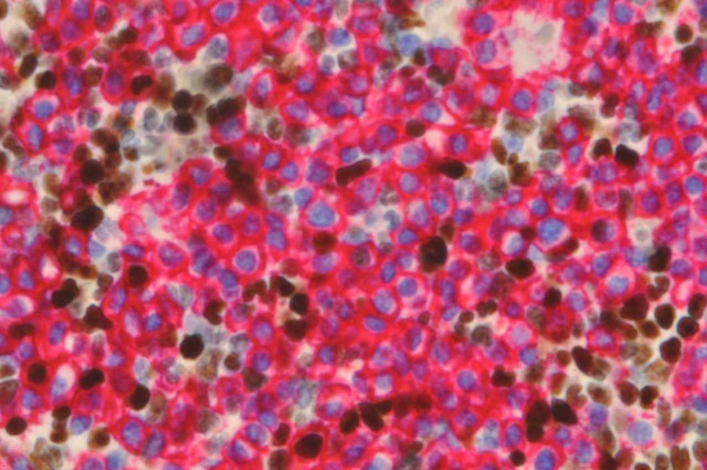
  • Mast cell neoplasms
  • Seminiomas
  • Small cell carcinomas / High grade neuroendocrine neoplasms
  • PNETs
  • Granulocytic sarcomas (small percentage, ~30%)
  • Melanomas
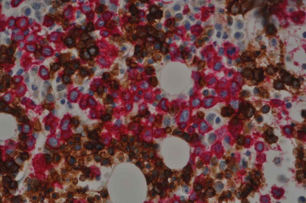
  • Myeolblasts (70% of CD34+ blasts in normal BMs); 
  • Plasma Cell Neoplasms
  • Basal like breast carcinomas (~31%)
  • Renal Tumors:  Chromophobe RCC (83-100%, Oncocytoma (71-100%), negative in other subtypes of RCC
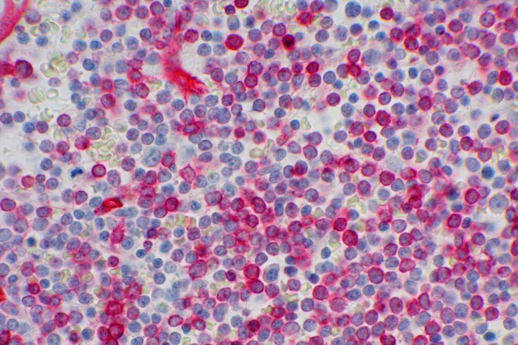
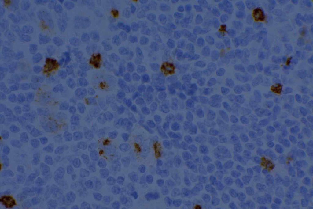
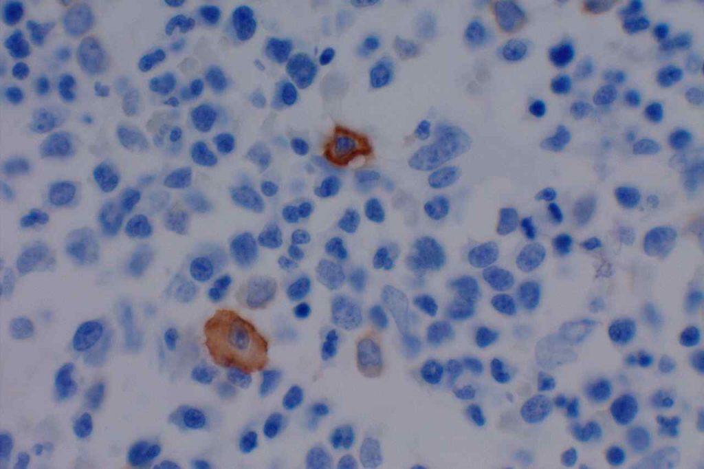
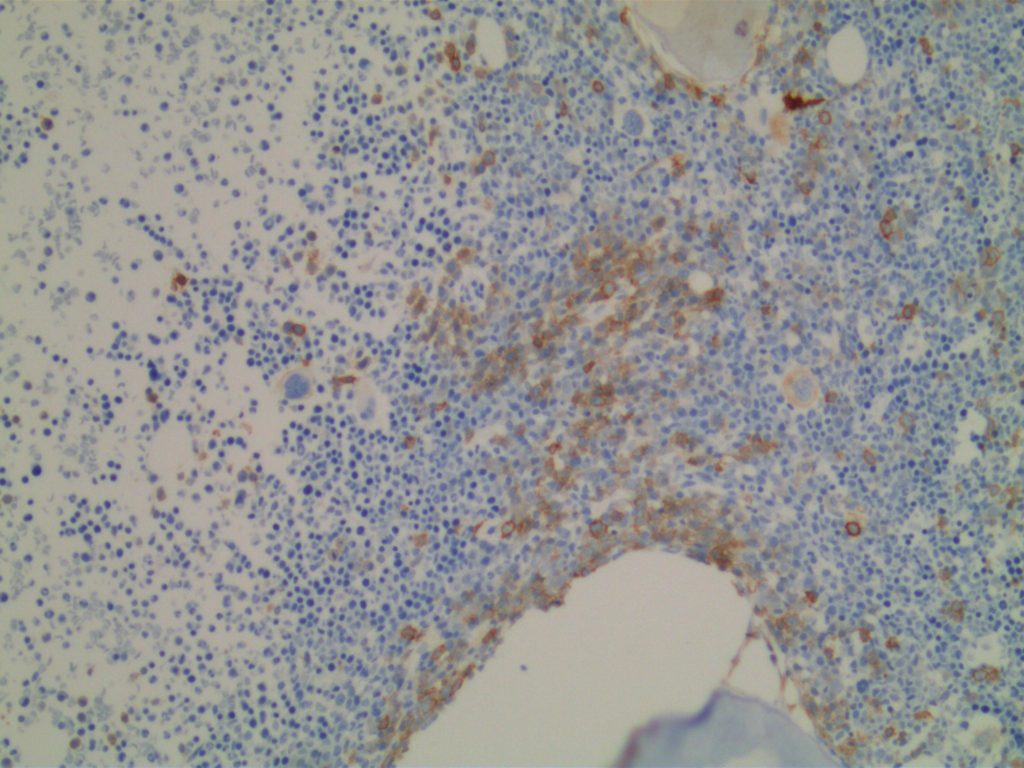
Hematopoietic
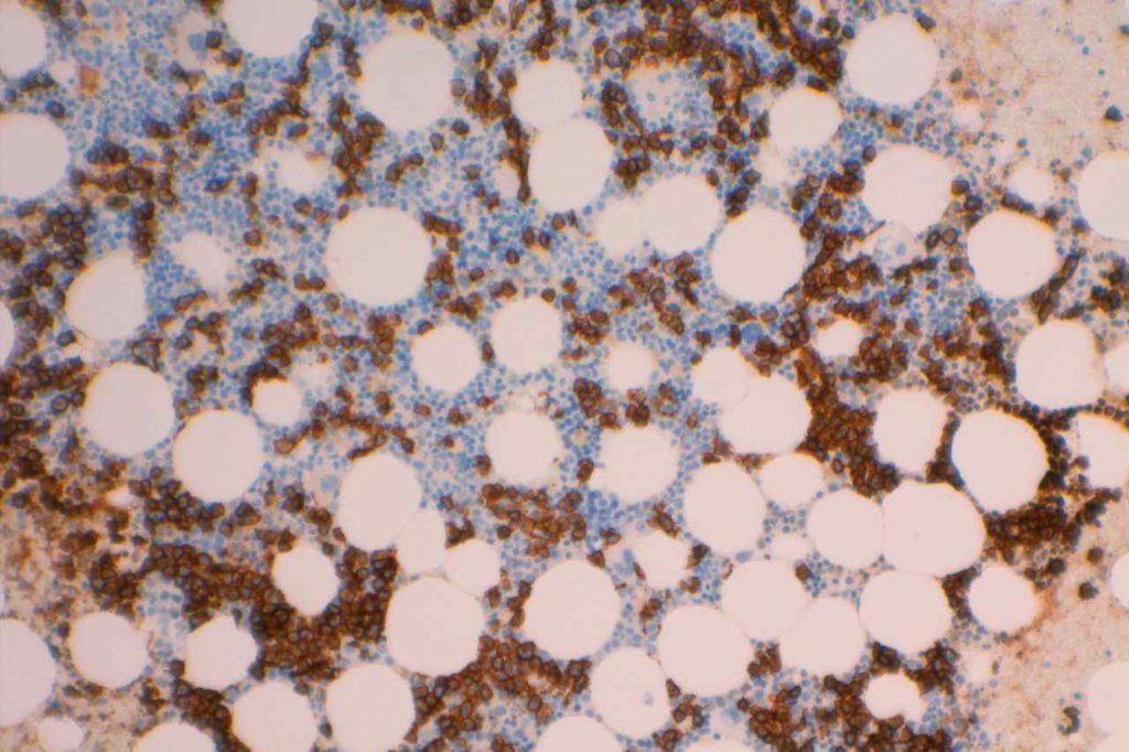
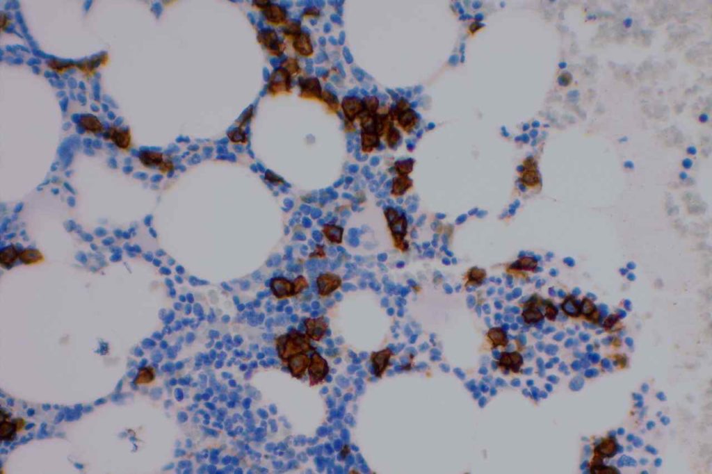
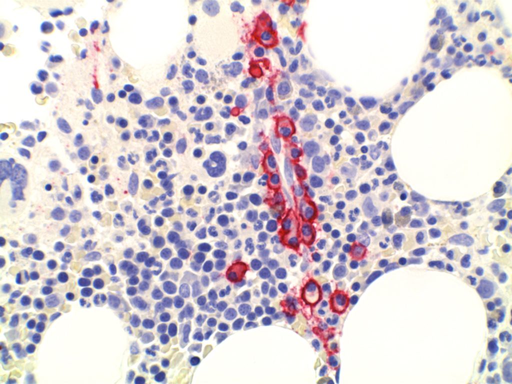
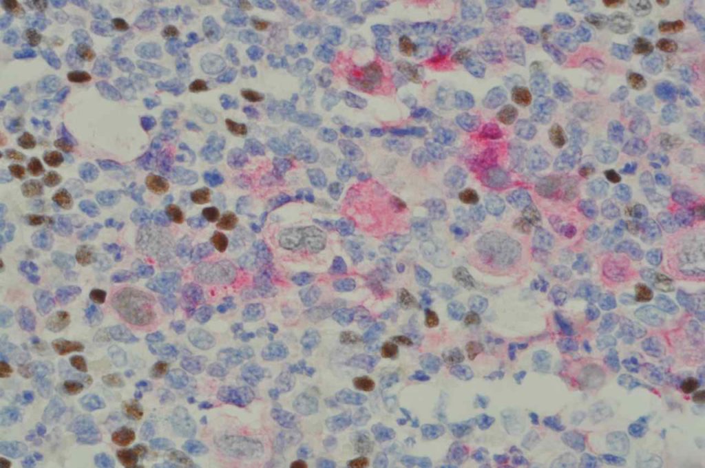
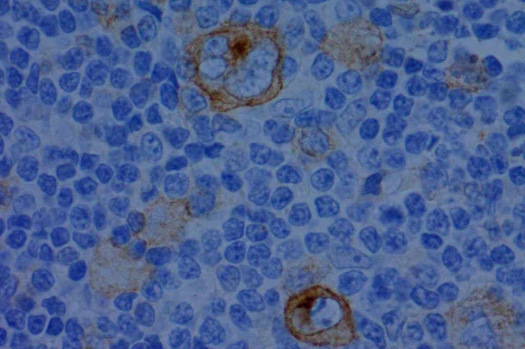
CD117 is most often used as a myeloblast marker in bone marrow biopsies, and to identify GISTs.  It is important to understand what else may stain with CD117 (bright staining of mast cells in BM bxs., compared to dim staining of blasts), so one does not misinterpret the expression pattern.  Erythroid precursor cell may also express CD117, which may cause confusion when evaluating for myeloblast percentage.  Ohgami recommends using CD117, E-cadherin, and CD34 in combination in this situation.  CD71 (erythroid precursor marker) may also be helpful.
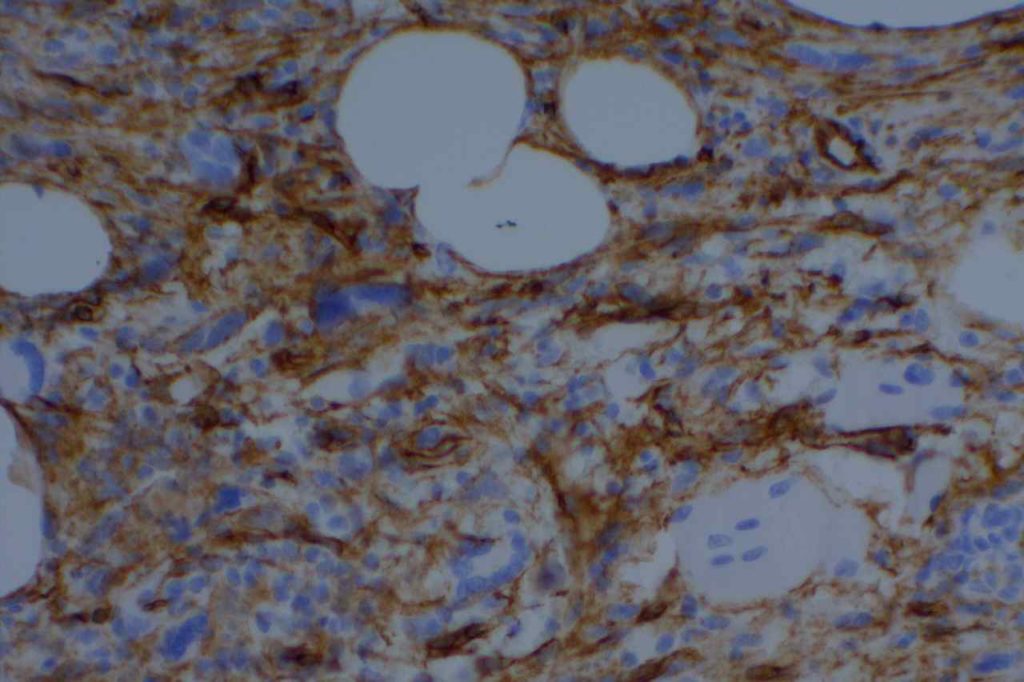
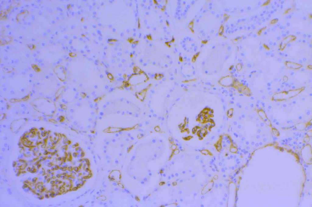
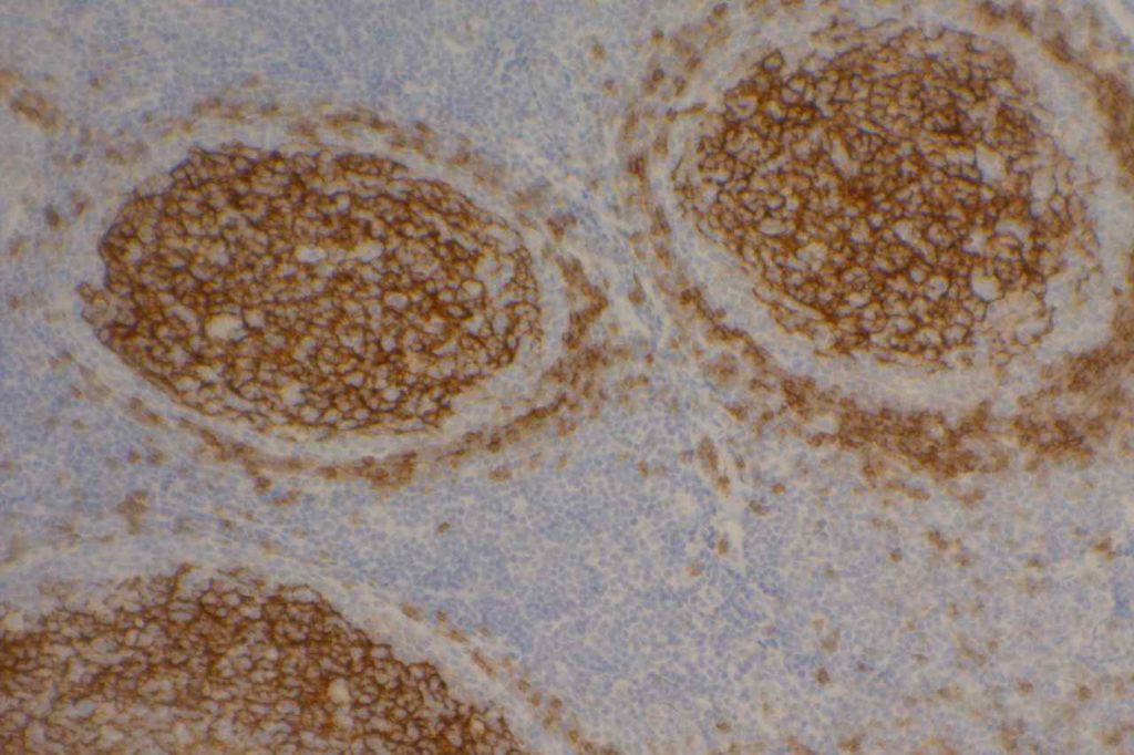
Renal Tumors
CD117 has been found to be expressed in 71-100% of oncocytomas and 83-100% of chromophobe renal cell carcinomas with other subtypes of renal cell carcinoma being non-reactive.  Therefore, CD117 may be a useful part of a larger panel (e.g. RCCMa, CD10, Vimentin, and cytokeratin) to diagnosis and subtype renal tumors (especially granular cell variant of RCC vs. chromophobe RCC or oncocytoma).
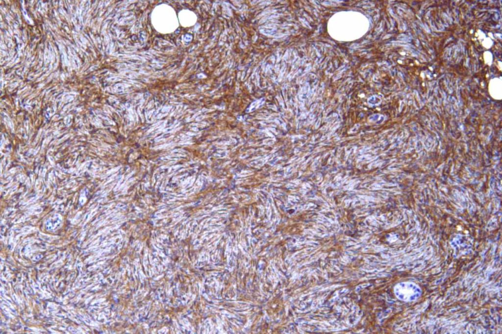
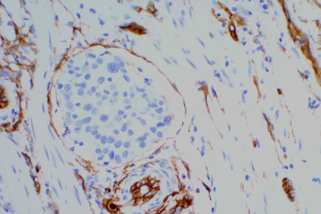
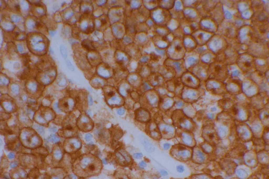
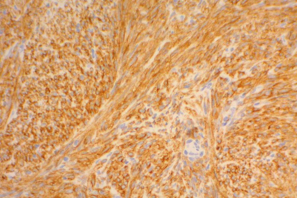
GIST
Approximately 95% of gastrointestinal stromal tumors (GIST) will react with CD117 by immunohistochemistry.  It is important to emphasize that CD117 expression does not necessarily correlate with response to imatinib therapy (tyrosine kinase inhibitor).  Lack of reactivity with CD117 should not exclude a patient from imatinib therapy.  CD117 negative (kit-negative) GIST often contain PDGFRA mutations, which may be sensitive to imatinib.  Sometimes weak expression of CD117 may be seen in non-GIST tumors, and this marker should be used as part of a larger panel incorporating the differential diagnosis (e.g. S-100, Desmin, CD34).  DOG-1 antibody may be expressed in CD117 negative GIST cases, which may serve as an alternative marker.
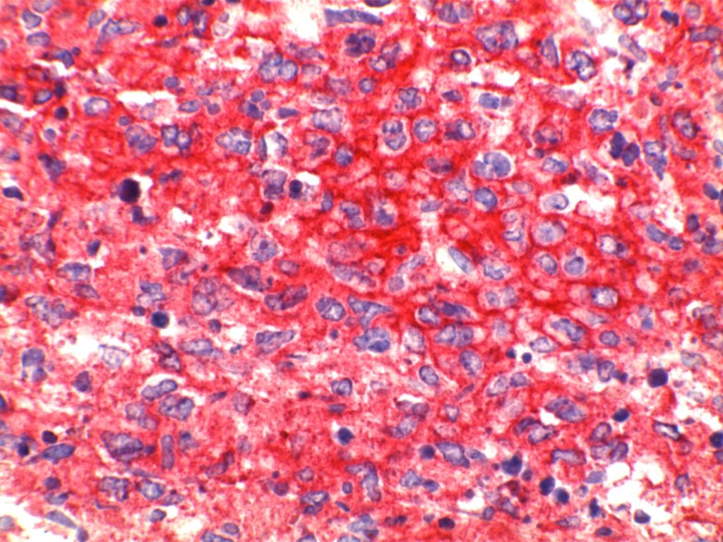
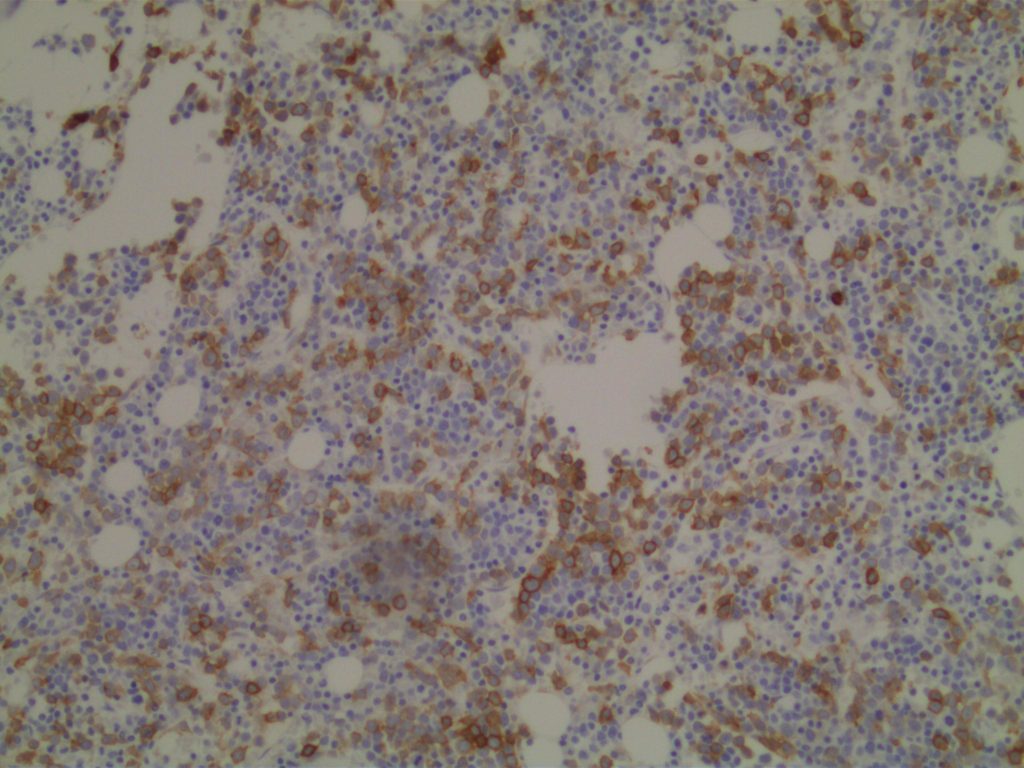
Photomicrographs
CD117 - GIST
CD117 expression in a gastrointestinal stromal tumor (GIST).
CD117 - Mast Cells
CD117 highlighting scattered mast cells in a bone marrow biopsy.
CD117 - CMML
CD117 highlighting areas of blasts in CMML.
CD117 - AML M6b
CD117 expression in myeloblasts in a case of AML M6b.

References:
Wick, MR. “Immunohistochemical approaches to the diagnosis of undifferentiated malignant tumor.”Annals of Diagnostic Pathology12(2008):72-84.
 
Bone Marrow IHC.  Torlakovic, EE, et. al. American Society for Clinical Pathology Pathology Press © 2009.  pp. 128.
 
Leidy, J., Khan, A., & Kandil, D. (2014). Basal-like breast cancer: update on clinicopathologic, immunohistochemical, and molecular features. Archives of Pathology & Laboratory Medicine, 138(1), 37–43. doi:10.5858/arpa.2012-0439-RA 
 
Huang, Q., Wu, H., Nie, L., Shi, J., Lebenthal, A., Chen, J., et al. (2013). Primary high-grade neuroendocrine carcinoma of the esophagus: a clinicopathologic and immunohistochemical study of 42 resection cases. The American Journal of Surgical Pathology, 37(4), 467–483. doi:10.1097/PAS.0b013e31826d2639 
 
Dunphy, C. H., O’Malley, D. P., Perkins, S. L., & Chang, C.-C. (2007). Analysis of immunohistochemical markers in bone marrow sections to evaluate for myelodysplastic syndromes and acute myeloid leukemias. Applied Immunohistochemistry & Molecular Morphology : AIMM / Official Publication of the Society for Applied Immunohistochemistry, 15(2), 154–159. doi:10.1097/PAI.0b013e318030dec7 
 
Patil, D. T., & Rubin, B. P. (2011). Gastrointestinal stromal tumor: advances in diagnosis and management. Archives of Pathology & Laboratory Medicine, 135(10), 1298–1310. doi:10.5858/arpa.2011-0022-RA 
 
Bénet, C., Gomez, A., Aguilar, C., Delattre, C., Vergier, B., Beylot-Barry, M., et al. (2011). Histologic and immunohistologic characterization of skin localization of myeloid disorders: a study of 173 cases. American Journal of Clinical Pathology, 135(2), 278–290. doi:10.1309/AJCPFMNYCVPDEND0 
 
Al-Ghawi, H., Asojo, O. A., Truong, L. D., Ro, J. Y., Ayala, A. G., & Zhai, Q. J. (2010). Application of Immunohistochemistry to the Diagnosis of Kidney Tumors. Pathology Case Reviews, 15(1), 25–34. doi:10.1097/PCR.0b013e3181d51c70 
 
Liegl-Atzwanger, B., Fletcher, J. A., & Fletcher, C. D. M. (2010). Gastrointestinal stromal tumors. Virchows Archiv : an International Journal of Pathology. doi:10.1007/s00428-010-0891-y 
 
Siegert, S. I., Diebold, J., Ludolph-Hauser, D., & Löhrs, U. (2004). Are gastrointestinal mucosal mast cells increased in patients with systemic mastocytosis? American Journal of Clinical Pathology, 122(4), 560–565. doi:10.1309/2880-LF7Q-6XK3-HA3Q
 
Antonescu, C. R. (2008). Targeted therapy of cancer: new roles for pathologists in identifying GISTs and other sarcomas. Modern Pathology : an Official Journal of the United States and Canadian Academy of Pathology, Inc, 21 Suppl 2, S31–6. doi:10.1038/modpathol.2008.9  
 
Medeiros, F., Corless, C. L., Duensing, A., Hornick, J. L., Oliveira, A. M., Heinrich, M. C., et al. (2004). KIT-negative gastrointestinal stromal tumors: proof of concept and therapeutic implications. The American Journal of Surgical Pathology, 28(7), 889–894. 
 
Wang, H.-Y., & Mills, S. E. (2005). KIT and RCC are useful in distinguishing chromophobe renal cell carcinoma from the granular variant of clear cell renal cell carcinoma. The American Journal of Surgical Pathology, 29(5), 640–646. 
 
Ohgami, R. S., Chisholm, K. M., Ma, L., & Arber, D. A. (2014). E-Cadherin Is a Specific Marker for Erythroid Differentiation and Has Utility, in Combination With CD117 and CD34, for Enumerating Myeloblasts in Hematopoietic Neoplasms. American Journal of Clinical Pathology, 141(5), 656–664. doi:10.1309/AJCP8M4QQTAZPGRP

CD138

CD138 (Syndecan-1) is expressed in mesenchymal and epithelial cells.  In the bone marrow hematopoietic cells, CD138 is a specific (and sensitive) marker for plasma cells (e.g. multiple myeloma).  The biggest pitfall in utilization of CD138 is plasmacytoid neoplasms showing expression of CD138 without consideration of a non-hematopoietic neoplasm expressing CD138!!  Always consider a metastatic process in the bone marrow, and utilize additional markers (AE1/AE3, kappa/lambda) to exclude/prove plasma cells origin.
 
The majority of epithelial neoplasms express CD138, and even a significant number of osteosarcomas, osteoid osteomas, and osteoblastomas react with CD138.  Recent literature suggests CD138 to be an adverse prognostic indicator in advanced and nonluminal subtype breast carcinomas.
Photomicrographs
CD138 - Bone Marrow
CD138 highlighting plasma cells in a bone marrow with multiple myeloma.
CD138-Ki67 - Bone Marrow
Combined CD138-Ki67 stain in a case of multiple myeloma.
CD138 - Bone Marrow
CD138 highlighting plasma cell in a bone marrow biopsy.
CD138 - Benign Plasma Cells
CD138 highlighting benign plasma cells with a perivascular cuffing pattern.

References
Bone Marrow IHC.  Torlakovic, EE, et. al. American Society for Clinical Pathology Pathology Press © 2009.  pp. 134.
 
Nunez, A. L., Siegal, G. P., Reddy, V. V. B., & Wei, S. (2012). CD138 (syndecan-1) expression in bone-forming tumors. American Journal of Clinical Pathology, 137(3), 423–428. doi:10.1309/AJCP6V4YPFBOCYXG 
 
Nguyen, T. L., Grizzle, W. E., Zhang, K., Hameed, O., Siegal, G. P., & Wei, S. (2013). Syndecan-1 overexpression is associated with nonluminal subtypes and poor prognosis in advanced breast cancer. American Journal of Clinical Pathology, 140(4), 468–474. doi:10.1309/AJCPZ1D8CALHDXCJ 
 
Joshi, R., Horncastle, D., Elderfield, K., Lampert, I., Rahemtulla, A., & Naresh, K. N. (2008). Bone marrow trephine combined with immunohistochemistry is superior to bone marrow aspirate in follow-up of myeloma patients. Journal of Clinical Pathology, 61(2), 213–216. doi:10.1136/jcp.2007.049130 

CD34

CD34 (human hematopoietic progenitor cell antigen) is expressed by endothelial cells and embryonic cells of the hematopoetic system.  CD34 is used in a wide variety of ways in diagnostic pathology.  It can generally be divided into hematopathology and non-hematopathology uses.  As with most situations in diagnostic immunohistochemistry, CD34 is often best used as part of a targeted panel considering the differential diagnosis at hand.
 
Hematopathology– CD34 will mark myeloblasts and a subset of lymphoblasts.  It is therefore not specific for myeloid differentiation (although fairy sensitive).  Normal myeloblasts will express CD34, and in cases of AML approximately 70% of cases will be positive.  Approximately 1/3rd of cases of ALL may express CD34.  It is also important to understand that not all blasts within a specific case may all express CD34, and correlation with flow cytometry data and aspirate count is critical.  CD34 also stains vascular endothelium, which makes for a nice internal control in bone marrow biopsies.  Granulocytic sarcomas (a.k.a. chloroma, soft tissue AML, or leukemia cutis) often have monocytic differentiation, and only ~5% of case demonstrate CD34 expression.
 
Non-Hematopathology– CD34 is expressed in cases of dermatofibrosarcoma protuberant (DFSP), hemangiopericytoma, solitary fibrous tumor, angiosarcoma & Kaposi’s sarcoma (>85%), epithelioid sarcoma (often), lymphocyte rich T cell lymphoma, gastrointestinal stromal tumors, and spindle cell lipomas.  CD34 is not expressed in dermatofibromas, desmoplastic mesothelioma, & endometrial stromal sarcoma.  CD34 is also an excellent vascular marker, which is helpful in identifying lymph-vascular invasion and tumors of vascular origin.
 
In hepatocellular carcinoma, CD34 highlights increased vascular structures associated with tumorigenesis.  Normal liver only shows CD34 expression in portal vasculature and sinusoids immediate adjacent to portal tracts.
 
Lymphatic & Vascular Invasion – CD34 is sometimes utilized to evaluate for “lymphovascular invasion” (LVI) in different tumors (e.g. breast more commonly).  CD34 and CD31 will stain both (blood) vascular endothelium (strong & sensitive) and also lymphatic endothelium (variable & less sensitive).  Sometimes CD34 will stain stromal cells, which can mimic endothelium (false positive).  
 
To determine the true nature of LVI (i.e. lymphatic vs. blood vascular invasion) additional markers specific for lymphatic endothelium (podoplanin or D2-40) need to be performed, and comparison made to CD31/CD34 for determination of invasion type:
  • Lymphatic invasion – CD34/CD31 +/-, D2-40/podoplanin +
  • Blood vascular invasion – CD34/CD31 +, D2-40/podoplanin –
CD34 Expression – Hematopathology
  • Myeloblasts (~70% of cases of AML)
  • Lymphoblasts (small subset)
  • Endothelial Cells (blood vascular and lymphatic)
  • Megakaryocytes (cytoplasmic)
CD34 Expression – Non-Hematopahtology
  • Skin – Dermatofibrosarcoma Protuberans – molecular analysis for COL1A1-PDGFB can be performed in difficult cases.
  • Skin – Superficial Acral Fibromyxoma (digital fibroma) – CD34+/CD99+/EMA+/- (cellular digital fibroma CD34+, CD99-/EMA-)
  • Skin – Superficial CD34+ Fibroblastic Tumor
  • Epithelioid Sarcoma (CD34+/CK+)
  • Hemangiopericytoma
  • Solitary Fibrous Tumor
  • Vascular Neoplasms (e.g. Kaposi Sarcoma, Angiosarcoma, etc.)
  • Normal Endothelium
  • Gastrointestinal Stromal Tumor (~70%, some smooth muscle neoplasms can express CD34 – up to 10%)
  • Spindle Cell Lipoma
  • Epithelioid Sarcoma
  • Subset of numerous other entities

Photomicrographs
CD34 - ALL
CD34 expression in a case of acute lymphoblastic leukemia/lymphoma (ALL).
CD34 - Spindle Cell Lipoma
CD34 expression in a spindle cell lipoma.
CD34 - GIST
CD34 expression in a gastrointestinal stromal tumor.
CD34 - DFSP
CD34 expression in a case of DFSP.
CD34-CD71 - AML
CD34 (red) – CD71 (brown) double stain in a case of AML.
CD34 - Lymphovascular Invasion
CD34 highlighting lymphovascular invasion of breast carcinoma.

References
Mohammed RAA, Martin SG, Gill MS, Green AR, Paish EC, Ellis IO. Improved methods of detection of lymphovascular invasion demonstrate that it is the predominant method of vascular invasion in breast cancer and has important clinical consequences. Am J Surg Pathol. 2007;31: 1825–1833. doi:10.1097/PAS.0b013e31806841f6
 
Yang H, Yu L. Cutaneous and Superficial Soft Tissue CD34(+) Spindle Cell Proliferation. Arch Pathol Lab Med. 2017;141: 1092–1100. doi:10.5858/arpa.2016-0598-RA
 
Rao N, Colby TV, Falconieri G, Cohen H, Moran CA, Suster S. Intrapulmonary solitary fibrous tumors: clinicopathologic and immunohistochemical study of 24 cases. Am J Surg Pathol. 2013;37: 155–166. doi:10.1097/PAS.0b013e31826a92f5
 
de Smet D, Trullemans F, Jochmans K, Renmans W, Smet L, Heylen O, et al. Diagnostic Potential of CD34+ Cell Antigen Expression in Myelodysplastic Syndromes. Am J Clin Pathol. 2012;138: 732–743. doi:10.1309/AJCPAGVO27RPTOTV
 
Rosado FGN, Itani DM, Coffin CM, Cates JM. Utility of immunohistochemical staining with FLI1, D2-40, CD31, and CD34 in the diagnosis of acquired immunodeficiency syndrome-related and non-acquired immunodeficiency syndrome-related Kaposi sarcoma. Arch Pathol Lab Med. 2012;136: 301–304. doi:10.5858/arpa.2011-0213-OA
 
Patil DT, Rubin BP. Gastrointestinal stromal tumor: advances in diagnosis and management. Arch Pathol Lab Med. 2011;135: 1298–1310. doi:10.5858/arpa.2011-0022-RA
 
Bénet C, Gomez A, Aguilar C, Delattre C, Vergier B, Beylot-Barry M, et al. Histologic and immunohistologic characterization of skin localization of myeloid disorders: a study of 173 cases. Am J Clin Pathol. 2011;135: 278–290. doi:10.1309/AJCPFMNYCVPDEND0
 
Dunphy CH, O’Malley DP, Perkins SL, Chang C-C. Analysis of immunohistochemical markers in bone marrow sections to evaluate for myelodysplastic syndromes and acute myeloid leukemias. Appl Immunohistochem Mol Morphol. 2007;15: 154–159. doi:10.1097/PAI.0b013e318030dec7
 
Dunphy CH, Polski JM, Evans HL, Gardner LJ. Evaluation of bone marrow specimens with acute myelogenous leukemia for CD34, CD15, CD117, and myeloperoxidase. Arch Pathol Lab Med. 2001;125: 1063–1069.
 
Wang HL, Kim CJ, Koo J, Zhou W, Choi EK, Arcega R, et al. Practical Immunohistochemistry in Neoplastic Pathology of the Gastrointestinal Tract, Liver, Biliary Tract, and Pancreas. Arch Pathol Lab Med. 2017;141: 1155–1180. doi:10.5858/arpa.2016-0489-RA
 
Wick, MR. “Immunohistochemical approaches to the diagnosis of undifferentiated malignant tumor.”Annals of Diagnostic Pathology12(2008):72-84.
 
Bone Marrow IHC.  Torlakovic, EE, et. al. American Society for Clinical Pathology Pathology Press © 2009.  pp. 88.

CD31

CD31 (PECAM-1) is thought of as a highly sensitive and specific marker for vascular endothelium.  It is helpful to identify tumors of vascular origin, and also to identify lympho-vascular invasion by tumors.  CD31 may also stain monocytes, megakaryocytic, and granulocytes in addition to endothelial cells.  Plasma cells may variably express CD31 (reactive PCs more common).  CD31 (like CD34) is more sensitive (and stronger staining) for blood vascular endothelium compared to lymphatic endothelium (less sensitive and variable staining).
Lymphatic & Vascular Invasion
CD31 is sometimes utilized to evaluate for “lymphovascular invasion” (LVI) in different tumors (e.g. breast more commonly).  CD34 and CD31 will stain both (blood) vascular endothelium (strong & sensitive) and also lymphatic endothelium (variable & less sensitive).  To determine the true nature of LVI (i.e. lymphatic vs. blood vascular invasion) additional markers specific for lymphatic endothelium (podoplanin or D2-40) need to be performed, and comparison made to CD31/CD34 for determination of invasion type:
  • Lymphatic invasion – CD34/CD31 +/-, D2-40/podoplanin +
  • Blood vascular invasion – CD34/CD31 +, D2-40/podoplanin –
Kaposi Sarcoma (KS)
Kaposi sarcoma is a vascular neoplasm associated with immunodeficiency (usually HIV/AIDS) and is caused by human herpesvirus 8 (HHV-8).  Vascular markers (CD31, CD34, D2-40, and FLI1) are helpful (in combination with HHV-8) for diagnosis.  CD31 is expressed in 58-75% of cases (strong and diffuse staining).  CD34 is considered more sensitive (92%).
Photomicrographs
CD31 - Kidney
CD31 highlighting vasculature in a normal section of kidney.

References
Wick, MR. “Immunohistochemical approaches to the diagnosis of undifferentiated malignant tumor.”Annals of Diagnostic Pathology12(2008):72-84.
 
Vanchinathan V, Mirzamani N, Mizramani N, Kantipudi R, Schwartz EJ, Sundram UN. The vascular marker CD31 also highlights histiocytes and histiocyte-like cells within cutaneous tumors. Am J Clin Pathol. 2015;143: 177–85– quiz 305. doi:10.1309/AJCPRHM8CZH5EMFD
 
Rosado FGN, Itani DM, Coffin CM, Cates JM. Utility of immunohistochemical staining with FLI1, D2-40, CD31, and CD34 in the diagnosis of acquired immunodeficiency syndrome-related and non-acquired immunodeficiency syndrome-related Kaposi sarcoma. Arch Pathol Lab Med. 2012;136: 301–304. doi:10.5858/arpa.2011-0213-OA
 
Morgado JMT, Sánchez-Muñoz L, Teodósio CG, Jara-Acevedo M, Álvarez-Twose I, Matito A, et al. Immunophenotyping in systemic mastocytosis diagnosis: ‘CD25 positive’ alone is more informative than the “CD25 and/or CD2” WHO criterion. Mod Pathol. 2012;25: 516–521. doi:10.1038/modpathol.2011.192
 
Mohammed RAA, Martin SG, Gill MS, Green AR, Paish EC, Ellis IO. Improved methods of detection of lymphovascular invasion demonstrate that it is the predominant method of vascular invasion in breast cancer and has important clinical consequences. Am J Surg Pathol. 2007;31: 1825–1833. doi:10.1097/PAS.0b013e31806841f6
 
Bone Marrow IHC.  Torlakovic, EE, et. al. American Society for Clinical Pathology Pathology Press © 2009.  pp. 81.

CD30

CD30 (Ki-1 antigen) is a member of the tumor necrosis receptor factor family (TNRF) and is not a specific marker.  CD30 will mark activated benign lymphoid cells in addition to several characteristic lymphoproliferative disease subtypes (e.g. Classical Hodgkin lymphoma and anapestic large cell lymphoma) of both B-cell and T-cell lineages.
CD30 Expression Pattern
  • Benign “Activated” lymphoid cells
  • EBV-driven lymphoproliferative disorders and reactive proliferations (e.g. mononucleosis)
  • Classical Hodgkin Lymphoma
  • Anaplastic Large Cell Lymphoma (ALK+/ALK-)
  • Peripheral T-cell Lymphomas (subset)
  • Diffuse Large B-cell Lymphoma (subset)
  • Primary Mediastinal Large B-Cell Lymphoma
  • Embryonal Carcinoma
  • Germ Cell Tumors
 CD30 is an “activation” marker on T and B cells.  This marker is most often used in the work-up of cases of Hodgkin lymphoma, anaplastic large cell lymphoma (ALCL), and embryonal carcinoma.  The staining pattern has a membraneous and Golgi pattern.  B5 fixative has negative impact on staining.  CD30 is also helpful in the diagnosis of mediastinal large B cell lymphoma (focal/patchy and dim staining).
 
Unfortunately, CD30 lacks specificity, and in addition to the staining pattern in normal cells compartments, it may also be expressed in a variety of disease processes including:  peripheral T-cell lymphoma (occasional), primary effusion lymphoma, cutaneous CD30+ lymphoproliferative disorders, DLBCL (variable, focal), plasmablastic lymphoma, sinonasal NK/T cell lymphoma, embryonal carcinoma of the testis (100%), Yolk sac tumors (24%), &  mesothelioma (rare).
 
Nodular LP Hodgkin lymphoma, Adult T cell leukemia/lymphoma, pre B ALL, and systemic mastocytosis should not express CD30.
CD30+ DLBCL
Cases of DLBCL which co-express CD30 (~14%, n=903) had a more favorable 5-year survival (79% vs. 59%) in both germinal center and activated B-cell phenotypes.  Gene expression profiling (GEP) revealed down regulation of proliferation and B-cell receptor signaling with up regulation of nuclear factor kappaB activation and lymphocyte survival.  CD30+ EBV+ DLBCLis a unique subset of lymphomas with an aggressive clinical course.  The WHO classification separates EBV+ cases into a separate category.
 
CD30+ Diffuse Large B-Cell Lymphoma
CD30 expression in a diffuse large B-cell lymphoma. CD30+ cases are associated with a relatively good prognosis (excluding EBV+ cases).
CD30/PAX -5 - Classical Hodgkin Lymphoma
CD30 (red) showing variable expression and co-expression of PAX-5 (variable dim brown nuclear expression) in classical Hodgkin lymphoma
CD30 - Classical Hodgkin Lymphoma
CD30 expression in classical Hodgkin lymphoma (CHL).
CD30 - ALCL
Anaplastic Large Cell Lymphoma (ALCL) with strong diffuse expression of CD30.
Clinical Significance
Brentuximab vedotin (Adcetris®) is an anti-CD30 monoclonal antibody used to treat anaplastic large cell lymphoma and relapsed or refractory Hodgkin lymphoma.  Ongoing research with promise is active in other CD30 positive neoplastic processes.

References
Wick, MR. “Immunohistochemical approaches to the diagnosis of undifferentiated malignant tumor.”Annals of Diagnostic Pathology12(2008):72-84.
 
Bone Marrow IHC.  Torlakovic, EE, et. al. American Society for Clinical Pathology Pathology Press © 2009.  pp. 78-79.
 
Hu S, Xu-Monette ZY, Balasubramanyam A, Manyam GC, Visco C, Tzankov A, et al. CD30 expression defines a novel subgroup of diffuse large B-cell lymphoma with favorable prognosis and distinct gene expression signature: a report from the International DLBCL Rituximab-CHOP Consortium Program Study. Blood. 2013;121: 2715–2724. doi:10.1182/blood-2012-10-461848
 
Ansell SM. Brentuximab vedotin. Blood. 2014. doi:10.1182/blood-2014-06

CD25

CD25 is an IL-2 receptor.  CD25 is most often used as a marker for hairy cell leukemia, systemic mastocytosis, and adult T cell leukemia/lymphoma.  CD25 may stain activated B and T cells and cases of lymphoplasmacytic lymphoma (LPL).  CD25 is similar to CD30 in that it is an “activation” marker, and not specific.  Macrophages, and osteoblasts also often express CD25.
 
Non-neoplastic mast cells, marginal zone B cell lymphoma, T cell large granular leukemia/lymphoma, and variant hairy cell leukemia are usually negative for CD25.
CD25 Expression Pattern

Photomicrographs
CD25 expression in systemic mastocytosis.

References
Bone Marrow IHC.  Torlakovic, EE, et. al. American Society for Clinical Pathology Pathology Press © 2009.  pp. 75-76.
 
Diagnostic Immunohistochemistry:  Theranostic and Genomic Applications [edited by] David J. Dabbs. 3rd Edition.  pp. 165.
 
Lin, P., Molina, T. J., Cook, J. R., & Swerdlow, S. H. (2011). Lymphoplasmacytic lymphoma and other non-marginal zone lymphomas with plasmacytic differentiation. American Journal of Clinical Pathology, 136(2), 195–210. doi:10.1309/AJCP8FOIVTB6LBER
 
Karube, K., Suzumiya, J., Okamoto, M., Takeshita, M., Maeda, K., Sakaguchi, M., et al. (2007). Adult T-cell lymphoma/leukemia with angioimmunoblastic T-cell lymphomalike features: Report of 11 cases. The American Journal of Surgical Pathology, 31(2), 216–223. doi:10.1097/01.pas.0000213325.79368.2c
 
Siegert, S. I., Diebold, J., Ludolph-Hauser, D., & Löhrs, U. (2004). Are gastrointestinal mucosal mast cells increased in patients with systemic mastocytosis? American Journal of Clinical Pathology, 122(4), 560–565. doi:10.1309/2880-LF7Q-6XK3-HA3Q
 
Lin, P., & Medeiros, L. J. (2005). Lymphoplasmacytic lymphoma/waldenstrom macroglobulinemia: an evolving concept. Advances in Anatomic Advances in Anatomic Pathology, 12(5), 246–255. 

CD23

CD23 is a transmembrane glycoprotein expressed by different hematopoietic cells and is a low-affinity receptor for IgE.  It is also involved in promoting survival of B-cells in the germinal center.  CD23 is useful as a follicular cell dendritic cell marker and is classically expressed in cases of CLL/SLL.  CD23 has been identified in many types of lymphomas, but is most commonly used to differentiate between CLL/SLL (CD23+) and mantle cell lymphoma (CD23-).  This testing is typically performed by flow cytometry, but immunohistochemisty for CD23 is available.  Expression of CD23 has been associated with better prognosis (at least in limited published data) in follicular lymphoma, CLL/SLL, mantle cell lymphoma, and diffuse large B-cell lymphoma dependent upon expression characteristics.  CD23 is not commonly performed/used as a prognostic marker for B-cell lymphomas.
 
Rarely CD23 may be expressed in cases of Hairy cell leukemia (17%) and DLBCL (16%).  Approximately 70% of Mediastinal large B-cell lymphoma cases express CD23.  Practically, this IHC marker is used as a follicular dendritic cell marker and to help differentiate CLL/SLL from mantle cell lymphoma.  Follicular dendritic cell tumors will also express CD23 like CD21.  CD21 is more sensitive compared to CD23 as a follicular dendritic marker.
 
Follicular Lymphoma (FL) – CD23 has been found to be expressed in some cases of FL, especially from inguinal lymph nodes, and prognosis appears comparatively better.   Olteanuet. al found that 87% of inguinal lymph nodes expressed CD23, compared to 61% from other sites, and that survival was prolonged more in CD23+ cases. 
 
Diffuse Large B-Cell Lymphoma – A subset of DLBCLs may express CD23, which may have a better prognosis (CD23 is not commonly performed for this purpose).
 
Chronic Lymphocytic Leukemia/Small Lymphocytic Lymphoma (CLL/SLL) – CD23 expression is characteristic of CLL/SLL, particularly in comparison to another CD5+ lymphoma, mantle cell lymphoma.  Strong membrane expression has been associated with a better outcome.  DiRaimondo,et. al found ~6% of CLL cases to be CD23 negative (flow cytometry), and they had a worse prognosis.  Many of these cases may have been misdiagnosed mantle cell lymphomas.
 
Mantle Cell Lymphoma (MCL) – CD23 is characteristically negative in MCL, which helps to differentiate it from CLL/SLL.  However, ~21% of cases of MCL were found to be CD23+ by Gao,et. al, and other studies have shown CD23 expression in MCL ranging from 0% to 45% (most data appears to be based on flow cytometry).
CD23 Expression Pattern
  • CLL/SLL – characteristically expressed (6% may be negative, probably much lower)
  • Mantle cell lymphoma may be CD23+ (21%+, 0-45%)
  • B-cell Lymphomas (e.g. some DLBCL and follicular lymphomas may show expression)
  • Follicular Dendritc Cells (not as sensitive as CD21)
  • B-cells in mantle zone of lymphoid follicles

Photomicrographs

CD23 - Tonsil
CD23 expression highlighting follicular dendritic meshwork in a normal tonsil.


Reference
Bone Marrow IHC.  Torlakovic, EE, et. al. American Society for Clinical Pathology Pathology Press © 2009.  pp. 69.
 
Linderoth J, Jerkeman M, Cavallin-Stahl E, et al. Immunohistochemical expression of CD23 and CD40 may identify prognostically favorable subgroups of diffuse largeB-cell lymphoma: a Nordic Lymphoma Group Study.ClinCancer Res.2003;9:722-728.
 
Olteanu H, Fenske TS, Harrington AM, Szabo A, He P, Kroft SH. CD23 Expression in Follicular Lymphoma: Clinicopathologic Correlations. Am J Clin Pathol. 2011;135: 46–53. doi:10.1309/AJCP27YWLIQRAJPW
 
Gao J, Peterson L, Nelson B, Goolsby C, Chen Y-H. Immunophenotypic variations in mantle cell lymphoma. Am J Clin Pathol. 2009;132: 699–706. doi:10.1309/AJCPV8LN5ENMZOVY
 
Troxell ML, Schwartz EJ, van de Rijn M, Ross DT, Warnke RA, Higgins JP, et al. Follicular dendritic cell immunohistochemical markers in angioimmunoblastic T-cell lymphoma. Appl Immunohistochem Mol Morphol. 2005;13: 297–303.
 
Dalton RR, Admirand JH, Medeiros LJ. Small Lymphocytic Lymphoma. Pathology Case Reviews. 2004;9: 7.
 
DiRaimondo F, Albitar M, Huh Y, O’Brien S, Montillo M, Tedeschi A, et al. The clinical and diagnostic relevance of CD23 expression in the chronic lymphoproliferative disease. Cancer. 2002;94: 1721–1730. doi:10.1002/cncr.10401

CD19

CD19 is a B-cell marker.  Until recently this antibody has only been available for use in flow cytometry.  Now CD19 is available as an immunohistochemistry (IHC) marker, with similar sensitivity for B-cells to CD20.  The IHC expression pattern is cytoplasmic and membranous.  It is normally expressed on both Precursor B-cells and B-cells.  CD19 combined with TdT would be specific to detect precursor B-cells. CD20 is not expressed on precursor B-cells, but mature B-cell with surface light chain expression (usually).

Continue reading CD19

CD15 (Leu-M1)

CD15 (Leu-M1) is a hematopoietic antigen expressed by granulocytes, monocytes, Reed-Sternberg cells, and subsets of T and B-cells.  CD15 may also be expressed in glandular or neuroendocrine epithelial cells as well as part of a panel to distinguish between metastatic adenocarcinoma (positive) and mesothelioma (negative).
CD15 Expression
  • Neutrophils (~90%)
  • Monocytes (30-60%)
  • Promyelocytes
  • Adenocarcinoma (72%)
  • Classical Hodgkin Lymphoma cells
  • T-cell Lymphomas (subset, MF)
  • Acute Myelogenous Leukemia (particularly with monocytic differentiation)
Classical Hodgkin Lymphoma
CD15 stains strongly the cytoplasm of granulocytes.  CD15 is most commonly used as a diagnostic marker for classical Hodgkin lymphoma (CHL).  The combined staining of CD15 and CD30 in the appropriate morphologic setting is very specific for Hodgkin Lymphoma.  The biggest issue with this marker in the diagnosis of CHL is sensitivity.  In a well-optimized setting using a preferred clone, the sensitivity can be fairly high, ~70%.  In general when CHL cases are positive for CD15, it will stain a subset of the CD30 positive cells.
 
When this marker is not well optimized and/or using a suboptimal clone, then the negative results can not be trusted (even when expressed in background granulocytes).  Comparison analysis with a reference lab to compare IHC expression in validation cases is recommended for this antibody.  PAX-5 is also a helpful marker, when CD15 is negative and CHL is suspected, as it will be dimly expressed (compared to background B-cells) in ~90% of CHL cases.
Bone Marrow
CD15 will stain at least a subset of normal granulocytes(~90%), monocytes (30-60%), promyelocytes in the bone marrow.  A subset of cases of acute myelogenous leukemia (AML) will express CD15.  CD15 is a myeloid marker, and appears to be preferentially expressed in AML cases with monocytic differentiation (not entirely sensitive or specific).  Flow cytometry is more sensitive in detected CD15 expression than immunohistochemistry (IHC).  A subset of neoplastic T and B-cells may also express CD15.
Mesothelioma vs. Adenocarcinoma
CD15 has been used as part of a panel to differentiate between mesothelioma and adenocarcinoma.  CD15 is expressed in ~7% of epithelioid mesotheliomas and is not known to be expressed in sarcomatous mesothelioma while being expressed in a majority of adenocarcinomas (~72%).  CD15 is not typically one of the main markers used in the mesothelioma vs. adenocarcinoma panel (there are other markers with better sensitivity/specificity profiles), but may be helpful in certain circumstances.
CD15 Expression (Marchevsky)
  • Epithelial mesothelioma – 7%
  • Sarcomatous mesothelioma – 0%
  • Adenocarcinoma – 72%
  • Squamous cell carcinoma – 30%
  • Renal cell carcinoma – 25-100%
Photomicrographs

CD15 - Classical Hodgkin Lymphoma
CD15 – Classical Hodgkin Lymphoma.  Note dark staining granulocytes in the background.

CD15 - Classical Hodgkin Lymphoma
CD15 – Classical Hodgkin Lymphoma


References
Wick, MR. “Immunohistochemical approaches to the diagnosis of undifferentiated malignant tumor.”Annals of Diagnostic Pathology12(2008):72-84.
 
Bone Marrow IHC.  Torlakovic, EE, et. al. American Society for Clinical Pathology Pathology Press © 2009.  pp. 51.
 
Marchevsky, A. M. (2008). Application of immunohistochemistry to the diagnosis of malignant mesothelioma. Archives of Pathology & Laboratory Medicine, 132(3), 397–401. 
 
Dunphy CH, Polski JM, Evans HL, Gardner LJ. Evaluation of bone marrow specimens with acute myelogenous leukemia for CD34, CD15, CD117, and myeloperoxidase. Arch Pathol Lab Med. 2001;125: 1063–1069.