Category Archives: Lymph Node

In Situ Follicular Neoplasia (ISFN)

In Situ Follicular Neoplasia (ISFN), which has been previously referred to as Follicular Lymphoma Is Situ (FLIS) is found in approximately 2% of otherwise reactive lymph nodes.  ISFN is defined as partial to total colonization of germinal centers by B-cells containing the t(14;18) BCL-2/IgH translocation characteristic of follicular lymphoma.

ISFN found as an incidental finding have a risk of developing follicular lymphoma (FL) of <= 5%.  The amount of involvement of ISFN within a lymph node does not appear to be directly related to the risk of future FL.

If patients have other evidence of lymphadenopathy, then additional biopsies may be helpful to exclude a B-cell lymphoma at another site.

Morphology

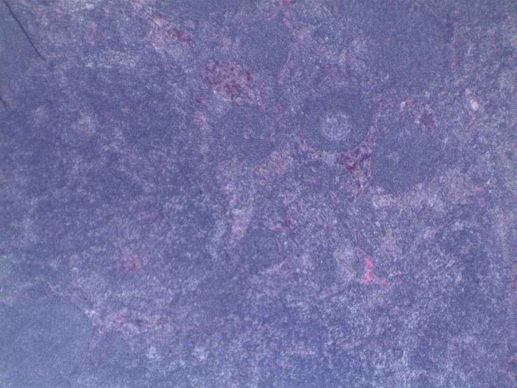
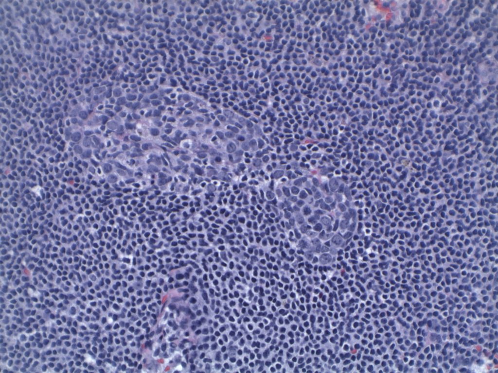
H&E staining demonstrates normal appearing lymph node architecture with well-formed and well-demarcated germinal centers.  Follicles with ISFN at first glance do not look significantly different from the adjacent reactive follicles.  Some areas of ISFN may more prominent and closely associated centrocytes (subtle).

Immunohistochemistry

  • CD10 – Strong expression
  • Bcl-6 – Positive
  • Bcl-2 – Positive (often stronger expression than surrounding lymphocytes)
  • Ki-67 – Usually significantly lower expression than that of adjacent reactive germinal centers
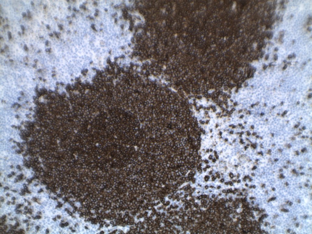
  • CD20 – Positive

Photomicrographs
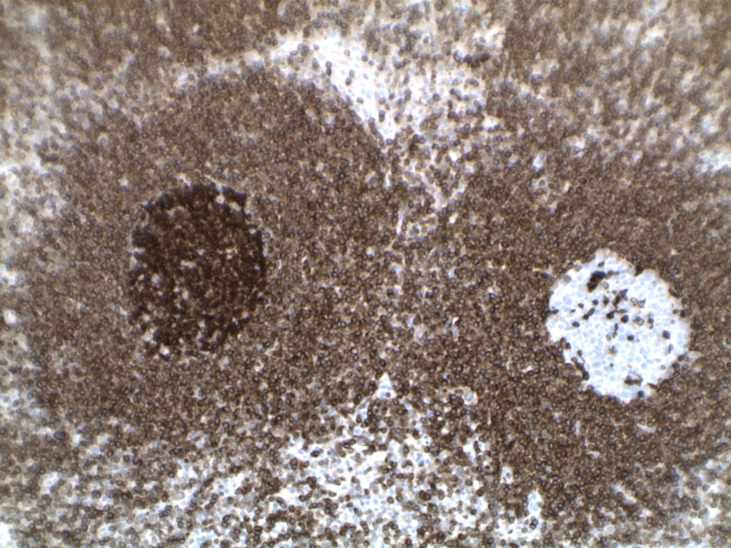
H&E section containing ISFN. Morphologically the node appears consistent with an otherwise reactive lymph node.
H&E section of ISFN.
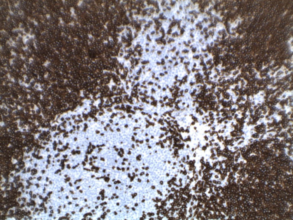
CD3 expression in ISFN.
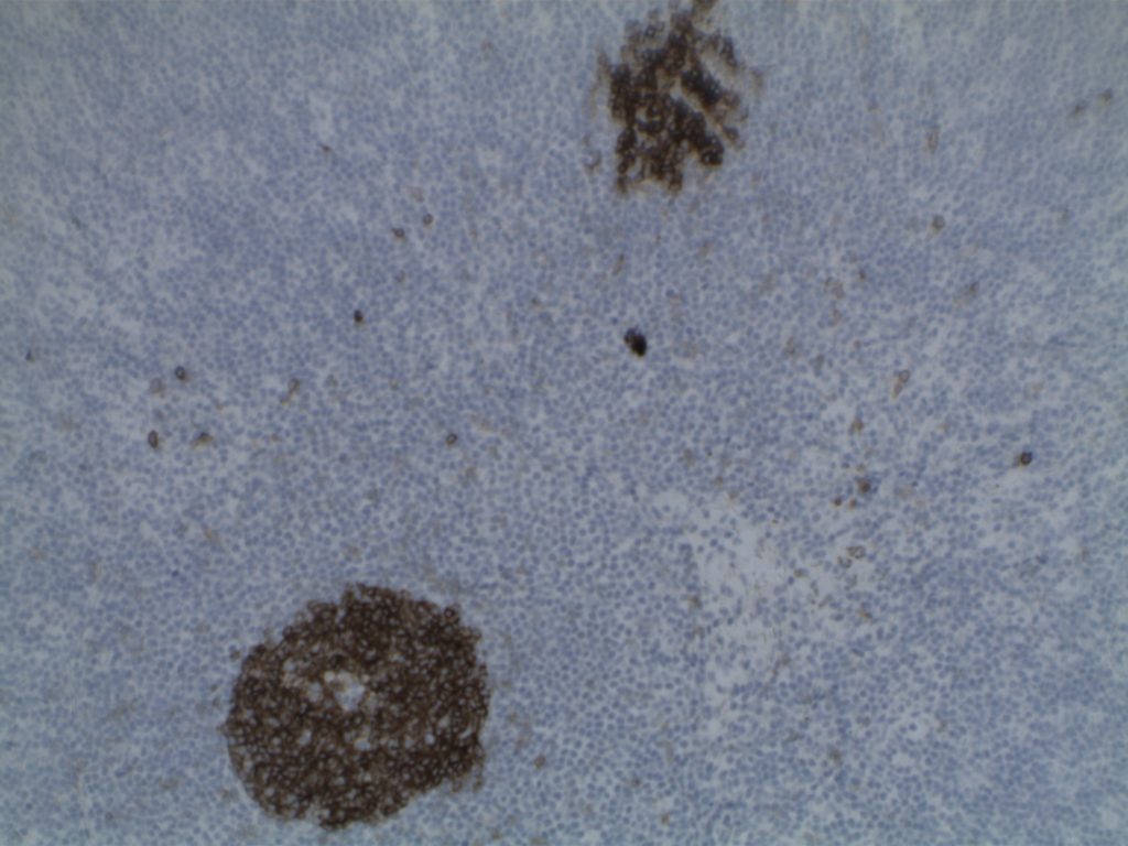
CD20 expression in a follicle containing ISFN.
CD10 expression in ISFN.
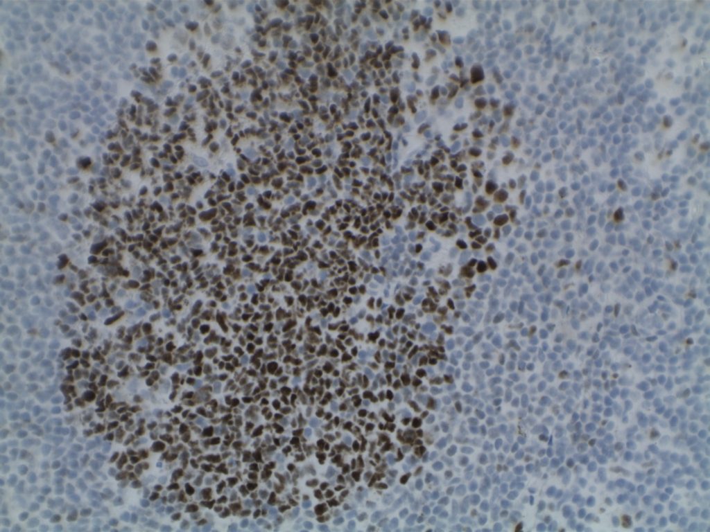
Bcl-6 expression in ISFN. Note the crowding of centrocytes highlighted by the Bcl-6 positive cells.
Strong Bcl-2 expression in the ISFN. Note the brighter expression compared to the surrounding T-cells and mantle zone. An adjacent reactive follicle is negative for Bcl-2.
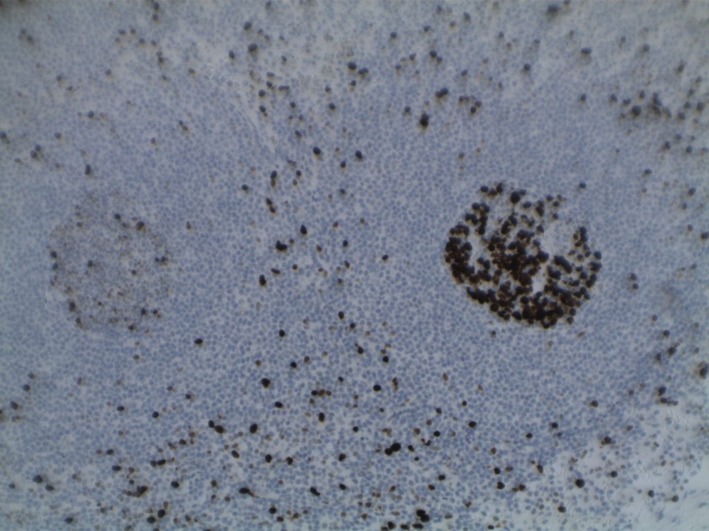
Low Ki-67 expression in ISFN (left side of image) compared to the high proportion of cell expression in the reactive follicle on the right.

Differentiating from partial involvement by follicular lymphoma

Sometimes it can be difficult to differentiate partial involvement of a lymph node by FL from ISFN.  The following features are helpful to differentiate partial involvement by FL from ISFN:

  • ISFN – Intact nodal architecture 
  • Partial involvement by FL – Altered architecture on H&E sections
  • ISFN – Normal sized follicles
  • Partial involvement by FL – Often enlarged follicles
  • ISFN – Sharp border between the follicle center and the surrounding mantle zone
  • Partial involvement by FL – Interface between the follicle center and mantle zone is often fuzzy or blurred.
  • ISFN – Bcl-2 is strongly positive (usually brighter than the surrounding T-cells and mantle zones); CD10 strongly positive
  • Partial involvement by FL – variable Bcl-2 and CD10 expression
  • ISFN – Atypical cells are confined to the follicle center
  • Partial involvement by FL – atypical cells (CD10/bcl2+) can be found outside of the follicle center areas

References

Swerdlow SH, Campo E, Harris, NL, Jaffe ES, Pileri SA, Stein H, Thiele J (Eds); WHO Classification of Tumours of Haematopoietic and Lymphoid Tissues (Revised 4th edition). IARC: Lyon 2017

DLBCL Associated with Chronic Inflammation

DLBCL associated with chronic inflammation is a special subtype of DLBCL first described in 1987 and subsequently recognized as a specific and separate entity in the 2008 WHO hematopathology classification.

Morphologically, it is recognized as a diffuse large B-cell lymphoma that arises in the setting of long-standing chronic inflammation and is associated with EBV infection. Commonly, these present as tumor masses involving body cavities. This is classically and originally described as pleural-based lesions in patients with chronic pyothorax (artificial pneumothorax for pulmonary tuberculosis or tuberculosis pleuritis).

Originally, these were designated as ‘pyothorax-associated lymphomas’ (PAL) and characteristically are associated with EBV.  It is thought that these lesions arise as a result of ‘local’ immunodeficiency, and seem to carry the following common characteristics (regardless of location):

  1. Association with EBV
  2. Confined space (often body cavity)
  3. Long standing/slow growing lesion associated with chronic inflammation
  4. Morphologic characteristics of diffuse large B-cell lymphoma
Immunophenotype

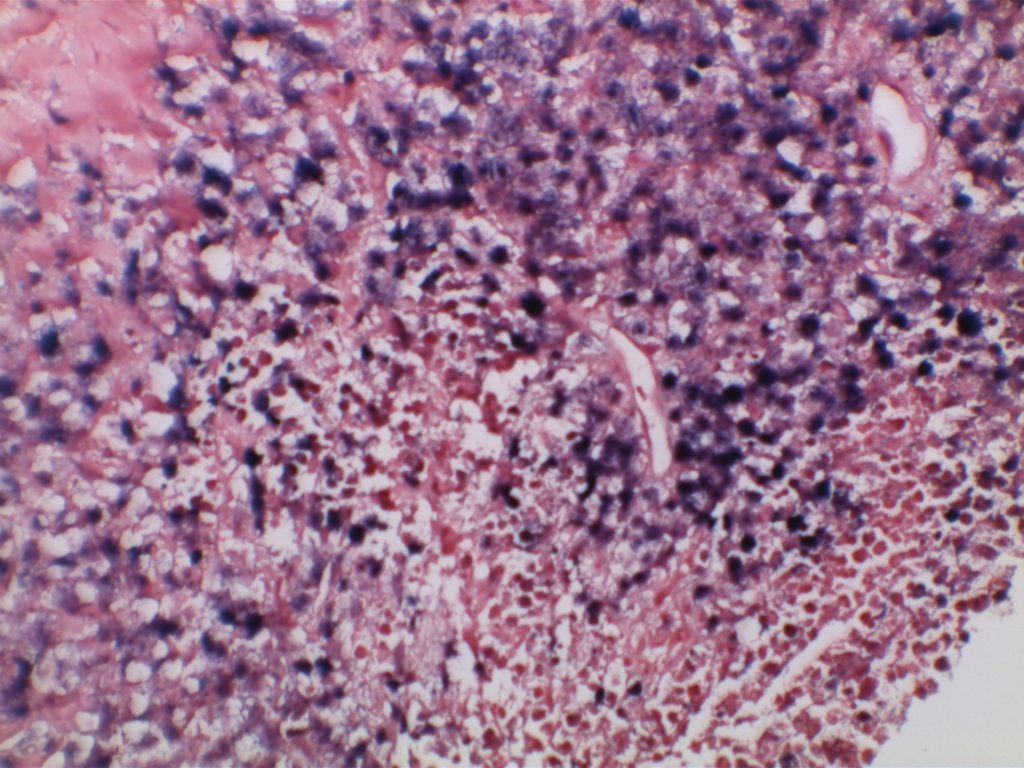
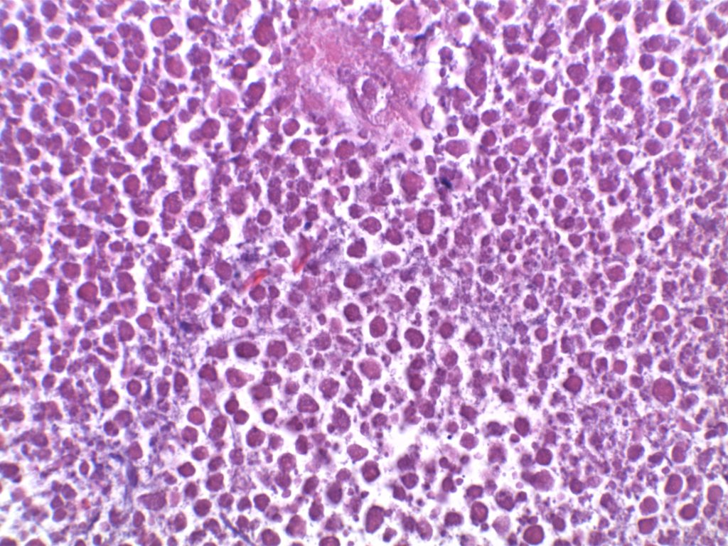
Often, these cases will have extensive necrosis, which may make diagnosis very difficult on small biopsy samples. Additionally, the immunophenotype may vary widely with variable loss and expression of both T and B cell markers.

  • CD20 +
  • CD79a +
  • MUM-1 +
  • CD138 +/-(these cases may be negative for CD20/CD79a)
  • Subset of cases with T-cell marker expression (CD2, CD3, CD4, and/or CD7)
  • EBV (EBER) +
Microscopic images
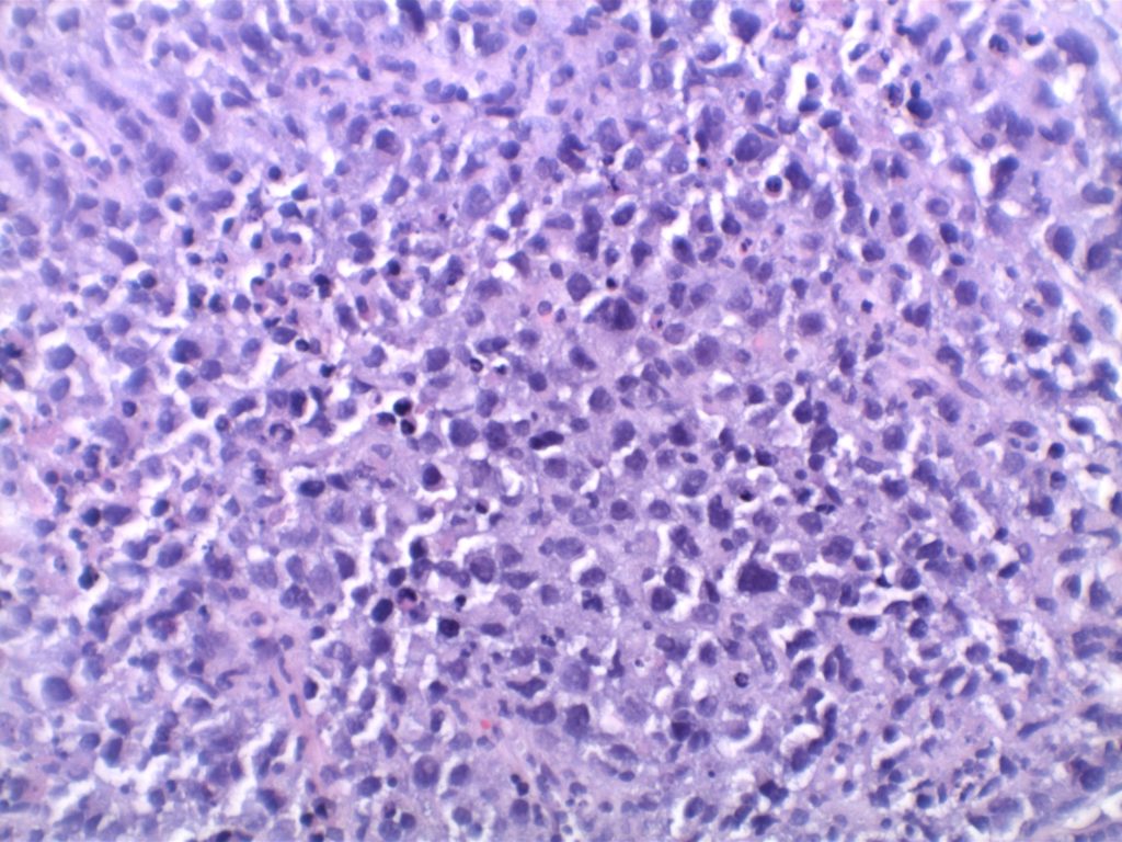
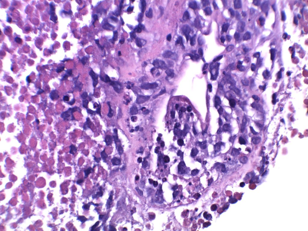
DLBCL associated with chronic inflammation
DLBCL associated with chronic inflammation – Extensive necrosis
DLBCL associated with chronic inflammation
Aberrant CD3 expression in DLBCL associated with chronic inflammation
Aberrant strong CD7 expression in DLBCL associated with chronic inflammation
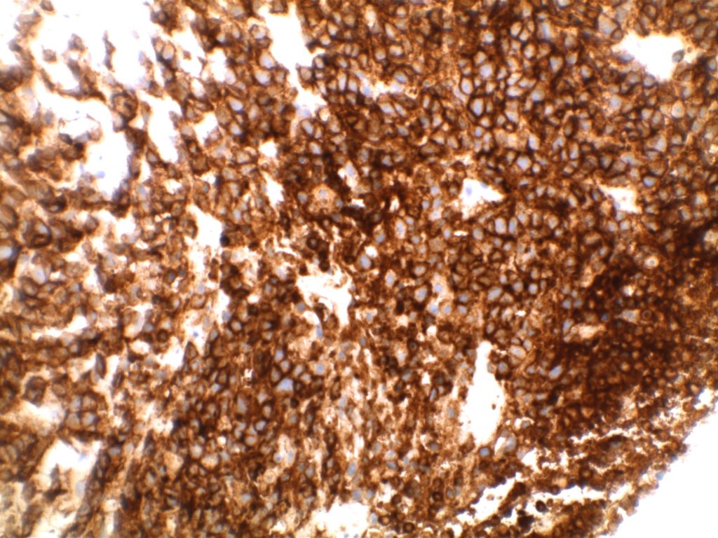
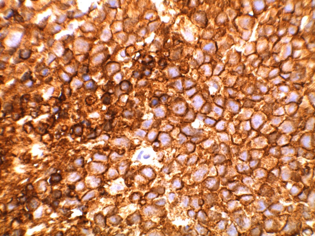
Strong CD20 expression in DLBCL associated with chronic inflammation
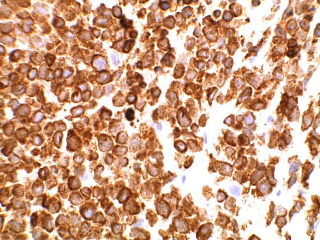
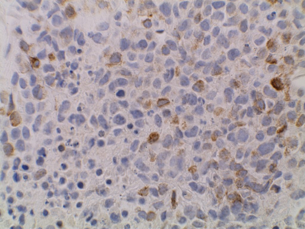
Dim subset expression of CD79a in DLBCL associated with chronic inflammation.
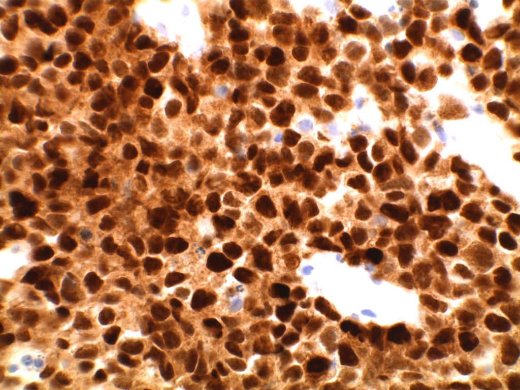
Strong diffuse MUM-1 expression in DLBCL associated with chronic inflammation
EBER expression (strong/diffuse) in neoplastic cells of DLBCL associated with chronic inflammation.
References

Loong F, Chan ACL, Ho BCS, Chau Y-P, Lee H-Y, Cheuk W, et al. Diffuse large B-cell lymphoma associated with chronic inflammation as an incidental finding and new clinical scenarios. Mod Pathol. 2010;23: 493–501. doi:10.1038/modpathol.2009.168


Swerdlow SH, Campo E, Harris, NL, Jaffe ES, Pileri SA, Stein H, Thiele J (Eds):  WHO Classification of Tumours of Haematopoietic and Lymphoid Tissues (Revised 4th edition). IARC: Lyon 2017
Large B-Cell Lymphoma with IRF4 Rearrangement

This is a special subtype of B-cell lymphoma, which may have an entirely diffuse, diffuse and follicular, or exclusive follicular pattern.  It is typically characterized by strong IRF4/MUM-1 expression by immunohistochemistry.  An IRF4 rearrangement is typically found.  This lymphoma most commonly occurs in children or young adults in the head and neck region (especially Waldeyer ring).
 
Morphologically these cases would otherwise be characterized as a diffuse large B-cell lymphoma, follicular lymphoma, or combined follicular and diffuse large B-cell lymphoma.   In the pure follicular pattern, some have referred to this as “Waldeyer Ring Follicular Lymphoma.”  Suspicion should be raised based on the patient’s age, disease location, and coexpression of CD10 and MUM-1.
Immunophenotype
  • CD20 positive
  • CD79a positive
  • PAX-5 positive
  • MUM-1 strongly expressed
  • BCL-6 positive
  • PRDM1 (BLIMP1) typically negative
  • CD10 usually positive (66%)
  • BCL-2 usually positive (66%)
  • Ki-67 high (lack of evidence of polarization in neoplastic follicles)
Cases with a phenotype of CD10 coexpression with MUM-1 should be considered for IRF4 rearrangements screening.  The prognosis is relatively good with appropriate treatment.
References
Swerdlow SH, Campo E, Harris, NL, Jaffe ES, Pileri SA, Stein H, Thiele J (Eds):  WHO Classification of Tumours of Haematopoietic and Lymphoid Tissues (Revised 4th edition). IARC: Lyon 2017
 
Fedoriw Y, Dogan A. The Expanding Spectrum of Follicular Lymphoma. Surg Pathol Clin. 2016;9: 29–40. doi:10.1016/j.path.2015.11.001
 
Salaverria I, Philipp C, Oschlies I, Kohler CW, Kreuz M, Szczepanowski M, et al. Translocations activating IRF4 identify a subtype of germinal center-derived B-cell lymphoma affecting predominantly children and young adults. Blood. 2011;118: 139–147. doi:10.1182/blood-2011-01-330795


DLBCL – Anaplastic Variant

The anaplastic variant of diffuse large B-cell lymphoma (DLBCL) is a morphologic variant under the DLBCL, NOS category in the WHO Classification.  Typical common features include:

Continue reading DLBCL – Anaplastic Variant

Plasma Cell Neoplasms

2016 WHO Classification

Continue reading Plasma Cell Neoplasms

Large B-Cell Lymphomas

Extranodal Marginal Zone Lymphoma of Mucosal Associated Lymphoid Tissue (MALT Lymphoma)

Extranodal marginal zone lymphoma of mucosa-associated lymphoid tissue (MALT) involves extra nodal tissue and shows infiltration of the marginal zones and inter follicular regions. 
  • Gastric MALT is associated with H. pylori infection in up to 90% of cases. 
  • MALT in the thyroid has been associated with Hashimoto’s thyroiditis (~20% of cases).
  • Ocular MALT lymphomas have been associated with various infections.

Continue reading Extranodal Marginal Zone Lymphoma of Mucosal Associated Lymphoid Tissue (MALT Lymphoma)