The use of immunohistochemistry (IHC) in many of the MPN/MDS neoplasms is limited. Identifying an increased blast population is one of the most useful, and may indicate a more aggressive course or transformation to acute myelogenous leukemia (AML). Helpful IHC markers may include:
CD117 is a specific myeloid marker, and marks a subset of myeloblasts. The expression is dim, and one often must look at 20-40X to clearly see expression. Mast cells (fried egg looking cell) will have very strong expression.
CD71 marks nucleated erythroid cells. This may be helpful in quantitating and differentiating erythroid cells from myeloid cells. This marker may be set-up as a double stain with CD34.
In the setting of hematopoietic cells, E-Cadherin marks immature erythroid cells. Like CD71, E-Cadherin may be useful to differentiate immature erythroid cells from immature myeloid cells.
TdT is a sensitive lymphoblast (~95%) marker. It is not entirely specific for lymphoblasts, but other markers can help clarify diagnostic difficulties (B and T-cell markers).
PB leukocytosis with increased PMNs and precursors (evidence of dysgranulopoiesis)
Neutrophil precursors (less mature than a band-form) make up ≥10% of leukocytes
Absence of significant basophilia (<2% or leukocytes)
No monocytosis (<10% of leukocytes)
Hypercellular bone marrow with granulocytic dysplasia (+/- dysplasia in other lineages)
<20% blasts in PB and BM
Does not meet diagnostic criteria for BCR-ABL1+ CML, PMF, PV, or ET (if you compare to the criteria for other MPNs, this is a circular argument)
No PDGFA, PDGFB, FGFR1, or JAK2-PCM1 rearrangement
In the past has been difficult to differentiate from CNL. This category has been somewhat better clarified in the 2016 WHO revision.
<10% have CSF3R mutation (strong association with CNL, if present should closely examine to exclude CNL – mutation may have therapeutic implications irregardless of diagnosis)
Not usually associated with JAK2, CALR, or MPL mutations
Up to 1/3rd of cases have SETBP1 and/or ETNK1 mutations
Immunohistochemistry
The use of immunohistochemistry (IHC) in many of the MPN/MDS neoplasms is limited. Identifying an increased blast population is one of the most useful, and may indicate a more aggressive course or transformation to acute myelogenous leukemia (AML). Helpful IHC markers may include:
CD117 is a specific myeloid marker, and marks a subset of myeloblasts. The expression is dim, and one often must look at 20-40X to clearly see expression. Mast cells (fried egg looking cell) will have very strong expression.
CD71 marks nucleated erythroid cells. This may be helpful in quantitating and differentiating erythroid cells from myeloid cells. This marker may be set-up as a double stain with CD34.
In the setting of hematopoietic cells, E-Cadherin marks immature erythroid cells. Like CD71, E-Cadherin may be useful to differentiate immature erythroid cells from immature myeloid cells.
TdT is a sensitive lymphoblast (~95%) marker. It is not entirely specific for lymphoblasts, but other markers can help clarify diagnostic difficulties (B and T-cell markers).
References
Arber DA, Orazi A, Hasserjian R, Thiele J, Borowitz MJ, Le Beau MM, et al. The 2016 revision to the World Health Organization classification of myeloid neoplasms and acute leukemia. Blood. 2016;127: 2391–2405. doi:10.1182/blood-2016-03-643544
WHO Classification of Tumors of Haematopoietic and Lymphoid Tissues. SH Swerdlow, et al. International Agency for Research on Cancer. Lyon, 2008.
Persistent peripheral blood monocytosis >1,000/μL (and accounts for ≥10% of the WBC count)
Does not meet diagnostic criteria for BCR-ABL1+ CML, PMF, PV, or ET (if you compare to the criteria for other MPNs, this is a circular argument)
No PDGFA, PDGFB, FGFR1, or JAK2-PCM1 rearrangement (specificity exclude if there is eosinophilia)
<20% blasts (blood or bone marrow); blasts defined as promonocytes, myeloblasts, or monoblasts
Myeloid dysplasia in one or more lineages, or
(If dysplasia is absent or minimal) presence of an acquired cytogenetic/molecular abnormality in the hematologic cell lineage
or, in the absence of the above criteria
At 3 months of persistent monocytosis (and)
Other causes have been excluded
CMML is unlikely if there is a previous diagnosis of a MPN, as such entities can progress to a phase with monocytosis (and this should be closely evaluated)
“Proliferative type” CMML – WBC ≥13,000/μL
“Dysplastic type” CMML – SBC <13,000/μL
CMML-0
<2% blasts in PB and
<5% blasts in BM
CMML-1
2-4% blasts in PB and/or
5-9% blasts in BM
CMML-2
5-19% blasts in PB
10-19% blasts in BM and/or
Presence of Auer rods
Immunohistochemistry
The use of immunohistochemistry (IHC) in many of the MPN/MDS neoplasms is limited. Identifying an increased blast population is one of the most useful, and may indicate a more aggressive course or transformation to acute myelogenous leukemia (AML). Helpful IHC markers may include:
CD117 is a specific myeloid marker, and marks a subset of myeloblasts. The expression is dim, and one often must look at 20-40X to clearly see expression. Mast cells (fried egg looking cell) will have very strong expression.
CD71 marks nucleated erythroid cells. This may be helpful in quantitating and differentiating erythroid cells from myeloid cells. This marker may be set-up as a double stain with CD34.
In the setting of hematopoietic cells, E-Cadherin marks immature erythroid cells. Like CD71, E-Cadherin may be useful to differentiate immature erythroid cells from immature myeloid cells.
TdT is a sensitive lymphoblast (~95%) marker. It is not entirely specific for lymphoblasts, but other markers can help clarify diagnostic difficulties (B and T-cell markers).
References
Arber DA, Orazi A, Hasserjian R, Thiele J, Borowitz MJ, Le Beau MM, et al. The 2016 revision to the World Health Organization classification of myeloid neoplasms and acute leukemia. Blood. 2016;127: 2391–2405. doi:10.1182/blood-2016-03-643544
WHO Classification of Tumors of Haematopoietic and Lymphoid Tissues. SH Swerdlow, et al. International Agency for Research on Cancer. Lyon, 2008.
The use of immunohistochemistry (IHC) in many of the myeloproliferative neoplasms is limited. Identifying an increased blast population is one of the most useful, and may indicate a more aggressive course or transformation to acute leukemia. Helpful IHC markers may include:
CD117 is a specific myeloid marker, and marks a subset of myeloblasts. The expression is dim, and one often must look at 20-40X to clearly see expression. Mast cells (fried egg looking cell) will have very strong expression.
CD71 marks nucleated erythroid cells. This may be helpful in quantitating and differentiating erythroid cells from myeloid cells. This marker may be set-up as a double stain with CD34.
In the setting of hematopoietic cells, E-Cadherin marks immature erythroid cells. Like CD71, E-Cadherin may be useful to differentiate immature erythroid cells from immature myeloid cells.
TdT is a sensitive lymphoblast (~95%) marker. It is not entirely specific for lymphoblasts, but other markers can help clarify diagnostic difficulties (B and T-cell markers).
Mastocytosis is a clonal (neoplastic) proliferation of mast cells. It can be a heterogeneous disorder ranging from skin lesion, which spontaneously regress, to aggressive systemic disease with a short survival. Mastocytosis is divided into two generalized categories: cutaneous mastocytosis (CM) and systemic mastocytosis (SM). CM is limited strictly to the skin, but SM has at least one extracutaneous organ involved (+/- skin involvement).
In SM the bone marrow is almost always involved. Approximately 50% of SM patients will have skin involvement.
Strongly reactive (must be used in combination with MPO)
MPO
Negative
Tryptase
Positive, not specific. Positivity of spindle cells in the bone marrow (BM) is considered specific for mast cells. Positivity of round cells in the BM may represent one of three entities: (1) mast cells (CD117+, chymase +), (2) neoplastic basophils, or (3) AML.
Chymase
Subset Positive (highly specific but not sensitive)
Expressed in a subset of neoplastic mast cells (specific). Must differentiate from T-cells. Commonly expressed in neoplastic mast cells involving the GI tract.
The use of immunohistochemistry (IHC) in many of the myeloproliferative neoplasms is limited. Identifying an increased blast population is one of the most useful, and may indicate a more aggressive course or transformation to acute leukemia. Helpful IHC markers may include:
CD117 is a specific myeloid marker, and marks a subset of myeloblasts. The expression is dim, and one often must look at 20-40X to clearly see expression. Mast cells (fried egg looking cell) will have very strong expression.
CD71 marks nucleated erythroid cells. This may be helpful in quantitating and differentiating erythroid cells from myeloid cells. This marker may be set-up as a double stain with CD34.
In the setting of hematopoietic cells, E-Cadherin marks immature erythroid cells. Like CD71, E-Cadherin may be useful to differentiate immature erythroid cells from immature myeloid cells.
TdT is a sensitive lymphoblast (~95%) marker. It is not entirely specific for lymphoblasts, but other markers can help clarify diagnostic difficulties (B and T-cell markers).
Does not meet the WHO criteria for another myeloid neoplasm
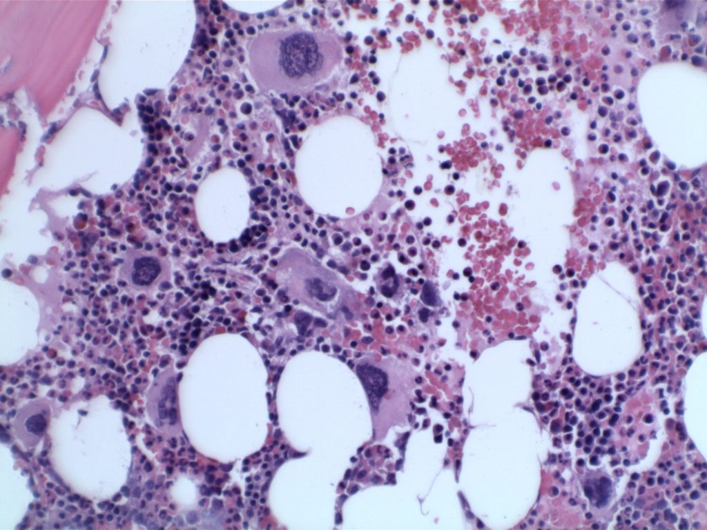
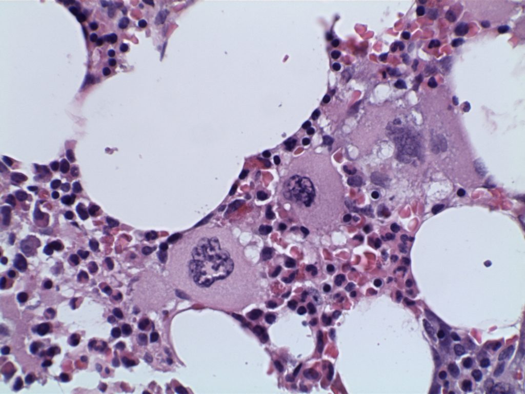
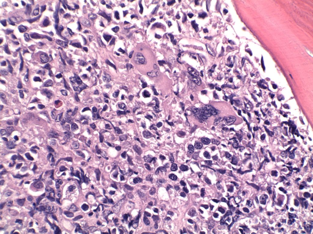
BM biopsy showing megakaryocytic proliferation
Enlarged mature megakaryocytes
Hyperlobated nuclei (not as pleomorphic or bizzare as in PMF)
Granulopoiesis and erythropoiesis is not increased or left shifted
No more than MF1 reticulin fibrosis
Minor Criteria
Evidence of a clonal marker or exclusion of reactive thrombocytosis
Diagnosis of ET is confirmed by all four major criteria, or if there is no evidence of a JAK2, CALR, or MPL mutation (major criterion #2), then the presence of the minor criterion.
Frequency of Molecular Abnormalities
JAK2 – 63% (V617F mutation)
CALR – 18%
MPL – 2-3%
Overall, approximately 83% of cases of ET have either a JAK2, CALR, or MPL mutation (n=79).
The use of immunohistochemistry (IHC) in many of the myeloproliferative neoplasms is limited. Identifying an increased blast population is one of the most useful, and may indicate a more aggressive course or transformation to acute leukemia. Helpful IHC markers may include:
CD117 is a specific myeloid marker, and marks a subset of myeloblasts. The expression is dim, and one often must look at 20-40X to clearly see expression. Mast cells (fried egg looking cell) will have very strong expression.
CD71 marks nucleated erythroid cells. This may be helpful in quantitating and differentiating erythroid cells from myeloid cells. This marker may be set-up as a double stain with CD34.
In the setting of hematopoietic cells, E-Cadherin marks immature erythroid cells. Like CD71, E-Cadherin may be useful to differentiate immature erythroid cells from immature myeloid cells.
TdT is a sensitive lymphoblast (~95%) marker. It is not entirely specific for lymphoblasts, but other markers can help clarify diagnostic difficulties (B and T-cell markers).
Arber DA, Orazi A, Hasserjian R, Thiele J, Borowitz MJ, Le Beau MM, et al. The 2016 revision to the World Health Organization classification of myeloid neoplasms and acute leukemia. Blood. 2016;127: 2391–2405. doi:10.1182/blood-2016-03-643544
Kim SY, Im K, Park SN, Kwon J, Kim J-A, Lee DS. CALR, JAK2, and MPL mutation profiles in patients with four different subtypes of myeloproliferative neoplasms: primary myelofibrosis, essential thrombocythemia, polycythemia vera, and myeloproliferative neoplasm, unclassifiable. Am J Clin Pathol. 2015;143: 635–644. doi:10.1309/AJCPUAAC16LIWZMM
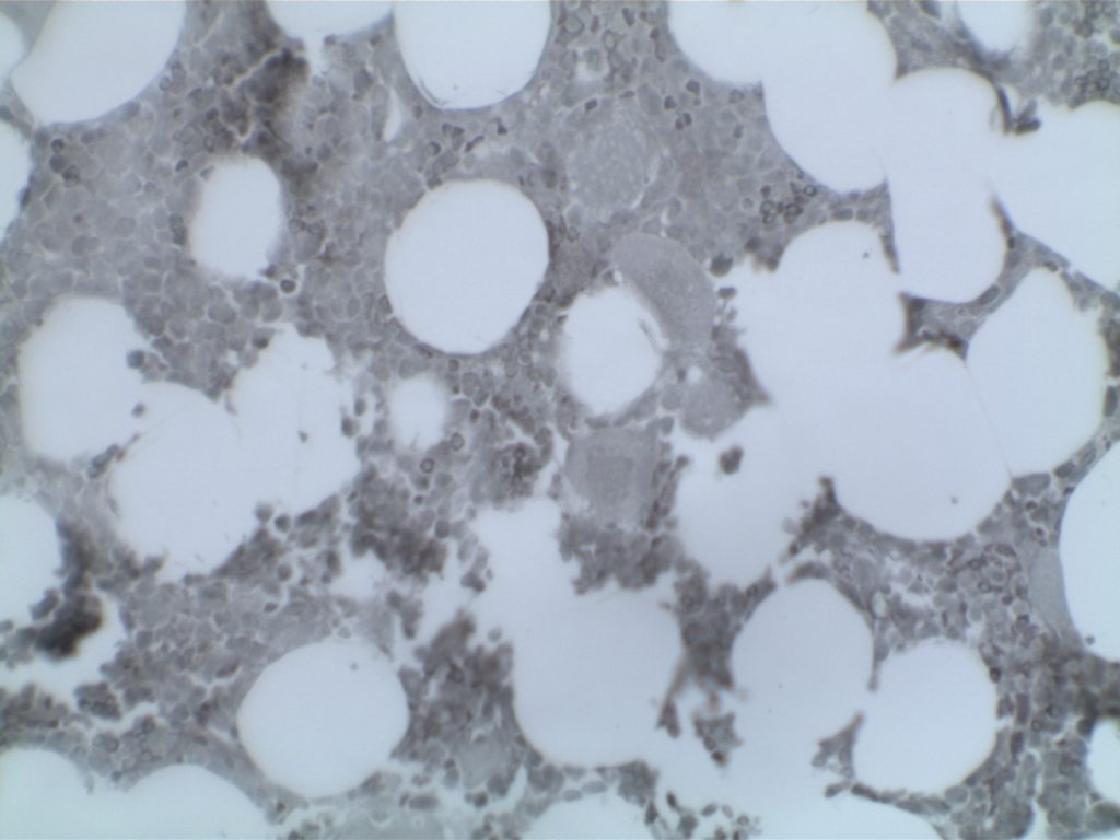
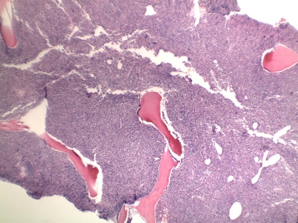
Diffuse increase of reticulin fibers with increased density and numerous intersections. Increased thick bundles of fibers consistent with collagen fibrosis. Osteosclerosis usually present.
In cases of MF2 or MF3, it is recommended to perform trichrome stain to evaluate for collagen fibrosis.
MF – Accelerated phase = 10-19% blasts in the peripheral blood and/or the bone marrow.
MF – Acute Transformation = ≥20% blasts in the blood or bone marrow.
The use of immunohistochemistry (IHC) in many of the myeloproliferative neoplasms is limited. Identifying an increased blast population is one of the most useful, and may indicate a more aggressive course or transformation to acute leukemia. Helpful IHC markers may include:
CD117 is a specific myeloid marker, and marks a subset of myeloblasts. The expression is dim, and one often must look at 20-40X to clearly see expression. Mast cells (fried egg looking cell) will have very strong expression.
CD71 marks nucleated erythroid cells. This may be helpful in quantitating and differentiating erythroid cells from myeloid cells. This marker may be set-up as a double stain with CD34.
In the setting of hematopoietic cells, E-Cadherin marks immature erythroid cells. Like CD71, E-Cadherin may be useful to differentiate immature erythroid cells from immature myeloid cells.
TdT is a sensitive lymphoblast (~95%) marker. It is not entirely specific for lymphoblasts, but other markers can help clarify diagnostic difficulties (B and T-cell markers).
References
Swerdlow SH, Campo E, Harris, NL, Jaffe ES, Pileri SA, Stein H, Thiele J (Eds): WHO Classification of Tumours of Haematopoietic and Lymphoid Tissues (Revised 4th edition). IARC: Lyon 2017
Arber DA, Orazi A, Hasserjian R, Thiele J, Borowitz MJ, Le Beau MM, et al. The 2016 revision to the World Health Organization classification of myeloid neoplasms and acute leukemia. Blood. 2016;127: 2391–2405. doi:10.1182/blood-2016-03-643544
Kim SY, Im K, Park SN, Kwon J, Kim J-A, Lee DS. CALR, JAK2, and MPL mutation profiles in patients with four different subtypes of myeloproliferative neoplasms: primary myelofibrosis, essential thrombocythemia, polycythemia vera, and myeloproliferative neoplasm, unclassifiable. Am J Clin Pathol. 2015;143: 635–644. doi:10.1309/AJCPUAAC16LIWZMM
Tefferi A, Lasho TL, Finke CM, Knudson RA, Ketterling R, Hanson CH, et al. CALR vs JAK2 vs MPL-mutated or triple-negative myelofibrosis: clinical, cytogenetic and molecular comparisons. Leukemia. 2014;28: 1472–1477. doi:10.1038/leu.2014.3