Category Archives: Organ Systems

Breast – Invasive Carcinoma

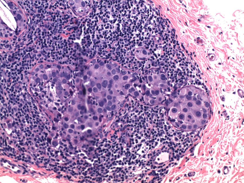
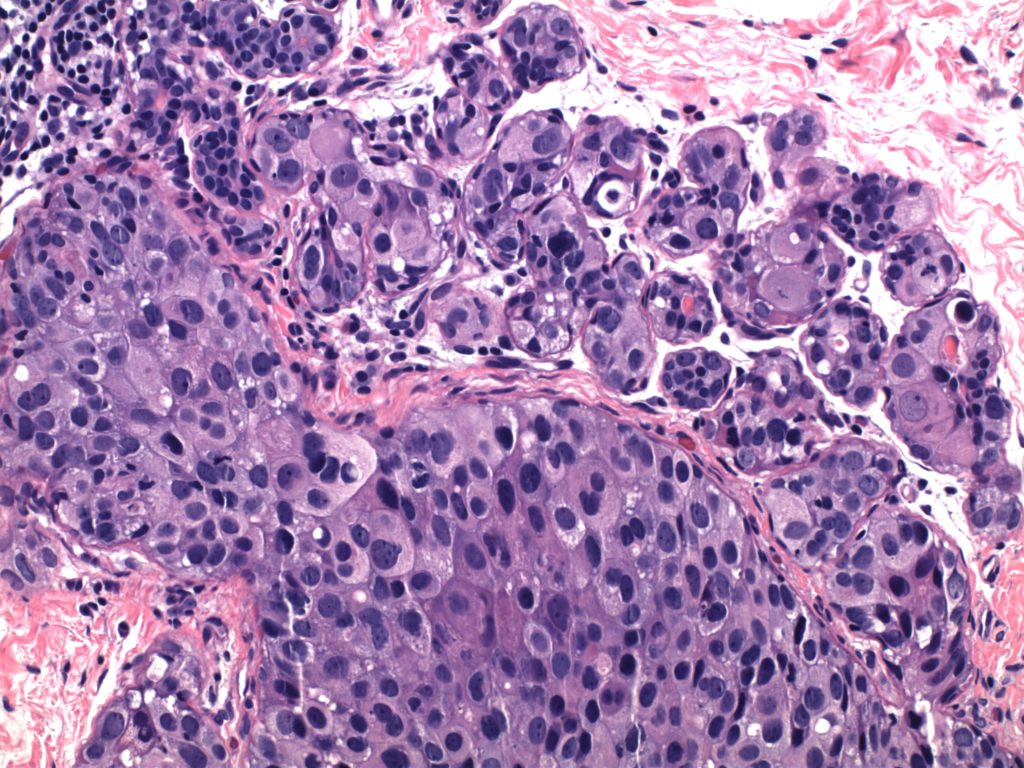
Invasive Breast Carcinoma
  • Invasive breast carcinomas not segregated out as a special subtype are classified as invasive ductal carcinoma, no special type (NST).  These tumors (and most of the special types) have prognosis and treatment plans based on the receptor status (and sometimes additional molecular profiling).
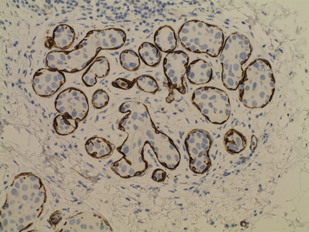
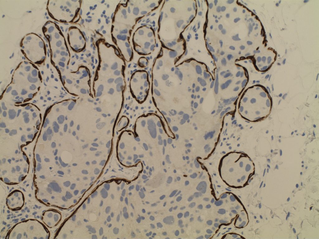
  • Receptor testing includes ER/PR/Her-2 and Ki-67 (proliferation marker)

Continue reading Breast – Invasive Carcinoma

Breast Carcinoma – Risk Factors

  • Most common cancer in women (~225,000 cases/yr. with ~40,000 deaths/yr.)
  • Second most common cause of cancer death in women (lung #1)
  • Three major subtypes
    • ER+/Her-2 negative (50-65%)
    • Her-2 + (10-20%)
    • Triple negative – ER/PR/Her-2 negative (10-20%)
  • Breast cancers in African American women are more aggressive biologically, and are more likely to be ER-negative and high nuclear grade

Continue reading Breast Carcinoma – Risk Factors

Breast – DCIS

Ductal Carcinoma In Situ (DCIS)
  • Neoplastic proliferation resembling small ducts with expanded duct and acinar structures
  • Myoepithelial cell layer is intact surrounding the neoplastic proliferation (non-invasive tumor)
  • Typically express E-Cadherin
  • Bilateral in 10-20% of cases
  • Detected by mammography (not clinically evident)
  • Represents 15-30% of neoplasms identified in screening populations
  • Typically identified by abnormal calcification, sometimes as abnormal densities
Comedo DCIS
  • High grade pleomorphic nuclei
  • Central necrosis
Non-comedo DCIS
  • Lacks either high grade nuclei or central necrosis
  • Subtypes/patterns
    • Solid DCIS
    • Micropapillary DCIS
    • Cribiform DCIS
  • DCIS grading
    • Low-grade DCIS
      • 1%/year risk of developing an invasive carcinoma
    • Intermediate-grade DCIS
    • High-grade DCIS
Paget Disease
  • Nipple manifestation of disease (1-4% of cases) – looks like eczema on the nipple
  • Tumor cells extend into the epidermis of the skin overlying the nipple from underlying DCIS within the ductal system of the breast.
  • 50-60% of women will have an underlying palpable mass
    • Vast majority will have an invasive carcinoma (often ER neg./Her-2 pos.)
    • Women without a palpable mass will usually only have DCIS
Photomicrographs
Breast - High Grade DCIS
High power view of high grade breast DCIS.
Breast - High Grade DCIS
High power view of breast high grade DCIS.
Smooth Muscle Myosin - High Grade DCIS
Smooth Muscle Myosin – High Grade DCIS
Smooth Muscle Myosin - High Grade DCIS
Smooth Muscle Myosin – High Grade DCIS
Smooth Muscle Myosin - High Grade DCIS
Smooth Muscle Myosin – High Grade DCIS
References

Kumar, Vinay, Abul K. Abbas, and Jon C. Aster. Robbins and Cotran Pathologic Basis of Disease. Ninth edition. Philadelphia, PA: Elsevier/Saunders, 2015. 

JAK2, CALR, & MPL Testing in Myeloproliferative Neoplasms (MPN)

Molecular Testing Specimen Adequacy Summary
  • JAK2, CALR, and MPL testing is often performed on peripheral blood specimens in an outpatient setting.
  • JAK2 mutations are preferably analyzed in granulocytes.
  • Peripheral blood and bone marrow specimens are equally adequate for the identification of JAK2 mutations.
  • By extrapolation, CALR and MPL testing on peripheral blood specimens should be equally adequate.

Continue reading JAK2, CALR, & MPL Testing in Myeloproliferative Neoplasms (MPN)

Extranodal Marginal Zone Lymphoma of Mucosal Associated Lymphoid Tissue (MALT Lymphoma)

Extranodal marginal zone lymphoma of mucosa-associated lymphoid tissue (MALT) involves extra nodal tissue and shows infiltration of the marginal zones and inter follicular regions. 
  • Gastric MALT is associated with H. pylori infection in up to 90% of cases. 
  • MALT in the thyroid has been associated with Hashimoto’s thyroiditis (~20% of cases).
  • Ocular MALT lymphomas have been associated with various infections.

Continue reading Extranodal Marginal Zone Lymphoma of Mucosal Associated Lymphoid Tissue (MALT Lymphoma)