
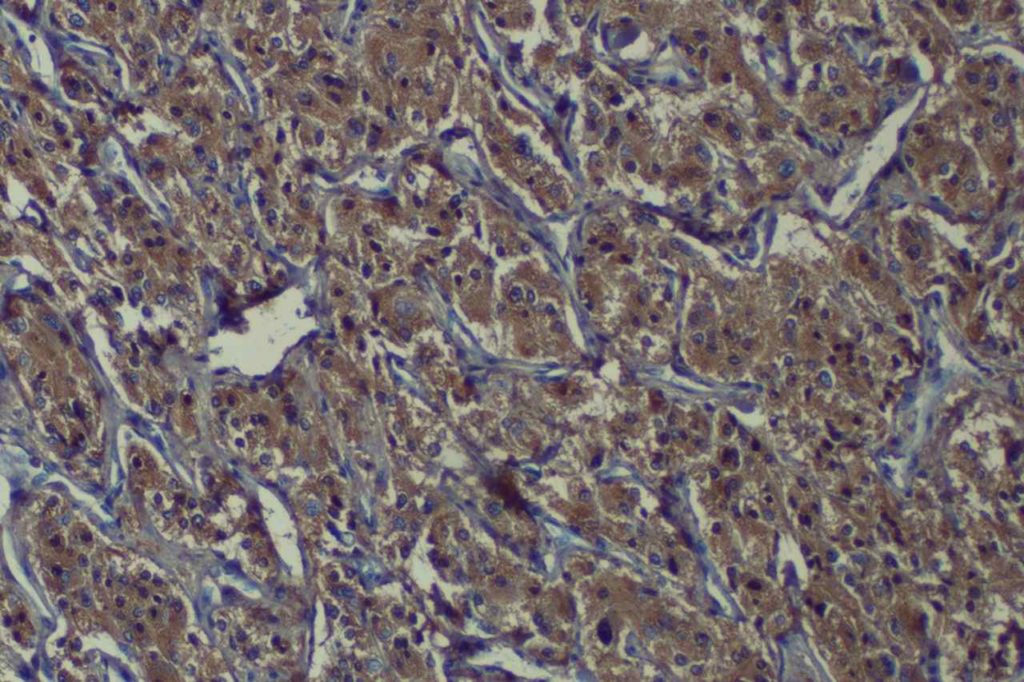
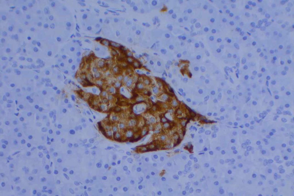
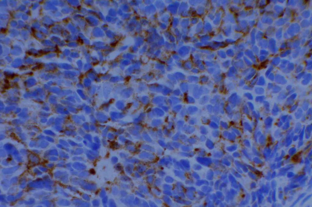
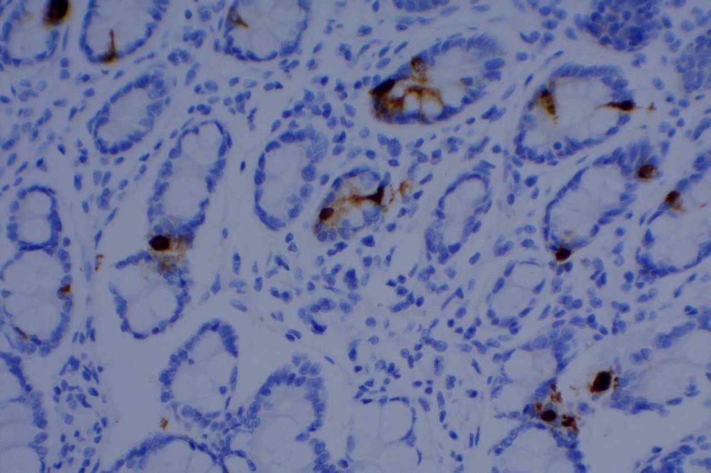
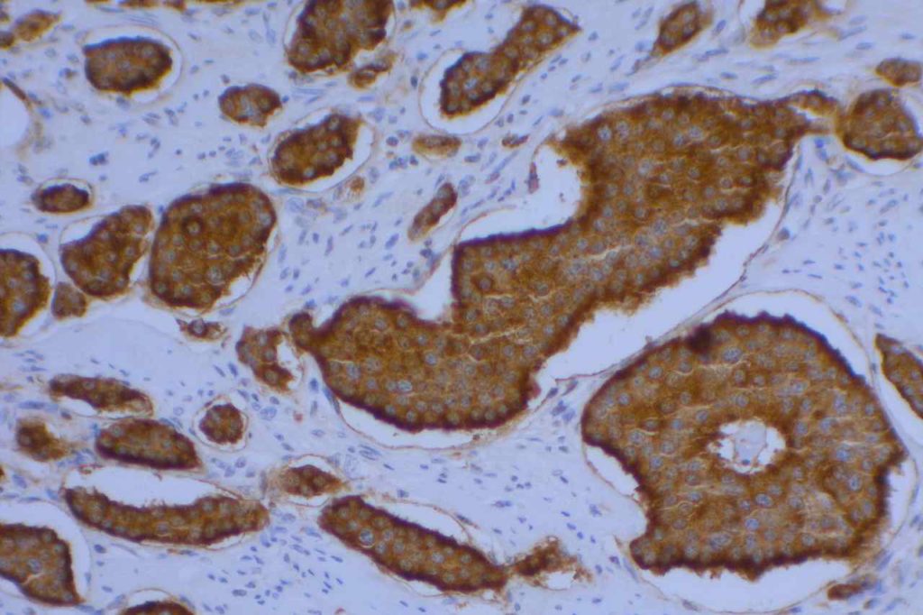
Chromogranin A is a very specific marker for neuroendocrine cell differentiation (reacts with cytoplasmic neurosecretory granules). Unfortunately, it is not highly sensitive. It is often used as part of a panel to identify neuroendocrine neoplasms (e.g. synaptophysin, CD56). It is more often positive in well-differentiated lesions, and stains the neurosecretory granules in the cell cytoplasm.
Chromogranin A is expressed in approximately 30% of small cell carcinomas and neuroblastomas.
Photomicrographs
Reference
Wick, MR. “Immunohistochemical approaches to the diagnosis of undifferentiated malignant tumor.”Annals of Diagnostic Pathology12(2008):72-84.