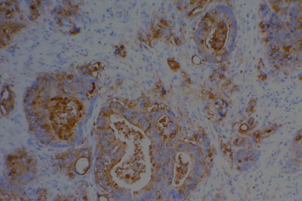
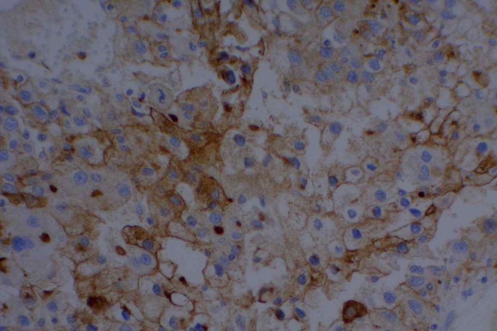
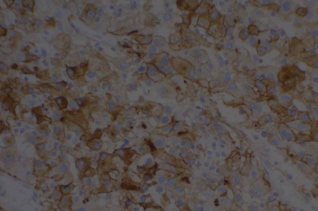
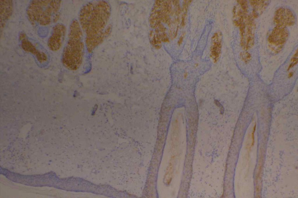
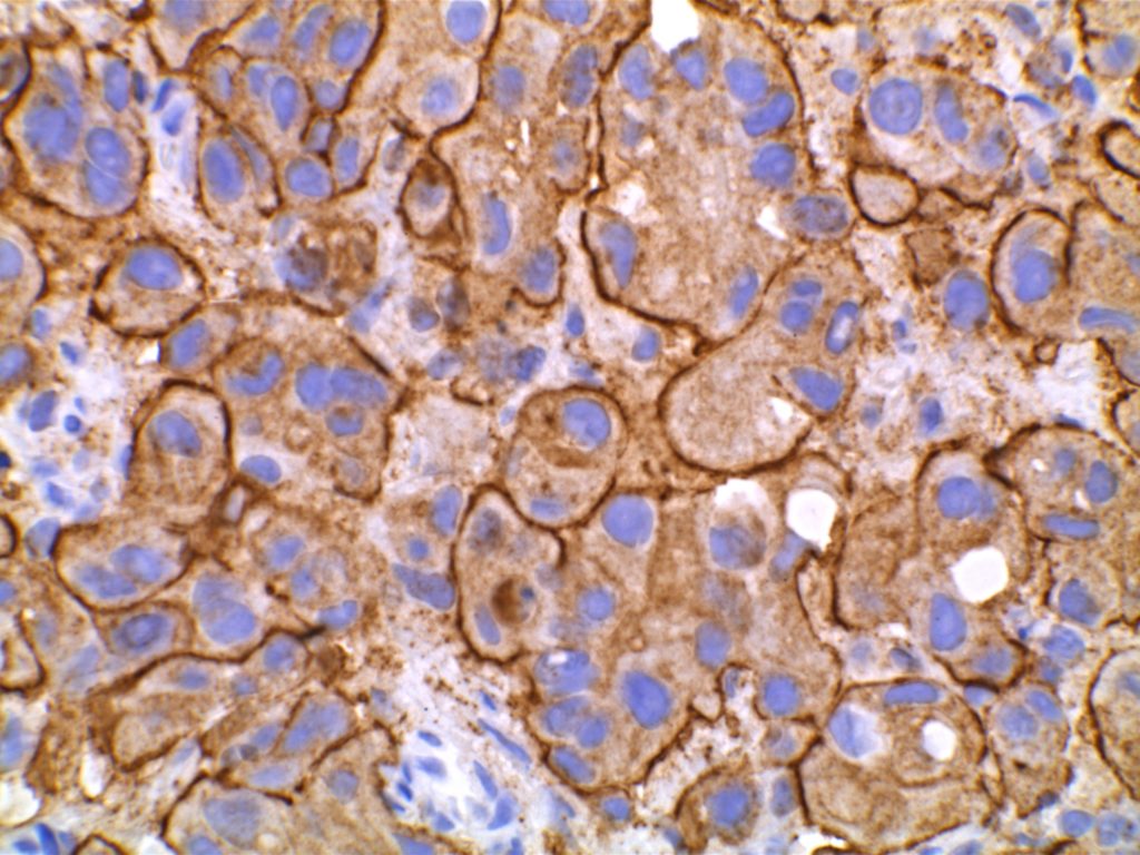
Epithelial Membrane Antigen (EMA), originally described in 1979, targets a complex membrane glycoprotein originally isolated from milk fat globules. It is also known as MUC-1. EMA is helpful in identifying epithelial differentiation, but is not entirely specific. In general EMA expression is similar to cytokeratin expression, although hepatocellular carcinomas, adrenocortical neoplasms, and germ cell neoplasms do not express EMA. Some non-epithelial processes, such s ALCL, plasmacytomas, epithelioid sarcoma, synovial sarcoma, meningioma, and T-cell lymphomas may show at least focal positivity. (Wick, M.R.)
In a recent study by Minato, H. et al, EMA was found to be expressed in 79% (n=34) of cases of mesothelioma compared to 13% (n=40) of cases of reactive mesothelial cells.
Photomicrographs
References
Wick, M. R. (2008). Immunohistochemical approaches to the diagnosis of undifferentiated malignant tumors. Annals of Diagnostic Pathology, 12(1), 72–84. doi:10.1016/j.anndiagpath.2007.10.003
Minato, H., Kurose, N., Fukushima, M., Nojima, T., Usuda, K., Sagawa, M., et al. (2014). Comparative Immunohistochemical Analysis of IMP3, GLUT1, EMA, CD146, and Desmin for Distinguishing Malignant Mesothelioma From Reactive Mesothelial Cells. American Journal of Clinical Pathology, 141(1), 85–93. doi:10.1309/AJCP5KNL7QTELLYI
