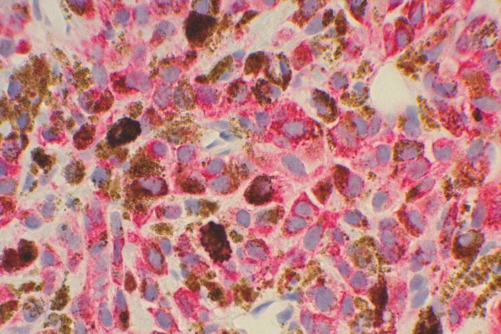
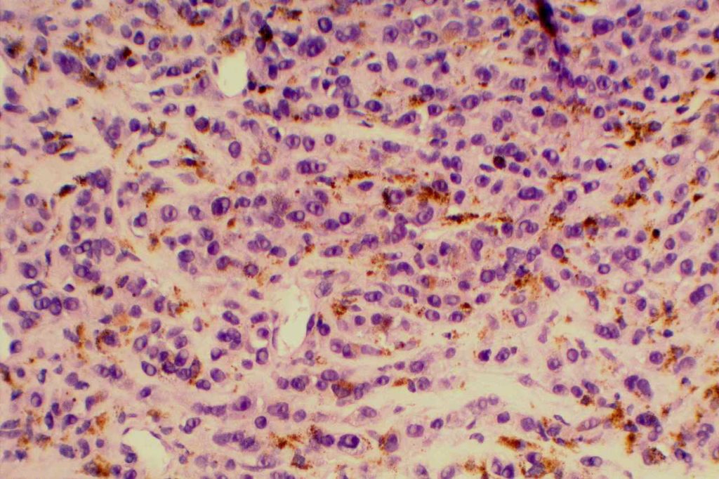
HMB-45 (Human Melanoma Black) is a monoclonal antibody to melanosomal glycoprotein gp100, and is relatively specific for melanocytes. The staining pattern is cytoplasmic. Similar to MART1, HMB-45 is less sensitive for melanoma when it has a spindle cell pattern. S-100 (less specific) is a better screening marker than HMB-45 or MART1. HMB-45 is better as a confirmatory marker.
HMB45 will stain: angiomyolipoma, melanoma, soft part sarcoma, sugar tumor of lung, lymphangiomyomatosis, pheochromocytoma (30%), pigmented nerve sheath tumors, and benign nevi / melanocytes. It is also important to understand that some histiocytes in lymph nodes may stain with HMB-45, which must be differentiated from metastatic melanoma cells. At least some histiocytes stained with HMB-45 in 50% of lymph nodes in a study by Hutchens, KA, et al.
HMB-45 will mark melanocytes and melanocytic derived neoplasms, but is not diagnostic in and of itself of anything abnormal. Dysplasia/neoplasia can only be diagnosed on the H&E slide after confirming morphology and/or melanocytic cell distribution with IHC analysis.
Photomicrograph


Reference
Hutchens, K. A., Heyna, R., Mudaliar, K., & Wojcik, E. (2013). The new AJCC guidelines in practice: utility of the MITF immunohistochemical stain in the evaluation of single-cell metastasis in melanoma sentinel lymph nodes. The American Journal of Surgical Pathology, 37(6), 933–937. doi:10.1097/PAS.0b013e3182815574
Kucher, C., Zhang, P. J., Acs, G., Roberts, S., & Xu, X. (2006). Can Melan-A replace S-100 and HMB-45 in the evaluation of sentinel lymph nodes from patients with malignant melanoma? Applied Immunohistochemistry & Molecular Morphology : AIMM / Official Publication of the Society for Applied Immunohistochemistry, 14(3), 324–327.
