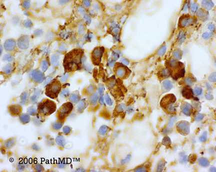
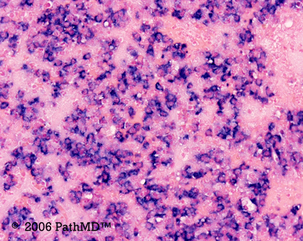
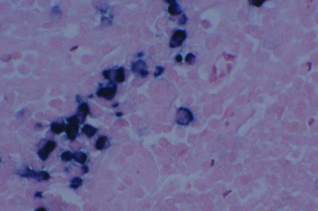
Kappa/Lambda (K/L) may be performed by immunohistochemistry (IHC) or in situ hybridization (ISH). K/L studies is primary used in the evaluation of plasma cell disorders or lymphoproliferative disorders containing some level of plasmacytoid differentiation. In tissue sections IHC for K/L often produces a large amount of background staining, which makes its use more difficult on a day-to-day basis compared to ISH (much less background).
The second big issue in K/L staining is that it is often performed on decalcified bone marrow biopsy core sections. The decalcification process may cause significant issues in the sensitivity of both IHC and ISH. As an alternative, always making a clot section on bone marrow specimens may greatly increase diagnostic yield and reliability.
Determination of monoclonality of plasma cells can sometimes be challenging. Historically, most consider a K:L ratio <0.5 or >4.0 to be indicative of monoclonality. Like almost everything in pathology, holding to a specific and tight threshold may put one in a perilous position in making a diagnosis. By flow cytometry, Samoszuk, et. al found that K:L ratios <0.7 and >5.5 were optimum for discriminating between lymphoma and benign hyperplasia (false negative rate= 27%, and false positive rate= 6%).
Evaluating K:L ratios in tissue sections is generally analogous to flow cytometry. Normally, there are 2-4 Kappa plasma cells to every Lamdda plasma cell. A ratio of K:L > 8:1 or a L:K ratio > 4:1 is strongly suggestive of a monoclonal plasma cell population.
Practically, if K:L ratio is no “obviously” monoclonal on ISH/IHC, it may be best to identify it as a kappa or lambda predominate population and correlate with other laboratory, radiologic and clinical findings. A significant pitfall may occur when either the kappa or lambda stain doesn’t work, and the case looks like a monoclonal population. Even in “obvious” monoclonal cases, one should be able to find a rare kappa or lambda staining plasma cell. If not, then the maker should be repeated.
Photomicrographs


References
Parker, A., et. al. “Best Practice in Lymphoma Diagnosis and Reporting.” British Committee for Standards in Haematology, Royal College of Pathologists. April, 2010.
