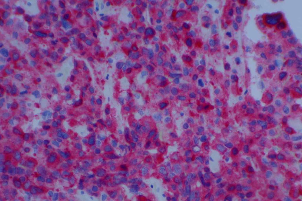
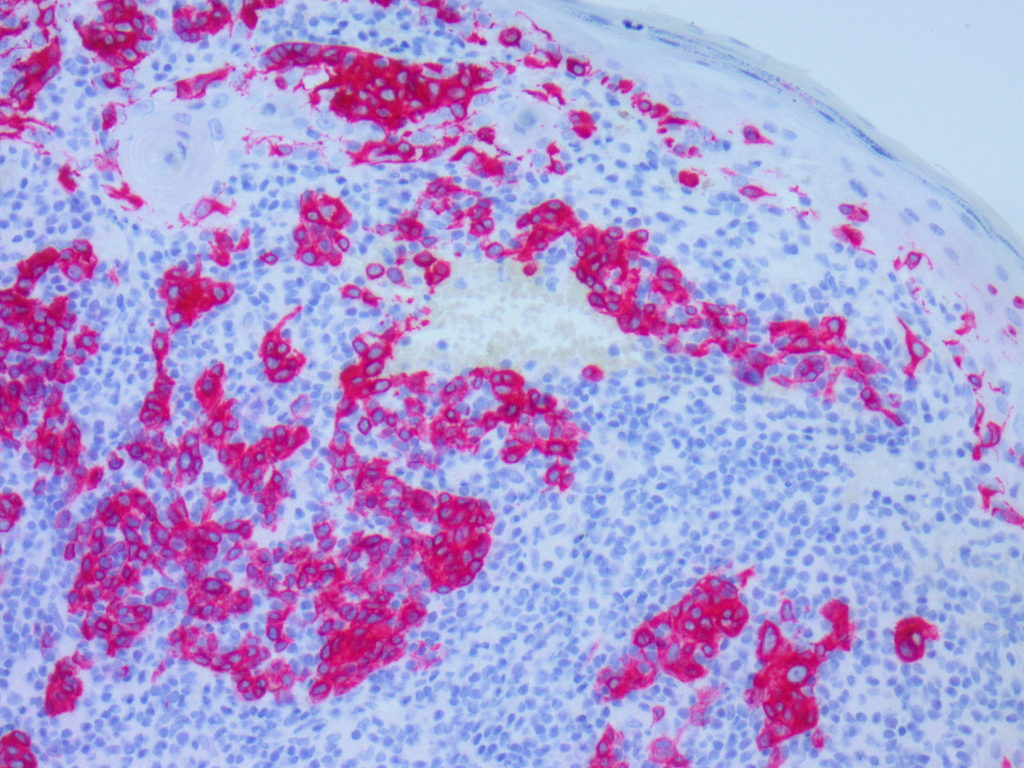
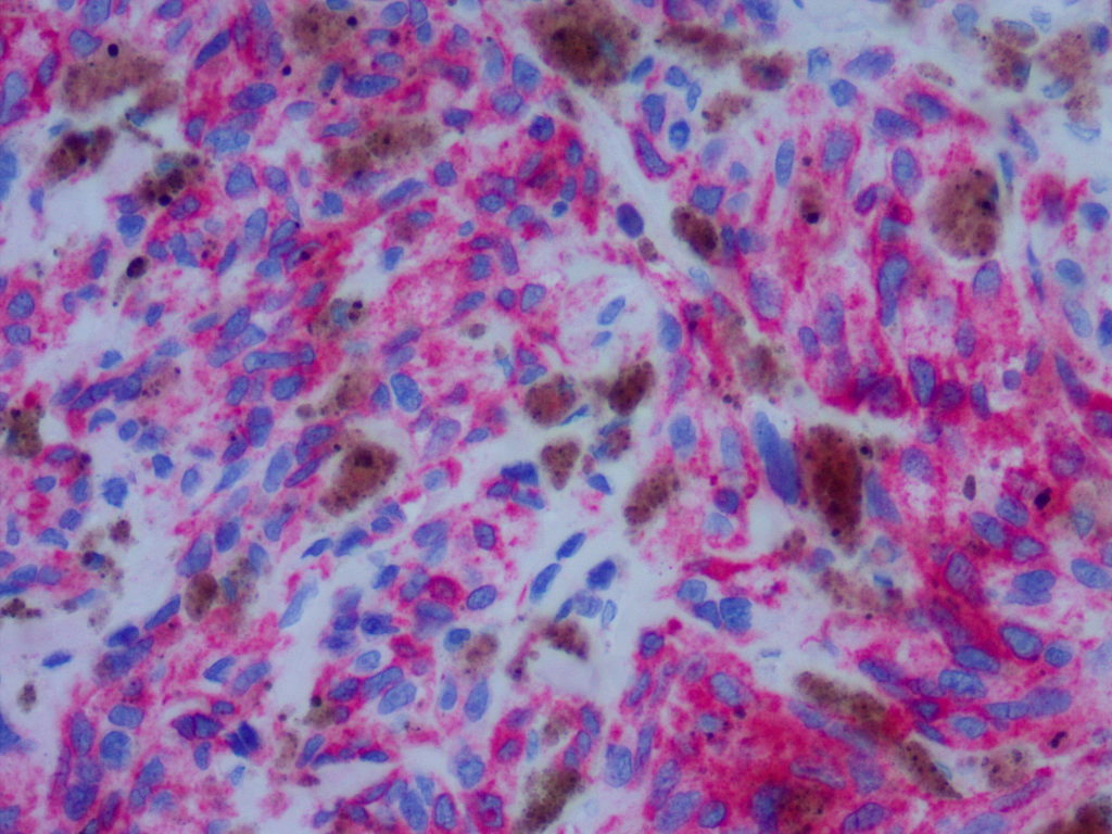
MART-1/Melan-A has an expression pattern very similar to HMB-45, and these stains are often used interchangeably. It is a sensitive and specific marker for melanocytes and melanocytic derived neoplasms. Like HMB-45, MART-1 is NOT as sensitive compared to S-100 in detecting melanomas with spindled morphology.
MART-1 is often used to detect isolated tumor cells in sentinel lymph nodes. Care must be taken in using MART-1 in this way, as histiocytes may present melanocyte antigens on their surface, which will cross-react with MART-1 (approximately 20%). Reports in the medical literature indicate that MART-1 performs better than HMB-45, which shows staining in approximately 50% of lymph nodes. Staining must be combined with morphology in evaluating this marker in lymph nodes. S-100 should not be used in lymph nodes, as there is too much background staining from dendritic cells.
MART-1 expression can be seen in most steroidogenic neoplasms and adrenal cortical carcinomas. HMB-45 does not appear to have this expression pattern. MART-1 and HMB-45 are expressed and helpful in diagnosing angiomyolipomas of the kidney. As a side piece of trivia, 89% of Xp11.2 translocation associated renal cell carcinomas expressed MART-1.
Photomicrographs


References
Hutchens KA, Heyna R, Mudaliar K, Wojcik E. The new AJCC guidelines in practice: utility of the MITF immunohistochemical stain in the evaluation of single-cell metastasis in melanoma sentinel lymph nodes. Am J Surg Pathol. 2013;37: 933–937. doi:10.1097/PAS.0b013e3182815574
Weissferdt A, Phan A, Suster S, Moran CA. Adrenocortical carcinoma: a comprehensive immunohistochemical study of 40 cases. Appl Immunohistochem Mol Morphol. 2014;22: 24–30. doi:10.1097/PAI.0b013e31828a96cf
Kucher C, Zhang PJ, Acs G, Roberts S, Xu X. Can Melan-A replace S-100 and HMB-45 in the evaluation of sentinel lymph nodes from patients with malignant melanoma? Appl Immunohistochem Mol Morphol. 2006;14: 324–327.
Shidham, VB, et al. AJSP 2001;25(8):1039-1046.
Wick, MR. “Immunohistochemical approaches to the diagnosis of undifferentiated malignant tumor.” Annals of Diagnostic Pathology 12(2008):72-84.
