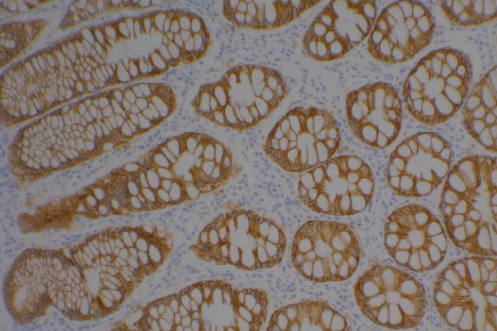
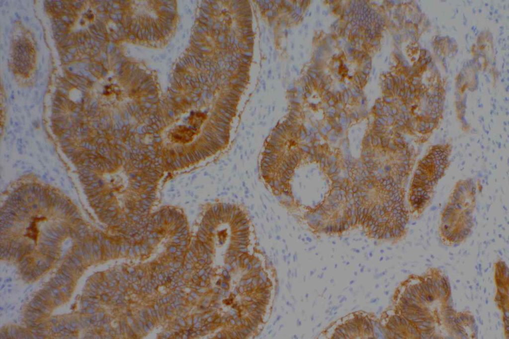
MOC-31 is a glycoprotein on the cell-membrane (epithelial glycoprotein 2/epithelial cell adhesion molecule – Ep-CAM) that is widely distributed on epithelial cells and tumor cells. MOC-31 is often used to differentiate adenocarcinoma (93% positive) from mesothelioma (93% negative). Other tumors also typically negative for MOC-31 include: hepatocellular carcinoma (HCC), germ cell tumors, and renal cell (some report up to 50%+) carcinomas. Some tumors may have characteristic staining patterns (e.g. membraneous vs. cytoplasmic or apical).
Mesothelioma vs. Adenocarcinoma
MOC-31 Expression (Positive)
- Adenocarcinomas (93%)
MOC-31 Negative
- Hepatocellular carcinoma
- Mesothelioma (93%)
- Germ cell tumors
- Renal cell carcinoma (some report expression in up to 50% of cases)
Photomicrographs


References
Ordóñez NG. Immunohistochemical diagnosis of epithelioid mesothelioma: an update. Arch Pathol Lab Med. 2005;129: 1407–1414.
Allende D, Yerian L. Immunohistochemical Markers in the Diagnosis of Hepatocellular Carcinoma. Pathology Case Reviews. 2009;14: 40–46.
Pan C-C, Chen PC-H, Tsay S-H, Ho DM-T. Differential immunoprofiles of hepatocellular carcinoma, renal cell carcinoma, and adrenocortical carcinoma: a systemic immunohistochemical survey using tissue array technique. Appl Immunohistochem Mol Morphol. 2005;13: 347–352.
Wick, MR. “Immunohistochemical approaches to the diagnosis of undifferentiated malignant tumor.” Annals of Diagnostic Pathology 12(2008):72-84.
