Napsin A is an aspartic proteinase, and is normally found in lung pneumocytes and alveolar macrophages, pancreatic acini and ducts, and renal tubules. Neoplastic tissue which stains with Napsin A includes: lung adencarcinoma, renal cell carcinoma (clear cell and papillary), thyroid (papillary) carcinoma, and ovarian clear cell carcinoma.
Napsin A is primary used in combination with TTF-1 in identifying lung adencarcinomas (TTF-1 appears to be a little more sensitive in poorly differentiated tumors). While approximately 97% of Napsin A expressing lung adenocarcinomas express TTF-1, up to 13% of TTF-1 negative cases express Napsin A. The more recent description of Napkin A expression in 100% of ovarian clear cell carcinomas by Kandalaft, et al. further emphasizes the importance of using an appropriate panel of IHC stains for a given differential diagnosis to obtain optimal sensitivity and specificity.
In the setting of ovarian tumors (Kandalaft, et al.), Napsin A was 100% sensitive for ovarian clear cell carcinoma. Endometrioid carcinomas of the ovary had focal Napsin A expression in 10% of cases, but no cases of high grade serous carcinoma or serous borderline tumor demonstrated expression.
Napsin A and TTF-1 expression in poorly differentiated non-small cell carcinomas (Mukjopadhyay, S, et al).
|
Tumor
|
Napsin A
|
TTF-1
|
|
Adenocarcinoma
|
11/19 (58%)
|
16/20 (80%)
|
|
Squamous Cell Carcinoma
|
0/15 (0%)
|
0/15 (0%)
|
|
Large Cell Carcinoma
|
0/4 (0%)
|
2/4 (50%)
|
Napsin A and TTF-1 expression in various tumor types (Bishop, JA, et al).
|
Tumor
|
Napsin A
(% positive)
|
TTF-1
(% positive)
|
|
Lung Tumors
|
|
|
|
Adneocarcinoma
|
79/95 (83%)
|
69/95 (73%)
|
|
– Well-differentiated
|
42/47 (89%)
|
38/47 (81%)
|
|
– Moderately-differentiated
|
27/32 (84%)
|
24/32 (75%)
|
|
– Poorly-differentiated
|
11/16 (69%)
|
7/16 (44%)
|
|
Squamous Cell Carcinoma
|
0/46 (0%)
|
0/48 (0%)
|
|
Large Cell Carcinoma
|
3/9 (33%)
|
4/9 (44%)
|
|
Small Cell Carcinoma
|
0/3 (0%)
|
1/3 (33%)
|
|
Atypical Carcinoid Tumor
|
0/1 (0%)
|
1/1 (100%)
|
|
Typical Carcinoid Tumor
|
0/2 (0%)
|
1/2 (50%)
|
|
Nonpulmonary Adenocarcinomas
|
|
|
|
Colon Adenocarcinoma
|
0/5 (0%)
|
0/5 (0%)
|
|
Pancreas Adenocarcinoma
|
0/31 (0%)
|
0/31 (0%)
|
|
Breast Adenocarcinoma
|
0/17 (0%)
|
0/17 (0%)
|
|
Mesothelioma (all types)
|
0/38 (0%)
|
0/38 (0%)
|
|
Renal Cell Carcinomas
|
|
|
|
Clear Cell
|
14/41 (34%)
|
0/41 (0%)
|
|
Papillary
|
34/43 (79%)
|
0/43 (0%)
|
|
Chromophobe
|
1/34 (3%)
|
0/34 (0%)
|
|
Thyroid Lesions
|
|
|
|
Papillary Carcinoma
|
2/38 (5%)
|
37/38 (97%)
|
|
Follicular Carcinoma
|
0/15 (0%)
|
15/15 (100%)
|
|
Follicular Adenoma
|
0/28 (0%)
|
28/28 (100%)
|
Microscopic Images
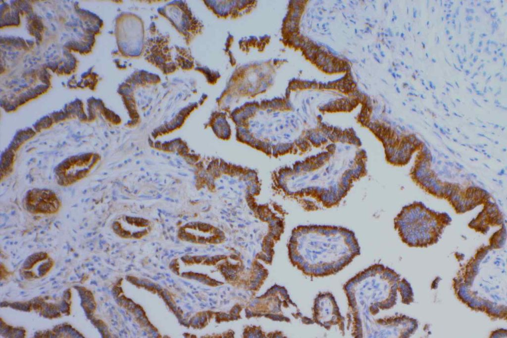
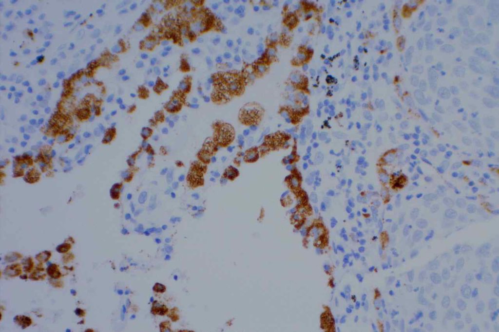
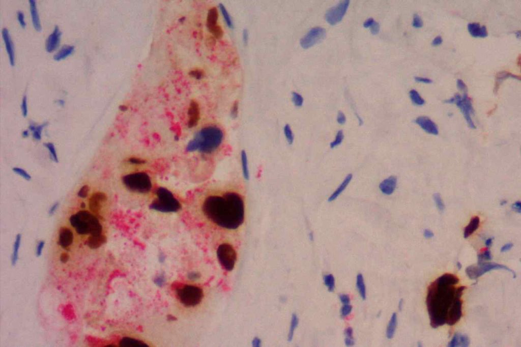
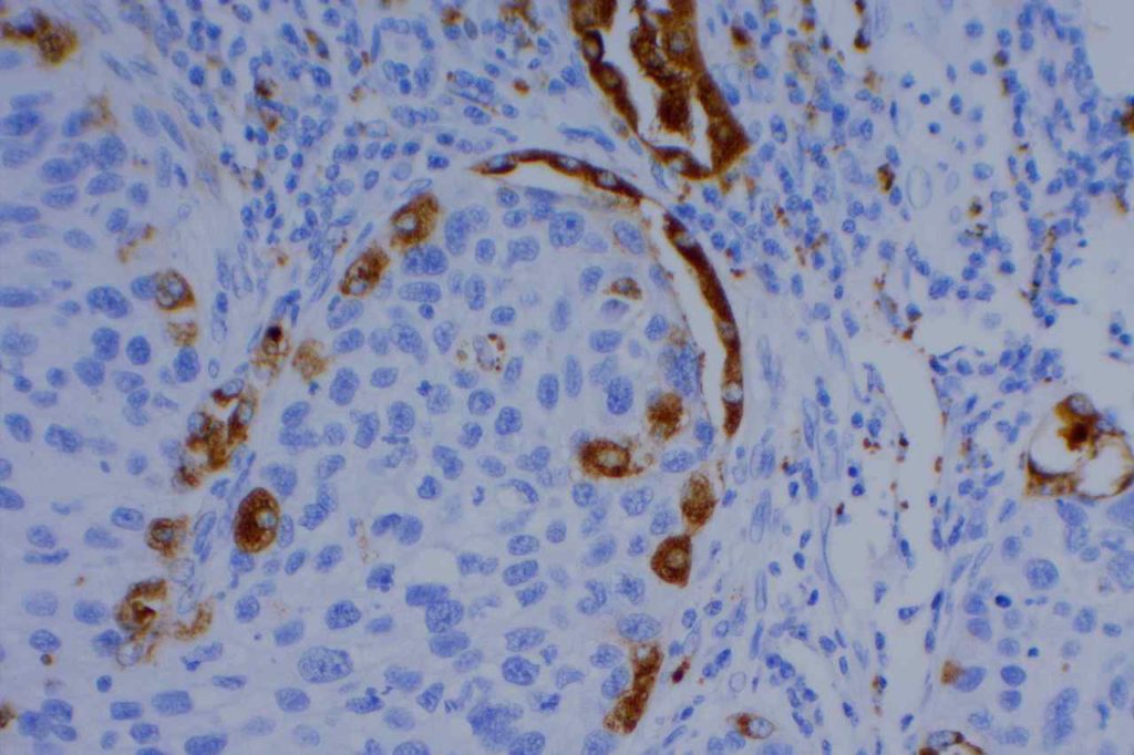
References
Eisen, RN, et al. Appl Immunohistochem Mol Morphol. Vol. 19, No. 6, December 2011.
Bishop, J. A., Sharma, R., & Illei, P. B. (2010). Napsin A and thyroid transcription factor-1 expression in carcinomas of the lung, breast, pancreas, colon, kidney, thyroid, and malignant mesothelioma. Human Pathology, 41(1), 20–25. doi:10.1016/j.humpath.2009.06.014
Mukhopadhyay, S., & Katzenstein, A.-L. A. (2011). Subclassification of non-small cell lung carcinomas lacking morphologic differentiation on biopsy specimens: Utility of an immunohistochemical panel containing TTF-1, napsin A, p63, and CK5/6. The American Journal of Surgical Pathology, 35(1), 15–25. doi:10.1097/PAS.0b013e3182036d05
Köbel M, Duggan MA. Napsin a: another milestone in the subclassification of ovarian carcinoma. Am J Clin Pathol. 2014;142(6):735–737. doi:10.1309/AJCPAVGZKA1A1HVC.
Kandalaft PL, Gown AM, Isacson C. The lung-restricted marker napsin a is highly expressed in clear cell carcinomas of the ovary. Am J Clin Pathol. 2014;142(6):830–836. doi:10.1309/AJCP8WO2EOIAHSOF.
Fadare O, Desouki MM, Gwin K, Hanley KZ, Jarboe EA, Liang SX, et al. Frequent Expression of Napsin A in Clear Cell Carcinoma of the Endometrium: Potential Diagnostic Utility. Am J Surg Pathol. 2013. doi:10.1097/PAS.0000000000000085
