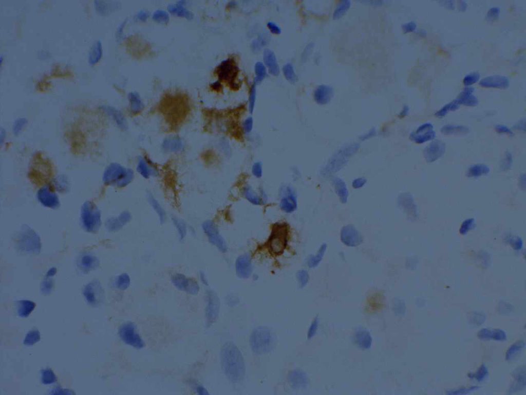
Renal Cell Carcinoma Marker (RCC Ma) is a cytoplasmic marker that stains the renal proximal tubule brush border antigen. The following table (McGregor, et al) shows the stain sensitivity (>10% tumor cell staining):
|
Tumor Type
|
Sensitivity
|
|
RCC Clear Cell
|
84%
|
|
RCC Papillary
|
96%
|
|
RCC Chromophobe
|
45%
|
|
RCC Sarcomatoid
|
25%
|
|
Collecting Duct Carcinoma
|
0%
|
|
Oncocytoma
|
0%
|
Sharma, S.G., et. al. (PAX-2 and RCCma expression in various papillary tumors)
|
Tumor Type
|
No.
|
PAX2
|
RCCma
|
|
Papillary RCC
|
24
|
67%
|
96%
|
|
Ovarian Papillary Serous Carcinoma
|
10
|
40%
|
80%
|
|
Uterine Papillary Serous Carcinoma
|
9
|
56%
|
44%
|
|
Papillary Thyroid Carcinoma
|
9
|
0%
|
100%
|
|
Papillary Urothelial Carcinoma
|
10
|
0%
|
10%
|
|
Intraductal Papillary Mucinous Tumor of the Pancreas
|
2
|
0%
|
50%
|
|
Chroroid Plexus Papilloma
|
1
|
0%
|
100%
|
|
Pituitary Adenoma with Papillary Features
|
1
|
0%
|
100%
|
|
Lung Adenocarcinoma with Papillary Features
|
2
|
0%
|
50%
|
RCC Ma has been shown to be relatively specific for RCC in the metastatic setting, but is known to stain cells of other histogenesis (e.g. breast carcinoma, embryonal carcinoma, parathyroid adenomas, etc.). RCC Ma is a useful marker, but not recommended to be used in isolation (especially papillary lesions). It is also important to confirm the performance of this antibody within one’s laboratory because it has shown performance variability from laboratory to laboratory (author’s experience). A useful panel for renal cell carcinoma includes CD10, vimentin, AE1/AE3, and RCC Ma.
Photomicrograph

Reference:
McGregor, D. K., Khurana, K. K., Cao, C., Tsao, C. C., Ayala, G., Krishnan, B., et al. (2001). Diagnosing primary and metastatic renal cell carcinoma: the use of the monoclonal antibody ‘Renal Cell Carcinoma Marker’. The American Journal of Surgical Pathology, 25(12), 1485–1492.
Sharma, S. G., Gokden, M., McKenney, J. K., Phan, D. C., Cox, R. M., Kelly, T., & Gokden, N. (2010). The utility of PAX-2 and renal cell carcinoma marker immunohistochemistry in distinguishing papillary renal cell carcinoma from nonrenal cell neoplasms with papillary features. Applied Immunohistochemistry & Molecular Morphology : AIMM / Official Publication of the Society for Applied Immunohistochemistry, 18(6), 494–498. doi:10.1097/PAI.0b013e3181e78ff8
Pan, Z., Grizzle, W., & Hameed, O. (2013). Significant variation of immunohistochemical marker expression in paired primary and metastatic clear cell renal cell carcinomas. American Journal of Clinical Pathology, 140(3), 410–418. doi:10.1309/AJCP8DMPEIMVH6YP
Zhai, Q. J. (2010). Application of Immunohistochemistry to the Diagnosis of Kidney Tumors. Pathology Case Reviews, 15(1), 25–34. doi:10.1097/PCR.0b013e3181d51c70
