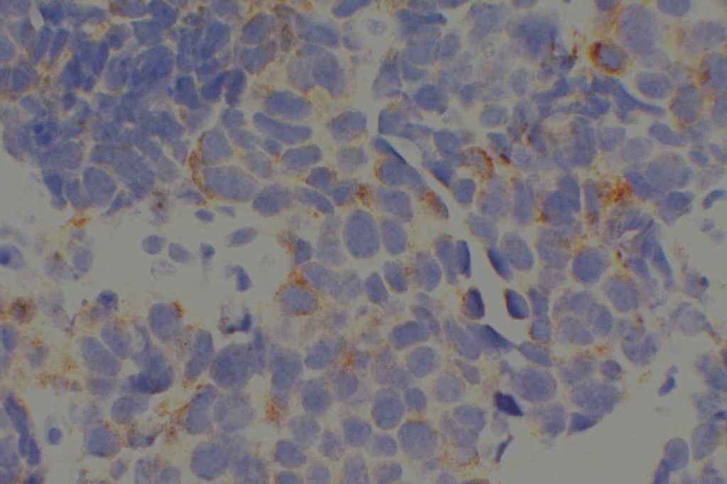
Synaptophysin reacts with the integral membrane glycoprotein of presynaptic vesicles. It is less specific than chromogranin, and it may mark neurofilaments and epithelial filaments. Some neuroendocrine neoplasms will mark with synaptophysin and not chromogranin A. The converse can also be true. Therefore, chromogranin A and synaptophysin should be used in tandem to identify neuroendocrine differentiation (Wick, MR). CD56 is often considered the most sensitive neuroendocrine marker, but it is not as specific as chromogranin and synaptophysin (Bahrami, A, et al).
Neuroendocrine differentiation may not be readily recognized until it is revealed by immunohistochemistry, but may be important as these tumors may be responsive to similar therapy used for small cell carcinoma (Bahrami, A, et al).
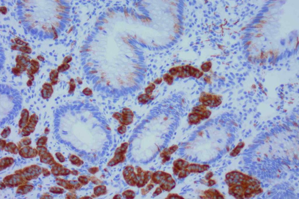
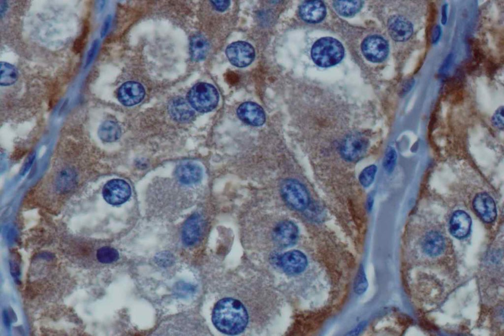
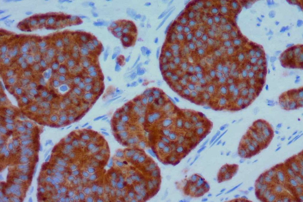
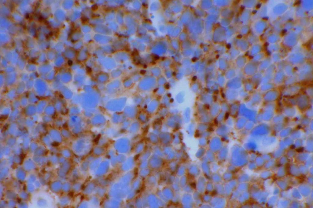
Microscopic Images
References
Wick, M. R. (2008). Immunohistochemical approaches to the diagnosis of undifferentiated malignant tumors. Annals of Diagnostic Pathology, 12(1), 72–84. doi:10.1016/j.anndiagpath.2007.10.003
Bahrami, A., Truong, L. D., & Ro, J. Y. (2008). Undifferentiated tumor: true identity by immunohistochemistry. Archives of Pathology & Laboratory Medicine, 132(3), 326–348.
