Tag Archives: breast

Breast – Atypical Ductal Hyperplasia (ADH)

ADH is a neoplastic proliferation that shares some characteristics of ductal carcinoma in situ (DCIS), but falls short quantitatively or qualitatively.  ADH is associated with a moderately increased risk of developing an invasive breast carcinoma (4-5x relative risk, 13-17% lifetime risk).
 
The morphology is similar to DCIS, but the findings do not entirely fill the duct spaces and/or don’t fulfill a quantitative size requirement for DCIS (some require 2 mm lesion).  In biopsy specimens it is important to perform an excision biopsy/lumpectomy because approximately 1/3rd of cases will have an associated higher grade lesion in the immediate vicinity (e.g. DCIS or an invasive carcinoma). 
Breast lesions and risk of developing an invasive carcinoma
Relative
Risk
Absolute
Risk
(lifetime)
Breast
Lesion
1
3%
1.5 – 2
5-7%
4 – 5
13-17%
8 – 10
25-30%
References

Robbins, p. 1050-1051

Breast – Intraductal Papilloma

Introductal papilloma can have varying appearances but characteristically contain an ductal epithelial proliferation on a fibrovascular core with an intact myoepithelial layer.  Larger duct lesions are located near the nipple and typically produce a discharge (~80%).  Small duct lesions are frequently multiple and located deeper in the breast.
 
Papilloma are are associated with a slight increased risk for breast carcinoma (1.5-2x increased relative risk).  The absence of a myoepithelial layer is not characteristic of a typical papilloma, and likely represents a different papillary lesion.
Breast lesions and risk of developing an invasive carcinoma
Breast Cancer Risk Chart

Reference 

Kumar, Vinay, Abul K. Abbas, and Jon C. Aster. Robbins and Cotran Pathologic Basis of Disease. Ninth edition. Philadelphia, PA: Elsevier/Saunders, 2015. p. 1048-1051

Breast – Complex Sclerosing Lesion (Radial Scar)

As the name indicates a complex sclerosing lesion is “complex” in that it has multiple parts, which contribute to is “scar-like” appearance.  These lesions can be detected frequently by mammography mimicking invasive carcinoma, and subsequently leads to biopsy.
 
These lesions contain a combination of different patterns discussed elsewhere, including sclerosing adenosis, papilloma, usual type hyperplasia (UTH), and fibrosis.  The architecture of this lesion is around a central area of compressed entrapped ducts within fibrosis, which radiate outward with varying dilation of duct structures and present of epithelial hyperplasia.
 
Complex scoring lesions are not neoplastic but are associated with a 1.5-2x increased relative risk for breast carcinoma.
Breast lesions and risk of developing an invasive carcinoma
Breast Cancer Risk Chart

References

Kumar, Vinay, Abul K. Abbas, and Jon C. Aster. Robbins and Cotran Pathologic Basis of Disease. Ninth edition. Philadelphia, PA: Elsevier/Saunders, 2015.

Breast – Sclerosing Adenosis

Sclerosing Adenosis (SA) is an increased density of duct-lobular acini, which characteristically are compressed in the central portion of the lesion with dense stroma/fibrosis.  Sometimes these lesions can closely mimic an invasive ducatal carcinoma, immunohistochemistry is necessary to separate lesions.
 
SA is not a neoplastic process, but is associated with a 1.5-2X increased relative risk for breast carcinoma.
Breast lesions and risk of developing an invasive carcinoma
Breast Cancer Risk Chart

References
Kumar, Vinay, Abul K. Abbas, and Jon C. Aster. Robbins and Cotran Pathologic Basis of Disease. Ninth edition. Philadelphia, PA: Elsevier/Saunders, 2015.


Breast – Usual Type Epithelial Hyperplasia (UTH)

Non-neoplastic luminal proliferation of ductal epithelial and myoepithelial cells, which can fill and expand ductal strucutures.  The morphology shows a mixed cellularity pattern with overlapping nuclei, which often have a “streaming” appearance.  In challenging cases immunohistochemistry can help differentiated these lesions from ADH or DCIS.
 
If present only focally, there is not an increased risk of breast carcinoma.  However, moderate to florid usual type hyperplasia is associated with a 1.5-2X increased relative risk of breast carcinoma.
 
 
Breast - Usual Type Hyperplasia
Usual type hyperplasia of the breast showing an intraductal proliferation of epithelial and myoepithelial cells with overlapping nuclei and streaming.
Breast lesions and risk of developing an invasive carcinoma
Breast Cancer Risk Chart

References

Kumar, Vinay, Abul K. Abbas, and Jon C. Aster. Robbins and Cotran Pathologic Basis of Disease. Ninth edition. Philadelphia, PA: Elsevier/Saunders, 2015. 

Breast – Fibrocystic Change

This is a non-proliferative lesion characterized clinically as small lumps, which do not carry an increased risk of breast carcinoma.  Fibrocystic changes have three characteristic features, which may be present in part or whole, and include:  
  • Adenosis – Increased ductal acini density.  Calcification may be present as well as other epithelial changes such as flat epithelial atypia (see separate discussion).
  • Cystic change – Cysts (typically of varying sizes) lines by flat atrophic epithelium or apocrine metasplasia are characteristic.  Calcification are also common.
  • FIbrosis – Areas of fibrosis are characteristically present, and are thought to originate from chronic inflammation secondary to ruptured cysts.
Breast - Fibrocystic Change
Dilated duct structures with apocrine changes (low power).
Breast - Fibrocystic Change
Fibrocystic change with apocrine metaplasia and micropapillary formation.
Breast - Fibrocystic Change
Duct dilation in fibrocystic change (low power).

References

Kumar, Vinay, Abul K. Abbas, and Jon C. Aster. Robbins and Cotran Pathologic Basis of Disease. Ninth edition. Philadelphia, PA: Elsevier/Saunders, 2015.

Micropapillary Breast Carcinoma

Key features
  •  Aggressive course
  •  High rate of axillary lymph node metastasis (66–100%)
  •  Morphology– tubulealveolar, morular clusters (spongelike), micropapillary
  •  Frequent calcifications, including psammoma bodies (42–67%)
  •  EMA (epithelial membrane antigen) has reverse membrane staining (outer border of cells)
  •  Receptor status
    • ER: 62–91% +
    • PR: 46–72% +
    • HER-2: 36-81% +
      • HER-2 FISH amplified tumors may have 1+ (negative) or 2+ (equivocal) staining by IHC, and it is recommended that IHC and FISH be used in cases with micropapillary pattern.

Continue reading Micropapillary Breast Carcinoma

Lobular Carcinoma In Situ (LCIS)

Lobular Carcinoma In Situ (LCIS)
  • Neoplastic proliferation within ducts/lobules (fill and expand lobules)
  • Associated with loss of expression of E-Cadherin
  • Incidental finding (not associated with calcifications or stromal reactions found by mammography)
  • LCIS is bilateral 20-40% of cases
  • Morphology
    • Mucin-positive signet ring cells often present
    • E-Cadherin negative cells
    • Uniform cells with oval/round nuclei
    • Pagetoid spread (cells present between the myoepithelial layer and over lying luminal epithelium 
    • Typically ER/PR positive
    • Her-2 is not overexpressed
  • 1%/year risk of developing an invasive tumor (similar to low-grade DCIS) – Lifetime risk is ~25-35% (20-30 year time period)
  • Cancer risk is equal in contralateral breast (unlike DCIS)
  • Pleomorphic variant of LCIS
    • High grade nuclei
    • May be ER negative
    • Some may overexpress Her-2
    • May be separate entity from typical LCIS
Photomicrographs
Breast - Lobular Carcinoma In Situ (LCIS)

LCIS characterized by filling and expansion of the lobular unit.

Breast - Lobular Carcinoma In Situ (LCIS)
LCIS characterized by filling and expansion of the lobular unit.
Breast - Lobular Carcinoma In Situ (LCIS)
LCIS characterized by filling and expansion of the lobular unit.

References

Kumar, Vinay, Abul K. Abbas, and Jon C. Aster. Robbins and Cotran Pathologic Basis of Disease. Ninth edition. Philadelphia, PA: Elsevier/Saunders, 2015.  p. 1045.

Breast – Predictive Markers

One of the most important aspects of breast cancer diagnosis is the evaluation of therapeutic markers (ER, PR, and HER2).  Ki-67 is often included in the panel as a prognostic marker.  ER expression determines eligibility to receive hormonal therapy (Tamoxifin), PR expression is a prognostic marker, and HER-2 over-expression determines eligibility to receive Herceptin®.  Diagnostically, the challenge is to consistently and accurately perform and interpret these IHC markers.
 
Estrogen Receptor (ER):
  • Nuclear Marker
  • Stain is reported as PERCENT STAINING OF TUMOR CELLS and STAIN INTENSITY (1+, 2+, 3+).
  • 1% or greater nuclear expression in tumor cells is considered positive, and therefore eligible to receive hormonal therapy.
  • CAP-ASCO recommendations are for <1 hr. from time of excision/biopsy to having a cut edge of tumor in 10% neutral bufferedormalin fixative.  Fixation window of 6-72 hrs.  These times should be noted in the pathology report (time of excision, time in gross room, and time in fixative).
  • Negative staining results in biopsy material without an internal control should be repeated on the excisional specimen using blocks with both tumor and benign breast parenchyma.
Progesterone Receptor (PR):
  • Nuclear Marker
  • Stain is reported as PERCENT STAINING OF TUMOR CELLS and STAIN INTENSITY (1+, 2+, 3+).
  • 1% or greater nuclear expression in tumor cells is considered positive.
  • PR expression is a prognostic marker, and not directly used for eligibility to receive a specific treatment.
  • PR expression without ER expression should raise significant concern that the ER and PR slides have been mixed up, or there is a problem with the ER assay.  Many scientists believe that ER expression is required for PR expression.
HER-2 Overexpression (HER-2):
  • Membraneous stain
  • Stain is interpreted by combining stain intensity and percentage of tumor involvement to classify as (0, 1+, 2+, or 3+).
    • 0 (negative) = No staining or cell membrane staining in <10% of tumor cells.
    • 1+ (negative) =  Faint membrane staining (partial membrane staining) in >10% of tumor cells.
    • 2+ (equivocal) =  Weak to moderate complete membrane staining in >10% of tumor cells, or strong complete staining in <10% of invasive tumor cells.
    • 3+ (positive) =  Strong complete membrane staining in >10% of tumor cells.
  • CAP-ASCO recommendations are for <1 hr. from time of excision/biopsy to having a cut edge of tumor in 10% neutral buffered fomalin fixative.  Fixation window of 6-72 hrs.  Over-fixation is probably not a clinically significant issue practically, but given the absence of relevant IHC data and the highly regulated environment surrounding HER2 testing, f/u FISH testing for negative results (outside the fixative window) is necessary.
  • Equivocal (2+) results should be followed-up with FISH testing, if IHC is used as the initial testing modality (most common).  Less than 1/3rd of equivocal cases show Her2 over-expression by FISH analysis.

References
 
Hammond ME, et. al.  “ASCO-CAP Guideline Recommendations for IHC Testing of ER and PR in Breast Cancer”.  Arch Pathol Lab Med-Vol. 134, June 2010.
 
Wolff, A. C., Hammond, M. E. H., Hicks, D. G., Dowsett, M., McShane, L. M., Allison, K. H., et al. (2013). Recommendations for Human Epidermal Growth Factor Receptor 2 Testing in Breast Cancer: American Society of Clinical Oncology/College of American Pathologists Clinical Practice Guideline Update. Archives of pathology & laboratory medicine. doi:10.5858/arpa.2013-0953-SA 
 
Tafe, L. J., Janjigian, Y. Y., Zaidinski, M., Hedvat, C. V., Hameed, M. R., Tang, L. H., et al. (2011). Human epidermal growth factor receptor 2 testing in gastroesophageal cancer: correlation between immunohistochemistry and fluorescence in situ hybridization. Archives of pathology & laboratory medicine, 135(11), 1460–1465. doi:10.5858/arpa.2010-0541-OA 
 
Arch Pathol Lab Med. 2001;125:746.

Breast – IHC Specific Markers

IHC stain expression pattern for various IHC antibodies in breast carcinoma.
 
IHC Stain
Comments
50-70%
30-60%
>80%
<10%
10-25%
0%
0%
0%
Mesothelin
<10%
Clin Cancer Rs 2005;11(10) May 15, 2005.
 
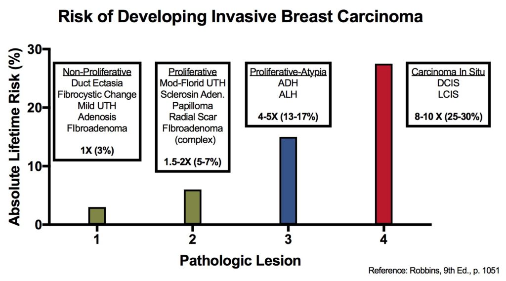
When evaluating for a possible breast primary, it is usually part of a larger differential diagnosis.  CK7 and CK20 is the common starting point for carcinomas of unknown primary site (CUPS), and breast has a characteristic CK7+/CK20= profile.  Unfortunately, this pattern is not uncommon, and more specific markers need to be performed.  Two specific breast markers are GCDFP-15 and ER., but their sensitivity while good is limited.  GATA-3 is a relatively newer antibody, which shows excellent sensitivity with good specificity, and should be strongly considered to be part of an antibody panel in work-up of potential breast carcinoma cases. 
  • GCDFP-15 is very specific for breast carcinoma in the setting of CUPS, but limited sensitivity.  
  • ER expression may be highly suggestive of a breast primary (especially epidemiologically), but it is not as specific.
  • Other female organs (ovary/uterus) not uncommonly express ER, and practically any tissue can occasionally excess ER.
  • GATA-3 appears to have excellent sensitivity (>90%) with good (not perfect) specificity.
Liu, et al (Biocare Medical, Concord, CA)
Tumor
GATA-3
GCDFP-15
MGB
Breast Carcinoma
94%
35-55%
65-70%
ER-negative breast ca.
69%
15%
35%
Urothelial Carcinoma
86%


Breast Metastasis vs. Ovarian Ca. Primary
Tumor
WT-1
CA-125
GCDFP-15
Primary Ovarian Ca.
(N=41)
76%
73%
0%
Metastatic Breast Ca.
(N-40)
3%
10%
43%
P-Value
<0.001
<0.001
<0.001
Chen, USCAP, 2004

References
Liu, H., Shi, J., Wilkerson, M. L., & Lin, F. (2012). Immunohistochemical evaluation of GATA3 expression in tumors and normal tissues: a useful immunomarker for breast and urothelial carcinomas. American Journal of Clinical Pathology, 138(1), 57–64. doi:10.1309/AJCP5UAFMSA9ZQBZ
 
Miettinen, M., McCue, P. A., Sarlomo-Rikala, M., Rys, J., Czapiewski, P., Wazny, K., et al. (2014). GATA3: A Multispecific But Potentially Useful Marker in Surgical Pathology: A Systematic Analysis of 2500 Epithelial and Nonepithelial Tumors. The American Journal of Surgical Pathology, 38(1), 13–22. doi:10.1097/PAS.0b013e3182a0218f 
 
Clin Cancer Rs 2005;11(10) May 15, 2005.
 
Chen, USCAP, 2004