Tag Archives: breast

Breast – DCIS vs. LCIS

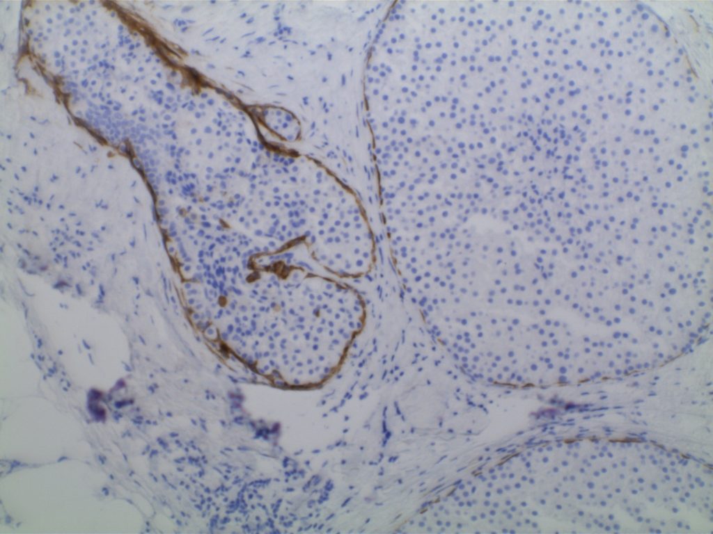
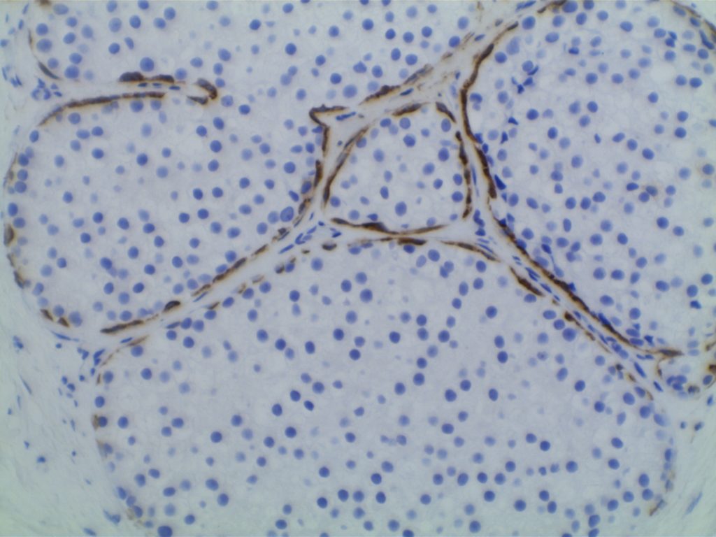
Lobular vs. ductal differentiation in breast lesions can on occasion be problematic.  E-cadherin is well described as a marker of ductal differentiation, but aberrant expression has be reported in 2-16% of cases of lobular neoplasia.  Some authors suggest not changing the interpretation/diagnosis based on expression of E-cadherin is the morphology is consistent with lobular carcinoma.  This author is not sure why one would do the stain to begin with if the morphologic characteristics are consistent with lobular carcinoma.  34betaE12 (CK903) has been described as a sensitive and specific in showing perinuclear dot-like expression in cases of lobular carcinoma and negativity in ductal carcinomas.  The combined panel of 34betaE12 and E-cadherin makes a nice complimentary panel to evaluate between lobular and ductal differentiation in breast carcinomas when morphology alone is not clear.
 
IHC Stain
DCIS
LCIS
E-Cadherin
+
=
34betaE12
=
+
References
Liu H. Application of immunohistochemistry in breast pathology: a review and update. Arch Pathol Lab Med. 2014;138(12):1629–1642. doi:10.5858/arpa.2014-0094-RA.
 
Bratthauer GL, Moinfar F, Stamatakos MD, et al. Combined E-cadherin and high molecular weight cytokeratin immunoprofile differentiates lobular, ductal, and hybrid mammary intraepithelial neoplasias. Hum Pathol. 2002;33(6): 620–627.

Breast – Stromal Invasion vs. In Situ or Benign Glands

Not infrequently in breast pathology, the differential diagnosis of micro-invasion vs. DCIS or a scerlosing lesion +/- tubular carcinoma will occur.  These type of cases can be very challenging based just on H&E morphology.  Fortunately, there are multiple IHC markers, which can be helpful in identifying the myoepithelial layer.  The main pitfall in interpretation of most of these markers, is that they will also mark myofibroblasts (on occasion) in the intervening stroma.  When this occurs, and myofibrilblasts abut the glands, the staining pattern can be misinterpreted as an intact myoepithelial layer.
 
Antibody
MEC
Myofibroblasts
++++
++
++++
++
SMA
++++
+++
p63 (nuclear)
++++
++++
+++
+
++
SMA = Smooth Muscle Action, SMM-HC = Smooth Muscle Myosin Heavy Chain.
 
The most commonly used stains include SMM-HC, calponin, CK5, and p63.  p63 can be combined with any of the other cytoplasmic stains as part of a double stain protocol.  SMA is the most proned of the stains to also mark fibroblasts.  S-100 is NOT recommended as a myoepithelial marker.  p63 has the least association with myofibroblast staining, but expression may be discontinuous in the myoepithelial cells layer, which can lower sensitivity when used alone.  In the opinion of many breast pathologists, SMM-HC as a single stain probably has the best combined sensitivity for the myoepithelial cells with minimal myofibroblastic staining, but many will use multiple markers in difficult cases.
 
Myofibroblast Staining
Actin > Calponin > SMM-HC
 

References
Hicks DG. Immunohistochemistry in the diagnostic evaluation of breast lesions. Appl Immunohistochem Mol Morphol. 2011;19(6):501–505. doi:10.1097/PAI.0b013e31822c8a48.
 
Liu H. Application of immunohistochemistry in breast pathology: a review and update. Arch Pathol Lab Med. 2014;138(12):1629–1642. doi:10.5858/arpa.2014-0094-RA.

Breast – UDH vs. ADH

A common differential diagnosis in breast lesions is between usual type hyperplasia (UDH) and atypical ductal hyperplasia (ADH)/ductal carcinoma in situ (DCIS).  In difficult cases there are some immunohistochemical patterns, which may be helpful to differentiate between diagnoses.  UDH has  mosaic expression pattern with HMWK  (high molecular weight keratins) (e.g. CK5 or CK5/6) whereas ADH/DCIS typically does not.  ADH/DCIS does typically has strong uniform up regulation of estrogen receptor (ER) in contrast to UDH.
 
DIAGNOSTIC FEATURES
Cellular Population
Florid UDH
ADH
True hyperplasia contains a mixture of cell types (streaming, slit-like spaces)
Clonal population of monotonous cells with rigid “punched-out” spaces
 
CK Expression
Florid UDH
ADH
Mixture of basal cells (CK5/14/17) and luminal cells (CK7/8/18)
Monotonous population of luminal cell types (CK 7/8/18)
 
ER Expression
Florid UDH
ADH
Variable patchy expression
Usually uniform strong expression
 
Breast Cancer Relative Risk
Florid UDH
ADH
Slightly increased (1.5-2 X)
Moderately increased (3.7-5.3 X)
 
 
Diagnostic Features
Florid UDH
ADH
Cellular Population
True hyperplasia contains a mixture of cell types (streaming, slit-like spaces)
Clonal population of monotonous cells with rigid “punched-out” spaces
CK Expression
Mixture of basal cells (CK5/14/17) and luminal cells (CK7/8/18)
Monotonous population of luminal cell types (CK 7/8/18)
EP Expression
Variable patchy expression
Usually uniform strong expression
Breast Cancer R.R.
Slightly increased (1.5-2X)
Moderately increased (3.7-5.3X)


References
Hicks DG. Immunohistochemistry in the diagnostic evaluation of breast lesions. Appl Immunohistochem Mol Morphol. 2011;19(6):501–505. doi:10.1097/PAI.0b013e31822c8a48.
 
Liu H. Application of immunohistochemistry in breast pathology: a review and update. Arch Pathol Lab Med. 2014;138(12):1629–1642. doi:10.5858/arpa.2014-0094-RA.

Breast – Normal IHC Expression

Normal breast ducts and lobules are lined by a 2-cell layer composed of luminal and myoepithelial cells.  There are also interspersed “basal” cells, which probably represent the epithelial progenitor cells.
 
IHC Marker
Luminal Cells
Myoepithelial Cells
LMWCKs
(CK7/8/18)
Positive
Negative
Variable Expression
Negative
HMWCKs
(CK5/14/17)
Negative
Positive
SMA
Negative
Positive
Negative
Positive
Negative
Positive
SMA=Smooth Muscle Actin, SMM-HC=Smooth Muscle Myosin Heavy Chain
 
An understanding of the normal IHC expression pattern in breast ductal tissue is important when considering IHC use in the differential diagnosis of breast pathology.

References
Hicks DG. Immunohistochemistry in the diagnostic evaluation of breast lesions. Appl Immunohistochem Mol Morphol. 2011;19(6):501–505. doi:10.1097/PAI.0b013e31822c8a48.
 
Liu H. Application of immunohistochemistry in breast pathology: a review and update. Arch Pathol Lab Med. 2014;138(12):1629–1642. doi:10.5858/arpa.2014-0094-RA.

Calponin

Calponin is an actin filament and is expressed in smooth muscle.  It is often used individually or combined with other IHC markers to identify the myoepithelial layer in breast epithelium.  It is important to remember, like other smooth muscle markers, that myofibroblasts may also express calponin.  Therefore, care should be taken not to mistake myofibroblast positivity for myoepithelial positivity. 
 
It is also expressed in cases of collagenous spherulosis (positive), but not adenoid cystic carcinoma of the breast.  Calponin is not a specific stain, and has been identified in numbers pathologic disease processes.  It is recommended to refer to specific medical literature for the IHC performance based on the differential diagnosis.
 
Positive Expression:
  • Atypical fibroxanthoma (30%) [Virchows Arch 200;437:58]
  • Benign Fibrous Histiocytoma (65%)
  • Collagenous Spherulosis[Mod Path 2006;19:1351]
  • DFSP (40%)
  • Fibromatosis [Am J Dermatopathol 2006;28:105]
  • Fibrosarcoma (60%)
  • Glomus Tumor [AJSP 2002;26:301]
  • Leiomyoma
  • Leiomyosarcoma
  • MFH of bone (47%) [J Clin Pathol 2002;55:853]
  • MPNST (40%)
  • Myoepithelioma-skin
  • Solitary fibrous tumor (70%)
  • Synovial Sarcoma [Histopathology 2003;42:588]
  • Myofibroblastic lesions
Photomicorgraphs
Calponin - Pleomorphic Lobular Carcinoma of Breast
Calponin – Pleomorphic Lobular Carcinoma of Breast
Calponin - Pleomorphic Lobular Carcinoma of Breast
Calponin – Pleomorphic Lobular Carcinoma of Breast
Calponin - Pleomorphic Lobular Carcinoma of Breast
Calponin – Pleomorphic Lobular Carcinoma of Breast
References
Zhao L, Yang X, Khan A, Kandil D. Diagnostic role of immunohistochemistry in the evaluation of breast pathology specimens. Arch Pathol Lab Med. 2014;138: 16–24. doi:10.5858/arpa.2012-0440-RA
 
Dewar R, Fadare O, Gilmore H, Gown AM. Best practices in diagnostic immunohistochemistry: myoepithelial markers in breast pathology. Arch Pathol Lab Med. 2011;135: 422–429. doi:10.1043/2010-0336-CP.1
 
Werling RW, Hwang H, Yaziji H, Gown AM. Immunohistochemical distinction of invasive from noninvasive breast lesions: a comparative study of p63 versus calponin and smooth muscle myosin heavy chain. Am J Surg Pathol. 2003;27: 82–90. 
 
Liu H. Application of immunohistochemistry in breast pathology: a review and update. Arch Pathol Lab Med. 2014;138: 1629–1642. doi:10.5858/arpa.2014-0094-RA