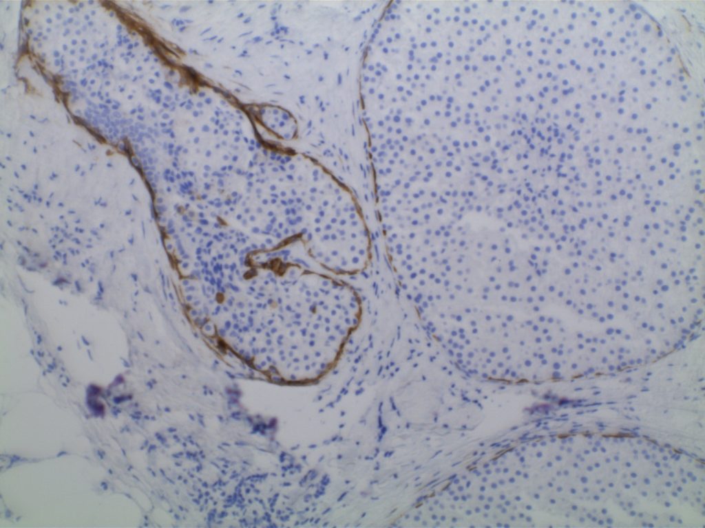
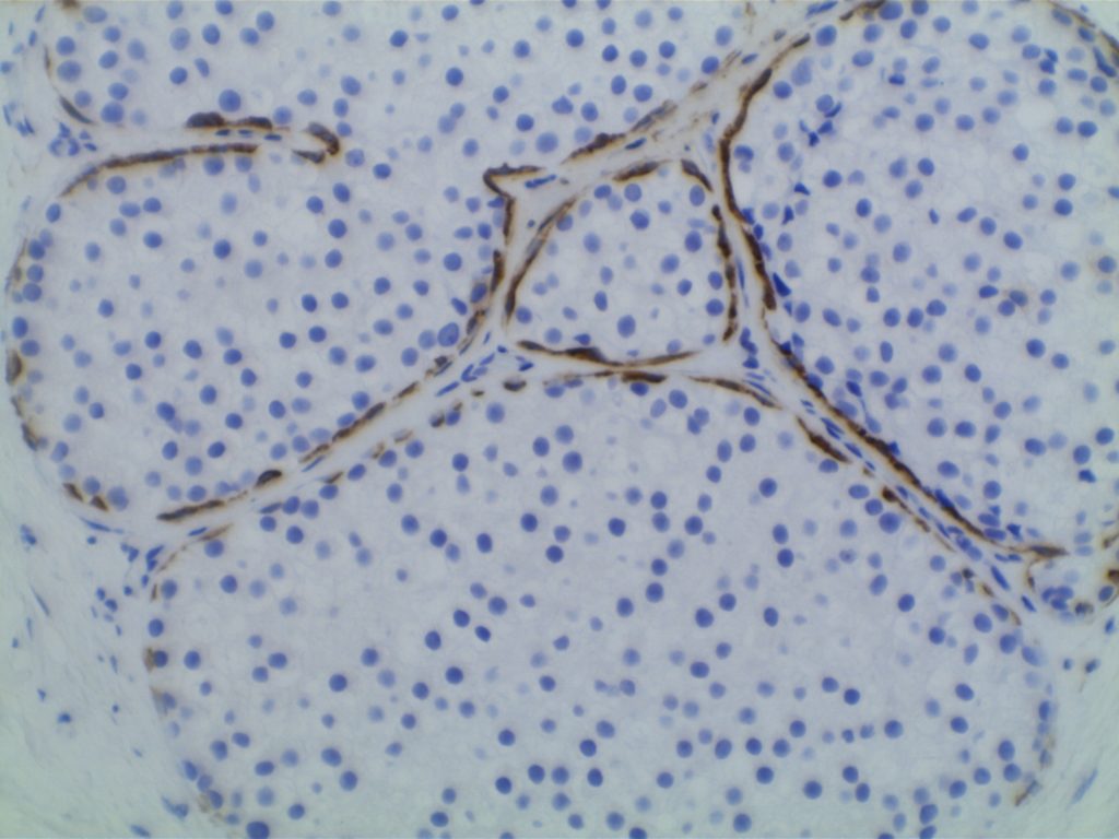
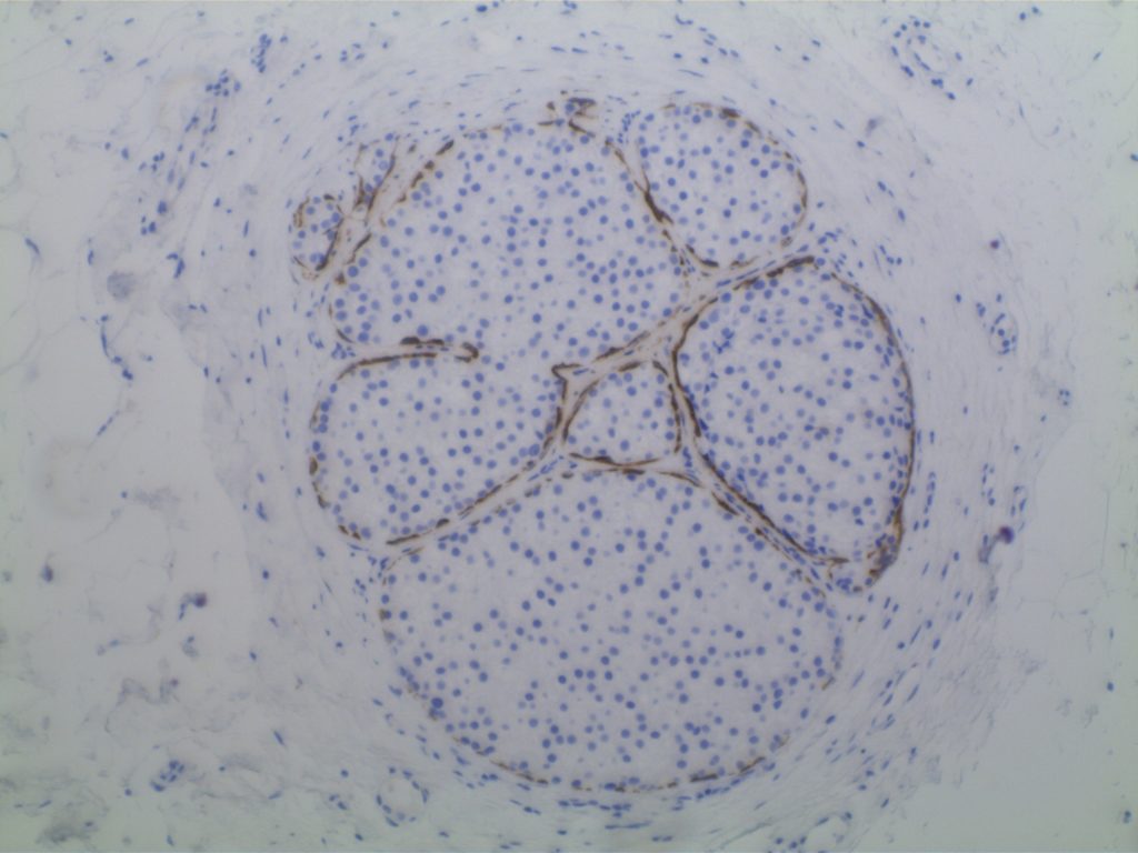
Calponin is an actin filament and is expressed in smooth muscle. It is often used individually or combined with other IHC markers to identify the myoepithelial layer in breast epithelium. It is important to remember, like other smooth muscle markers, that myofibroblasts may also express calponin. Therefore, care should be taken not to mistake myofibroblast positivity for myoepithelial positivity.
It is also expressed in cases of collagenous spherulosis (positive), but not adenoid cystic carcinoma of the breast. Calponin is not a specific stain, and has been identified in numbers pathologic disease processes. It is recommended to refer to specific medical literature for the IHC performance based on the differential diagnosis.
Positive Expression:
- Atypical fibroxanthoma (30%) [Virchows Arch 200;437:58]
- Benign Fibrous Histiocytoma (65%)
- Collagenous Spherulosis[Mod Path 2006;19:1351]
- DFSP (40%)
- Fibromatosis [Am J Dermatopathol 2006;28:105]
- Fibrosarcoma (60%)
- Glomus Tumor [AJSP 2002;26:301]
- Leiomyoma
- Leiomyosarcoma
- MFH of bone (47%) [J Clin Pathol 2002;55:853]
- MPNST (40%)
- Myoepithelioma-skin
- Solitary fibrous tumor (70%)
- Synovial Sarcoma [Histopathology 2003;42:588]
- Myofibroblastic lesions
Photomicorgraphs


References
Zhao L, Yang X, Khan A, Kandil D. Diagnostic role of immunohistochemistry in the evaluation of breast pathology specimens. Arch Pathol Lab Med. 2014;138: 16–24. doi:10.5858/arpa.2012-0440-RA
Dewar R, Fadare O, Gilmore H, Gown AM. Best practices in diagnostic immunohistochemistry: myoepithelial markers in breast pathology. Arch Pathol Lab Med. 2011;135: 422–429. doi:10.1043/2010-0336-CP.1
Werling RW, Hwang H, Yaziji H, Gown AM. Immunohistochemical distinction of invasive from noninvasive breast lesions: a comparative study of p63 versus calponin and smooth muscle myosin heavy chain. Am J Surg Pathol. 2003;27: 82–90.
Liu H. Application of immunohistochemistry in breast pathology: a review and update. Arch Pathol Lab Med. 2014;138: 1629–1642. doi:10.5858/arpa.2014-0094-RA
