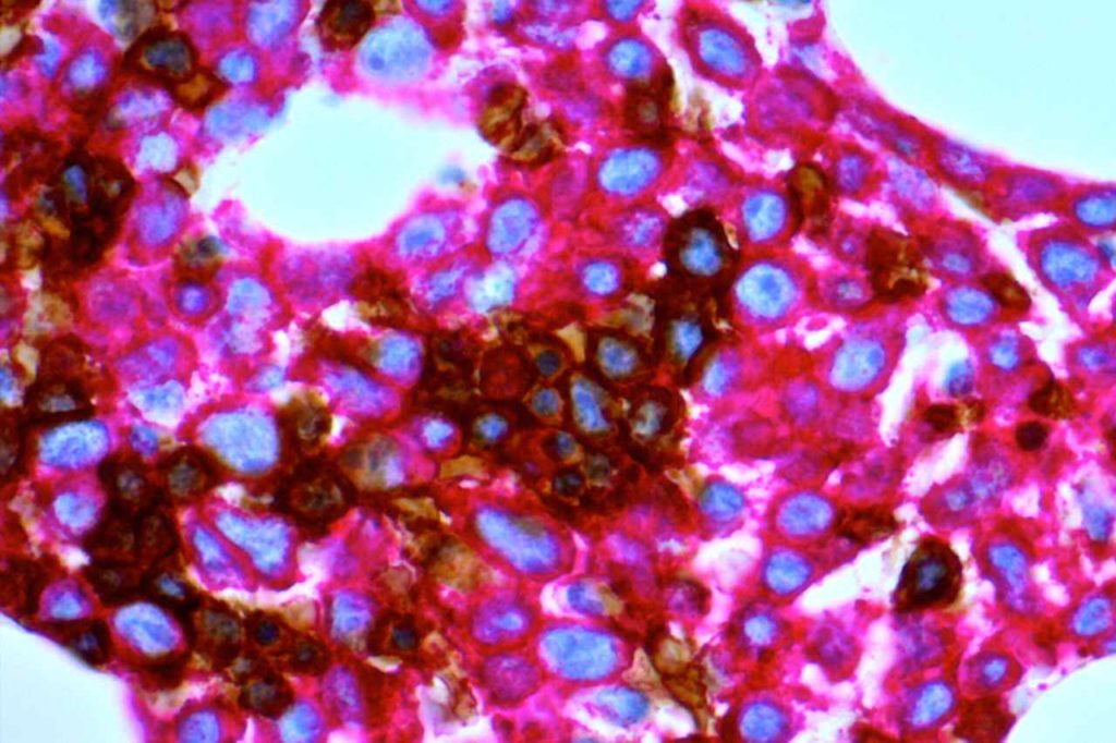
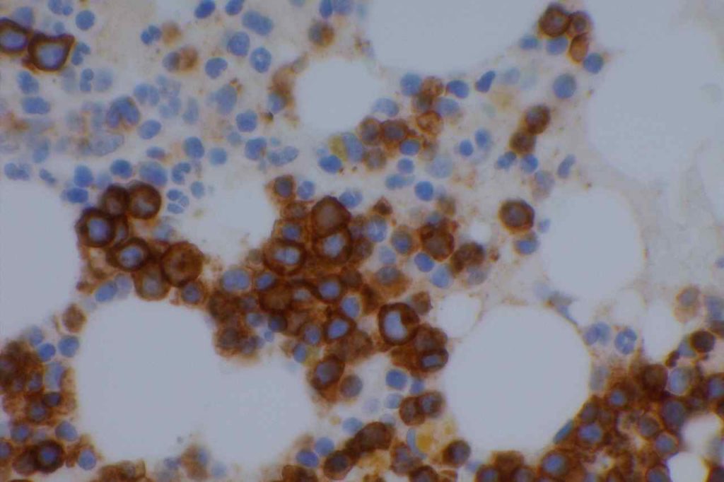
CD71 is one of the most useful, but least used antibodies in hematopathology. CD71 is an integral membrane protein, which is involved in the uptake of the transferrin-iron complex. Immunoreactivity is restricted to erythroid precursors with a membranous and cytoplasmic stain pattern.
Previously there have been other erythroid markers, but they have been difficult to interpret because they stain all RBCs and precursors. CD71 stains immature erythroid precursors, which are nucleated, and is not significantly expressed in mature RBCs. The utility of CD71 is to help differentiate between normoblasts and myeloblasts in bone marrow specimens. This is especially powerful when combined with CD34 (sensitive/not specific myeloblast marker) on a dual staining IHC platform (red and DAB chromogens).
CD71 can be a particularly useful tool to help accurately characterize immature cellular elements in bone marrow specimens, and decrease misidentification of normoblasts for myeloblasts and/or ALIP. E-Cadherin will also stain immature erythroid precursors (usually in a dimmer pattern compared to CD71).
Utilization of CD71 in gestational pathology has found it to be helpful to identify nucleated red blood cells (NRBCs) in partial molar pregnancies and spontaneous abortions in contrast to complete moles (absence of NRBCs).
Dim staining is expected in lymphoid cells (significantly different from nucleated red cell precursors), which can serve as a nice control (e.g. tonsil tissue).
Photomicrographs


References
Marsee, D. K., Pinkus, G. S., & Yu, H. (2010). CD71 (transferrin receptor): an effective marker for erythroid precursors in bone marrow biopsy specimens. American Journal of Clinical Pathology, 134(3), 429–435. doi:10.1309/AJCPCRK3MOAOJ6AT
Dong, H. Y., Wilkes, S., & Yang, H. (2011). CD71 is Selectively and Ubiquitously Expressed at High Levels in Erythroid Precursors of All Maturation Stages: A Comparative Immunochemical Study With Glycophorin A and Hemoglobin A. The American journal of surgical pathology, 35(5), 723–732. doi:10.1097/PAS.0b013e31821247a8
Luchini C, Parcesepe P, Nottegar A, Parolini C, Mafficini A, Remo A, et al. CD71 in Gestational Pathology: A Versatile Immunohistochemical Marker With New Possible Applications. Appl Immunohistochem Mol Morphol. 2016;24: 215–220. doi:10.1097/PAI.0000000000000175
Acs G, LiVolsi VA. Loss of membrane expression of E-cadherin in leukemic erythroblasts. Arch Pathol Lab Med. 2001;125: 198–201. doi:10.1043/0003-9985(2001)125<0198:LOMEOE>2.0.CO;2
Sadahira Y, Kanzaki A, Wada H, Yawata Y. Immunohistochemical identification of erythroid precursors in paraffin embedded bone marrow sections: spectrin is a superior marker to glycophorin. J Clin Pathol. 1999;52: 919–921.
