Tag Archives: DCIS

Breast – DCIS

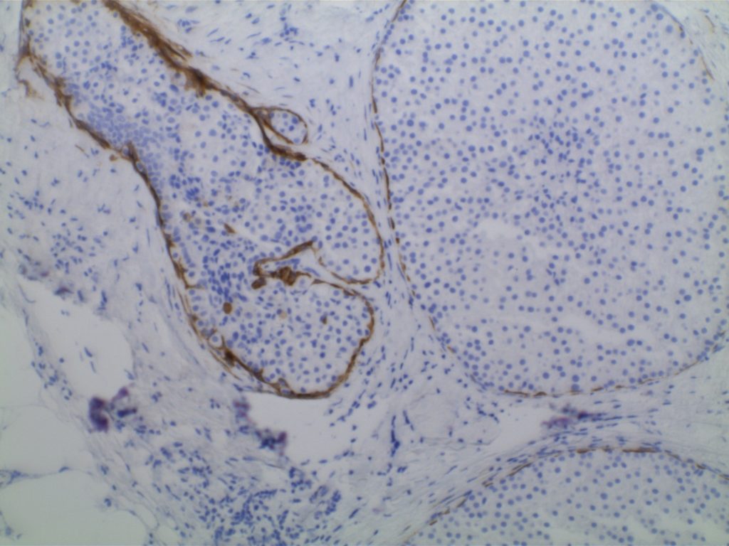
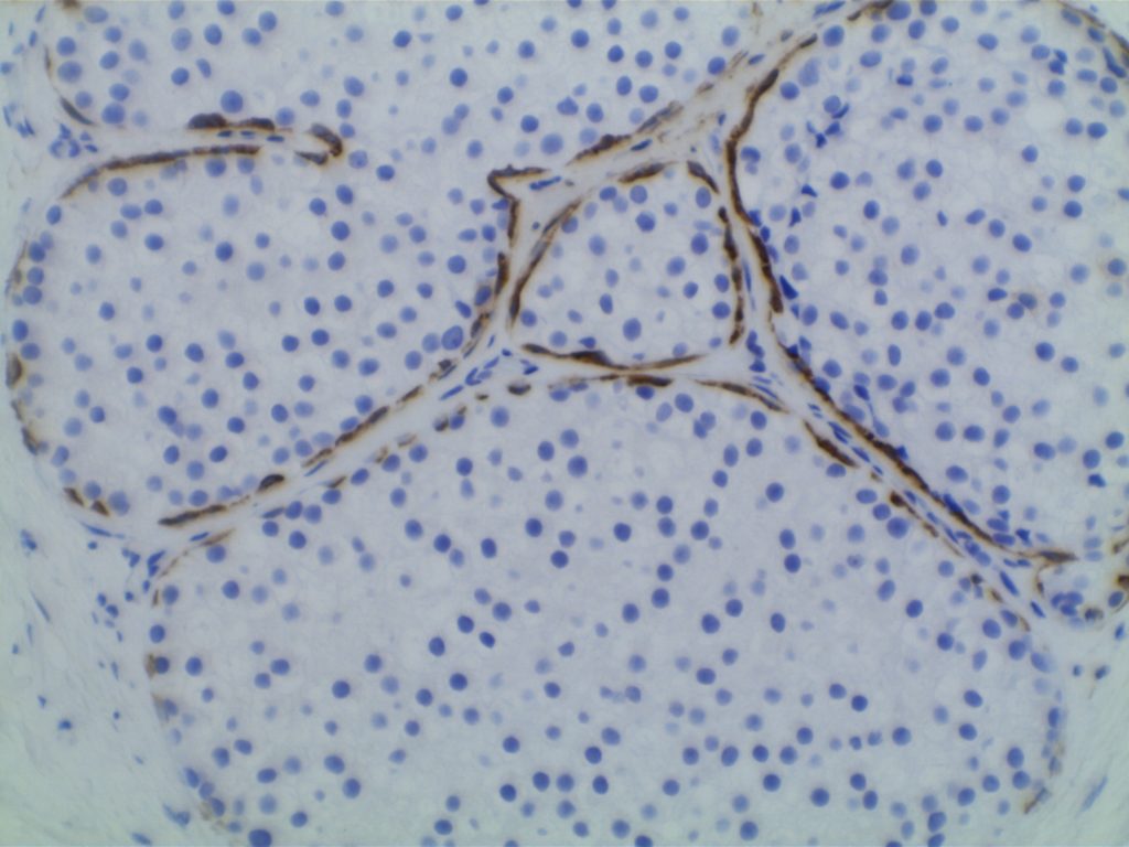
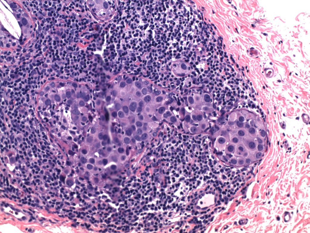
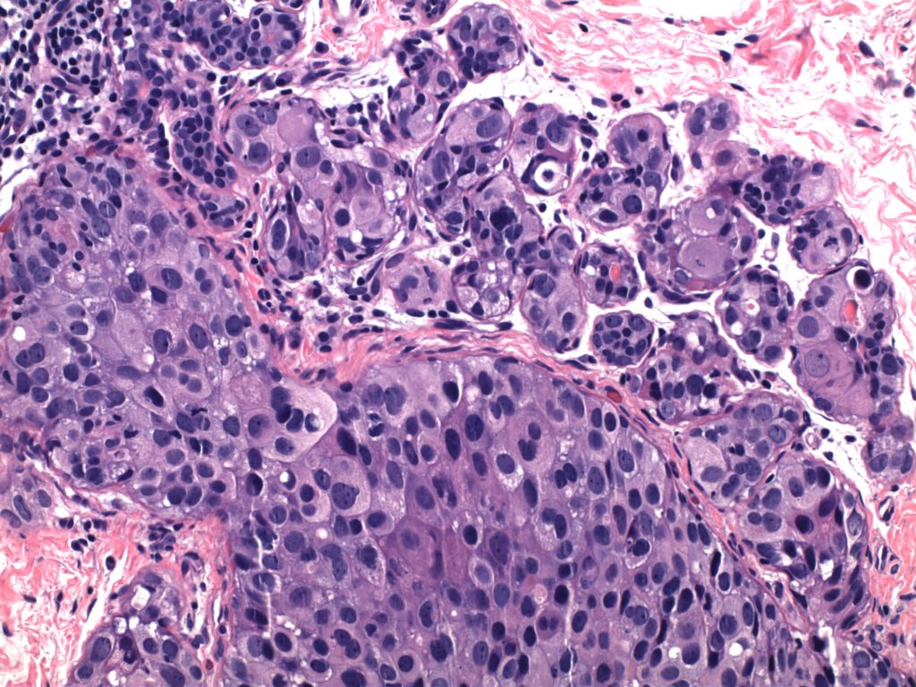
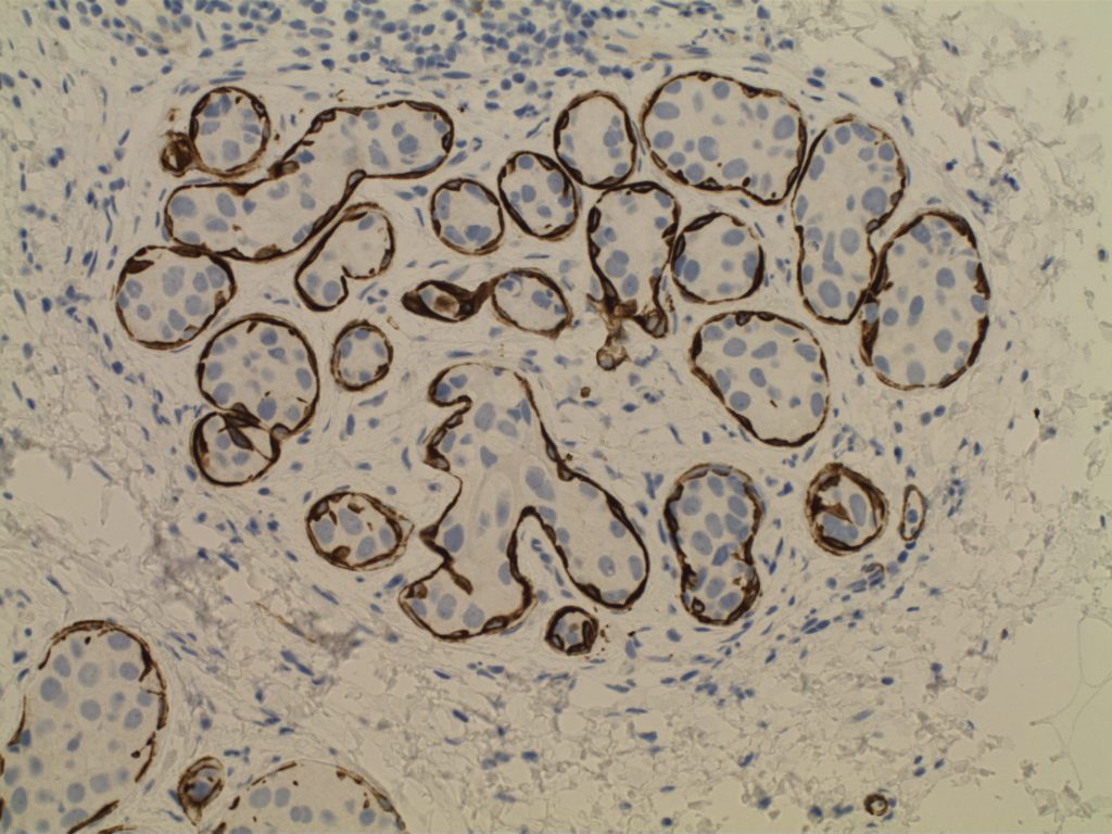
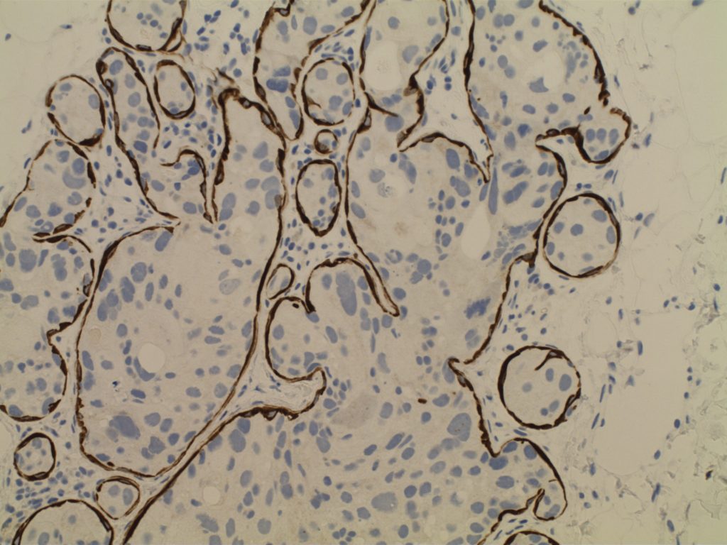
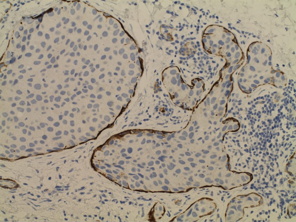
Ductal Carcinoma In Situ (DCIS)
  • Neoplastic proliferation resembling small ducts with expanded duct and acinar structures
  • Myoepithelial cell layer is intact surrounding the neoplastic proliferation (non-invasive tumor)
  • Typically express E-Cadherin
  • Bilateral in 10-20% of cases
  • Detected by mammography (not clinically evident)
  • Represents 15-30% of neoplasms identified in screening populations
  • Typically identified by abnormal calcification, sometimes as abnormal densities
Comedo DCIS
  • High grade pleomorphic nuclei
  • Central necrosis
Non-comedo DCIS
  • Lacks either high grade nuclei or central necrosis
  • Subtypes/patterns
    • Solid DCIS
    • Micropapillary DCIS
    • Cribiform DCIS
  • DCIS grading
    • Low-grade DCIS
      • 1%/year risk of developing an invasive carcinoma
    • Intermediate-grade DCIS
    • High-grade DCIS
Paget Disease
  • Nipple manifestation of disease (1-4% of cases) – looks like eczema on the nipple
  • Tumor cells extend into the epidermis of the skin overlying the nipple from underlying DCIS within the ductal system of the breast.
  • 50-60% of women will have an underlying palpable mass
    • Vast majority will have an invasive carcinoma (often ER neg./Her-2 pos.)
    • Women without a palpable mass will usually only have DCIS
Photomicrographs
Breast - High Grade DCIS
High power view of high grade breast DCIS.
Breast - High Grade DCIS
High power view of breast high grade DCIS.
Smooth Muscle Myosin - High Grade DCIS
Smooth Muscle Myosin – High Grade DCIS
Smooth Muscle Myosin - High Grade DCIS
Smooth Muscle Myosin – High Grade DCIS
Smooth Muscle Myosin - High Grade DCIS
Smooth Muscle Myosin – High Grade DCIS
References

Kumar, Vinay, Abul K. Abbas, and Jon C. Aster. Robbins and Cotran Pathologic Basis of Disease. Ninth edition. Philadelphia, PA: Elsevier/Saunders, 2015. 

Breast – DCIS vs. LCIS

Lobular vs. ductal differentiation in breast lesions can on occasion be problematic.  E-cadherin is well described as a marker of ductal differentiation, but aberrant expression has be reported in 2-16% of cases of lobular neoplasia.  Some authors suggest not changing the interpretation/diagnosis based on expression of E-cadherin is the morphology is consistent with lobular carcinoma.  This author is not sure why one would do the stain to begin with if the morphologic characteristics are consistent with lobular carcinoma.  34betaE12 (CK903) has been described as a sensitive and specific in showing perinuclear dot-like expression in cases of lobular carcinoma and negativity in ductal carcinomas.  The combined panel of 34betaE12 and E-cadherin makes a nice complimentary panel to evaluate between lobular and ductal differentiation in breast carcinomas when morphology alone is not clear.
 
IHC Stain
DCIS
LCIS
E-Cadherin
+
=
34betaE12
=
+
References
Liu H. Application of immunohistochemistry in breast pathology: a review and update. Arch Pathol Lab Med. 2014;138(12):1629–1642. doi:10.5858/arpa.2014-0094-RA.
 
Bratthauer GL, Moinfar F, Stamatakos MD, et al. Combined E-cadherin and high molecular weight cytokeratin immunoprofile differentiates lobular, ductal, and hybrid mammary intraepithelial neoplasias. Hum Pathol. 2002;33(6): 620–627.

Calponin

Calponin is an actin filament and is expressed in smooth muscle.  It is often used individually or combined with other IHC markers to identify the myoepithelial layer in breast epithelium.  It is important to remember, like other smooth muscle markers, that myofibroblasts may also express calponin.  Therefore, care should be taken not to mistake myofibroblast positivity for myoepithelial positivity. 
 
It is also expressed in cases of collagenous spherulosis (positive), but not adenoid cystic carcinoma of the breast.  Calponin is not a specific stain, and has been identified in numbers pathologic disease processes.  It is recommended to refer to specific medical literature for the IHC performance based on the differential diagnosis.
 
Positive Expression:
  • Atypical fibroxanthoma (30%) [Virchows Arch 200;437:58]
  • Benign Fibrous Histiocytoma (65%)
  • Collagenous Spherulosis[Mod Path 2006;19:1351]
  • DFSP (40%)
  • Fibromatosis [Am J Dermatopathol 2006;28:105]
  • Fibrosarcoma (60%)
  • Glomus Tumor [AJSP 2002;26:301]
  • Leiomyoma
  • Leiomyosarcoma
  • MFH of bone (47%) [J Clin Pathol 2002;55:853]
  • MPNST (40%)
  • Myoepithelioma-skin
  • Solitary fibrous tumor (70%)
  • Synovial Sarcoma [Histopathology 2003;42:588]
  • Myofibroblastic lesions
Photomicorgraphs
Calponin - Pleomorphic Lobular Carcinoma of Breast
Calponin – Pleomorphic Lobular Carcinoma of Breast
Calponin - Pleomorphic Lobular Carcinoma of Breast
Calponin – Pleomorphic Lobular Carcinoma of Breast
Calponin - Pleomorphic Lobular Carcinoma of Breast
Calponin – Pleomorphic Lobular Carcinoma of Breast
References
Zhao L, Yang X, Khan A, Kandil D. Diagnostic role of immunohistochemistry in the evaluation of breast pathology specimens. Arch Pathol Lab Med. 2014;138: 16–24. doi:10.5858/arpa.2012-0440-RA
 
Dewar R, Fadare O, Gilmore H, Gown AM. Best practices in diagnostic immunohistochemistry: myoepithelial markers in breast pathology. Arch Pathol Lab Med. 2011;135: 422–429. doi:10.1043/2010-0336-CP.1
 
Werling RW, Hwang H, Yaziji H, Gown AM. Immunohistochemical distinction of invasive from noninvasive breast lesions: a comparative study of p63 versus calponin and smooth muscle myosin heavy chain. Am J Surg Pathol. 2003;27: 82–90. 
 
Liu H. Application of immunohistochemistry in breast pathology: a review and update. Arch Pathol Lab Med. 2014;138: 1629–1642. doi:10.5858/arpa.2014-0094-RA