Lymphomatoid Granulomatosis (LYG) is a B-cell neoplasm of EBV + B-cells with characteristic anglocentric and angiodestructive features typically associated with some form of immunodeficiency.
LYG is an uncommon lymphoproliferative disorder and is not usually found in lymph nodes or spleen. Pulmonary involvement is present in >90% of cases, and other organs (e.g. brain, kidney, skin, liver) can be variably affected.
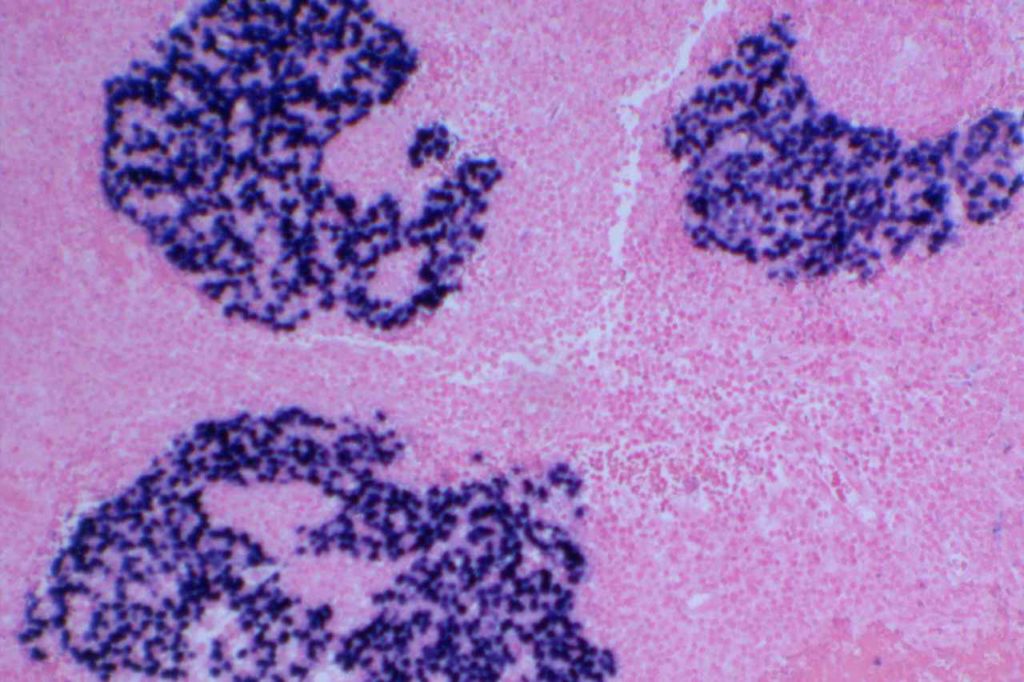
Morphology
LYG can have a varied appearance, but usually has a predominate background of small lymphocytes (T-cells), plasma cells, and histiocytes. Eosinophils and neutrophils are not a typical finding. Neoplastic EBV + B-cells can have a variable appearance ranging from immunoblast-like cells to morphologies similar to Hodgkin cells. The lymphoid infiltrate has an anglocentric and angiodestructive pattern, which may result in necrosis. If the pattern is of large cells in diffuse sheets, then the diagnosis of EBV+ diffuse large B-cell lymphoma should be made.
Differential diagnosis – Extranodal NK/T-cell lymphoma, nasal type is also an EBV+ tumor with an angiodestructive growth pattern.
Immunophenotype
Grading
- Grade 1 – < 5 EBV+ cells/HPF, polymorphic lymphoid infiltrate (large atypical cells rare/absent), No/focal necrosis.
- Grade 2 – 5-20 EBV+ cells/HPF (small clusters of B-cells by CD20), occasional large atypical/transformed cells.
- Grade 3 – > 50 EBV+ cells/HPF, numerous large atypical CD20+ B-cells (may form large aggregates)
References
WHO Classification of Tumors of Haematopoietic and Lymphoid Tissues. SH Swerdlow, et al. International Agency for Research on Cancer. Lyon, 2008. p. 247-249