Tag Archives: follicular lymphoma

In Situ Follicular Neoplasia (ISFN)

In Situ Follicular Neoplasia (ISFN), which has been previously referred to as Follicular Lymphoma Is Situ (FLIS) is found in approximately 2% of otherwise reactive lymph nodes.  ISFN is defined as partial to total colonization of germinal centers by B-cells containing the t(14;18) BCL-2/IgH translocation characteristic of follicular lymphoma.

ISFN found as an incidental finding have a risk of developing follicular lymphoma (FL) of <= 5%.  The amount of involvement of ISFN within a lymph node does not appear to be directly related to the risk of future FL.

If patients have other evidence of lymphadenopathy, then additional biopsies may be helpful to exclude a B-cell lymphoma at another site.

Morphology

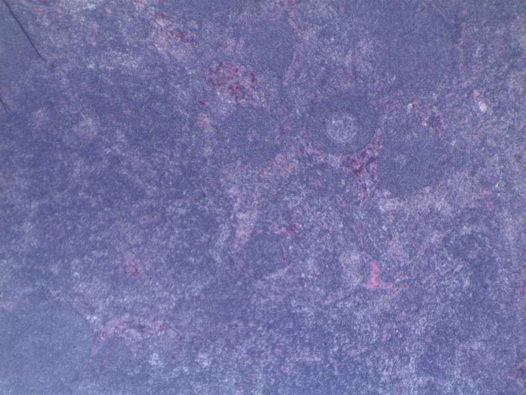
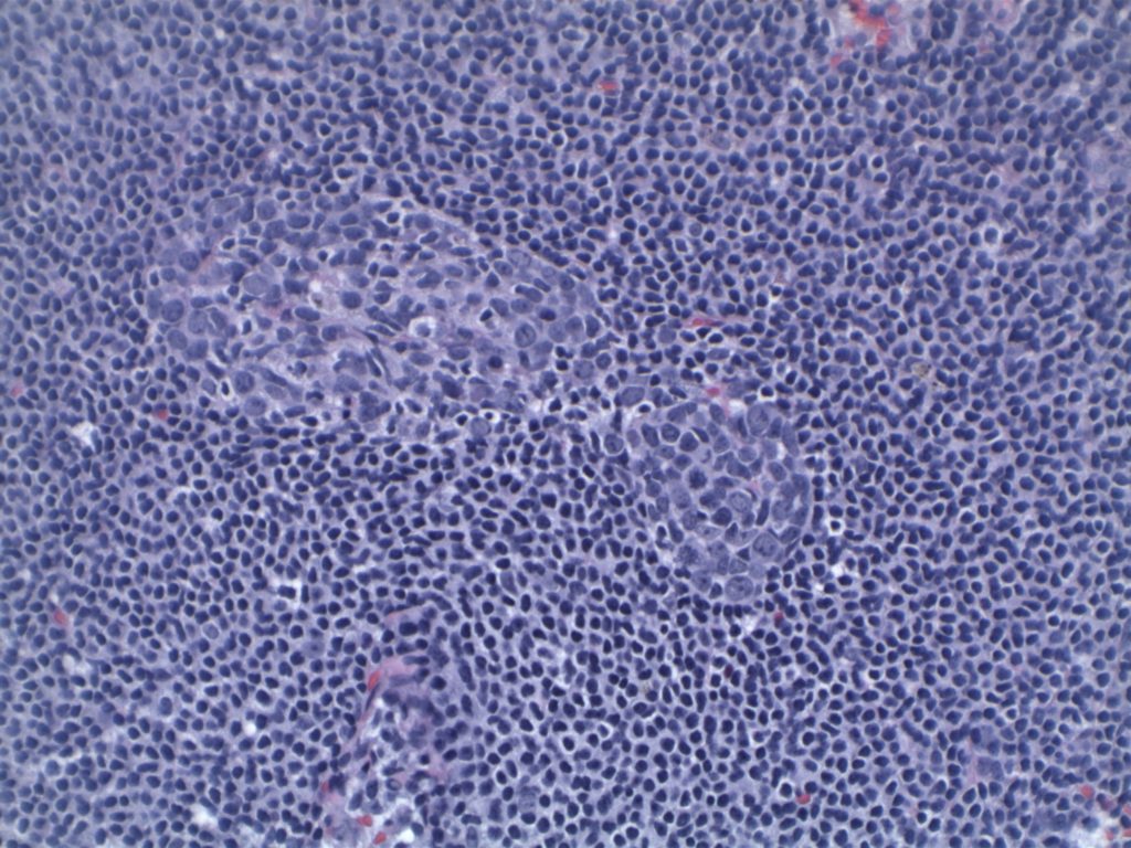
H&E staining demonstrates normal appearing lymph node architecture with well-formed and well-demarcated germinal centers.  Follicles with ISFN at first glance do not look significantly different from the adjacent reactive follicles.  Some areas of ISFN may more prominent and closely associated centrocytes (subtle).

Immunohistochemistry

  • CD10 – Strong expression
  • Bcl-6 – Positive
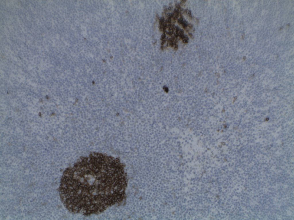
  • Bcl-2 – Positive (often stronger expression than surrounding lymphocytes)
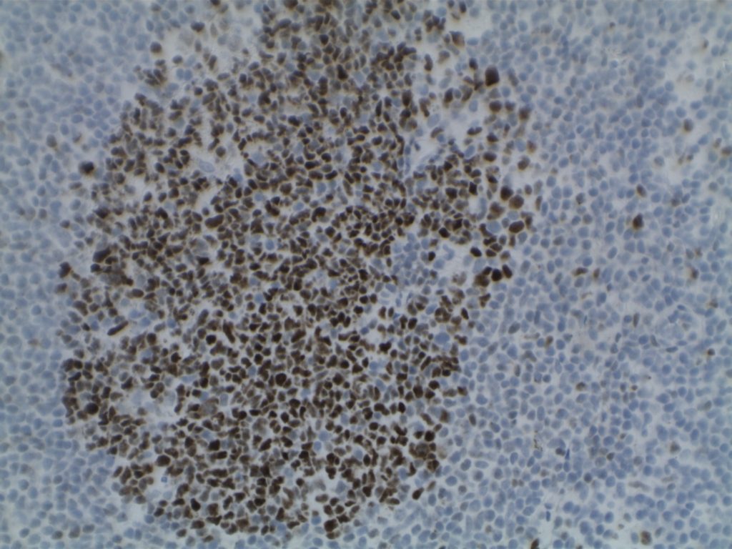
  • Ki-67 – Usually significantly lower expression than that of adjacent reactive germinal centers
  • CD20 – Positive

Photomicrographs

H&E section containing ISFN. Morphologically the node appears consistent with an otherwise reactive lymph node.
H&E section of ISFN.
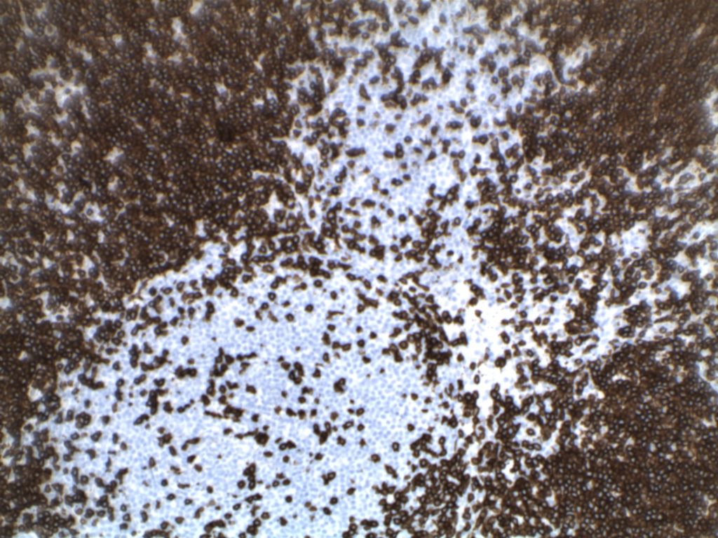
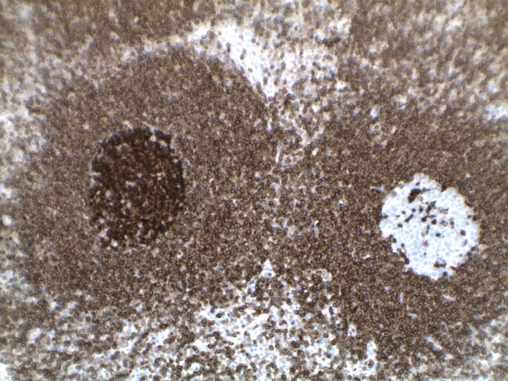
CD3 expression in ISFN.
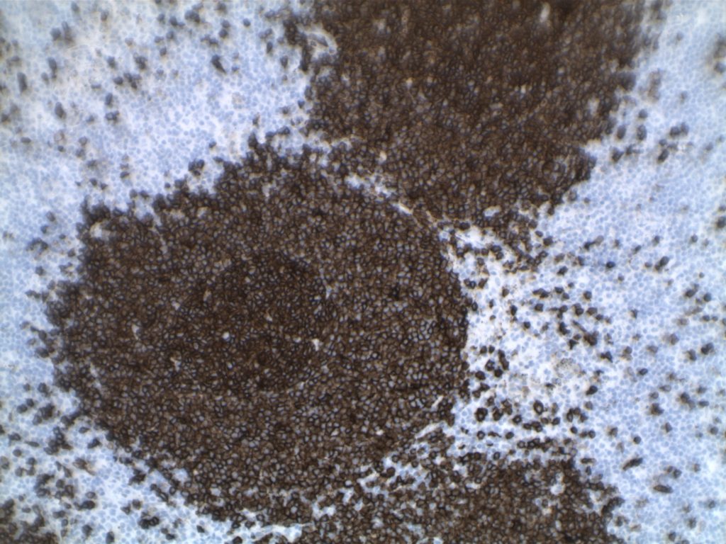
CD20 expression in a follicle containing ISFN.
CD10 expression in ISFN.
Bcl-6 expression in ISFN. Note the crowding of centrocytes highlighted by the Bcl-6 positive cells.
Strong Bcl-2 expression in the ISFN. Note the brighter expression compared to the surrounding T-cells and mantle zone. An adjacent reactive follicle is negative for Bcl-2.
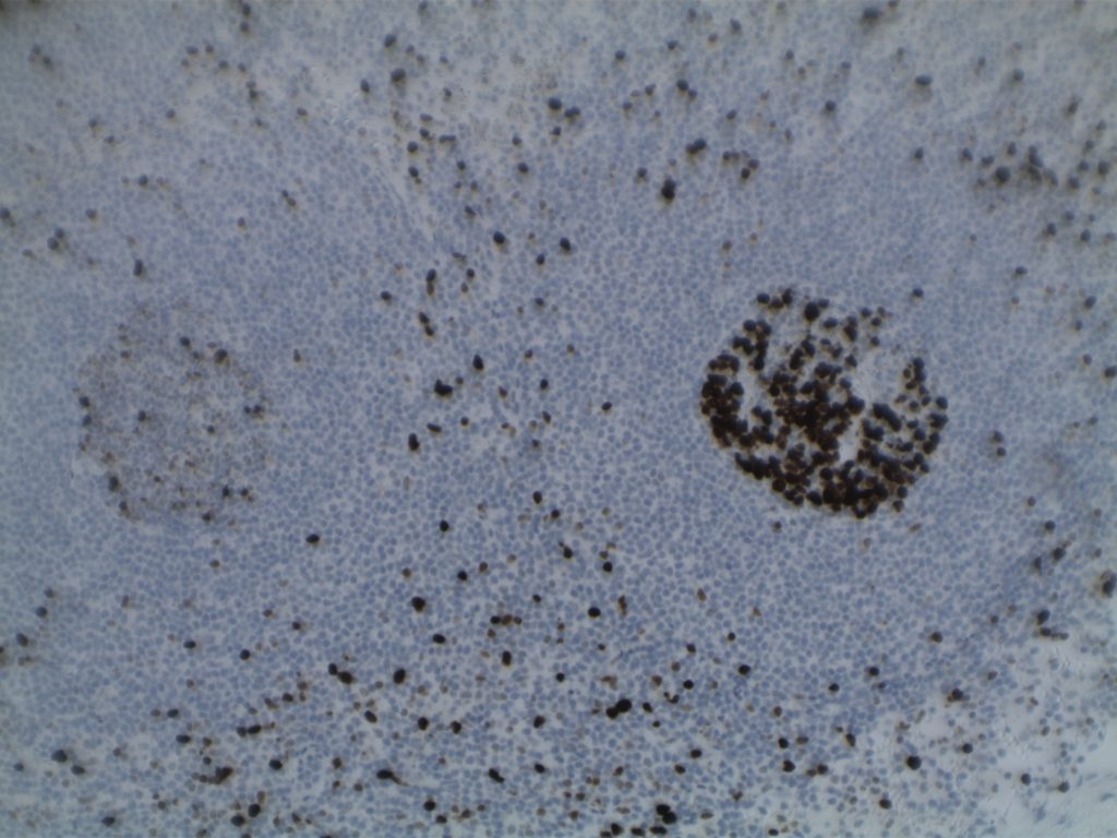
Low Ki-67 expression in ISFN (left side of image) compared to the high proportion of cell expression in the reactive follicle on the right.

Differentiating from partial involvement by follicular lymphoma

Sometimes it can be difficult to differentiate partial involvement of a lymph node by FL from ISFN.  The following features are helpful to differentiate partial involvement by FL from ISFN:

  • ISFN – Intact nodal architecture 
  • Partial involvement by FL – Altered architecture on H&E sections
  • ISFN – Normal sized follicles
  • Partial involvement by FL – Often enlarged follicles
  • ISFN – Sharp border between the follicle center and the surrounding mantle zone
  • Partial involvement by FL – Interface between the follicle center and mantle zone is often fuzzy or blurred.
  • ISFN – Bcl-2 is strongly positive (usually brighter than the surrounding T-cells and mantle zones); CD10 strongly positive
  • Partial involvement by FL – variable Bcl-2 and CD10 expression
  • ISFN – Atypical cells are confined to the follicle center
  • Partial involvement by FL – atypical cells (CD10/bcl2+) can be found outside of the follicle center areas

References

Swerdlow SH, Campo E, Harris, NL, Jaffe ES, Pileri SA, Stein H, Thiele J (Eds); WHO Classification of Tumours of Haematopoietic and Lymphoid Tissues (Revised 4th edition). IARC: Lyon 2017

Follicular Lymphoma

Follicular Lymphoma (FL) is a mature B-cell lymphoma, which recapitulates or resembles germinal center B-cells.  Most cases (~85%) harbor the characteristic t(14;18), which juxtaposes the BCL-2 gene on chromosome 18 with the IgH gene on chromosome 14 (and hence BCL-2 IHC protein expression).  Most patients (~80-85) will present with advanced disease (stage III/IV), and bone marrow involvement is found in ~40% of cases with characteristic paratrabecular aggregates (mantle cell lymphoma and lymphoplasmacytic lymphoma may also have paratrabecular lymphoid aggregates).  Most of the cases that lack the t(14;18) IgH/BCL-2 translocation (and are BCL-2 negative) are typically grade 3 FLs with a BCL-6 translocation (~10-15%).  BCL-6 translocations can be evaluated for by FISH analysis, but the finding is NOT specific for FL.
 
Over time 30-50% of cases transform to diffuse large B-cell lymphoma (DLBCL).  In a small subset of transformations, a second “hit” with a MYC translocation will occur resulting in a very aggressive high grade large B-cell lymphoma: the so-called “double hit” lymphoma. 
Morphology
FL usually has at least a component of nodularity (+/- diffuse areas).  There are two cell types that make up FL, centrocytes and centroblasts.  Centrocytes are small cleaved cells with folded irregular nuclei.  Centorblasts are large cells with more open chromatin, multiple nucleoli, and more cytoplasm compared to centrocytes.  
 
Sometimes FL can have patterns that resemble marginal zone lymphoma, and can even have plasmacytic differentiation.  Therefore, it is important that a panel of markers be used to identify (or exclude) evidence of germinal center differentiation.  Occasional cases can have Hodgkin-like cells.
Immunophenotype 
Marker
Comment
Negative
Positive
Positive
Positive
  • Grade 1 – ~90%
  • Grade 2 – ~70%
  • Grade 3 – ~60%
Positive (~90%), negative cases do not contain the t(14;18), which is more common in grade 3 cases
  • Grade 1 – >90% + for BCL-2
  • Grade 2 – >80% + for BCL-2
  • Grade 3 – 50-70% + BCL-2
Positive, (~88%)
CD35
Highlights the follicular dendritic meshwork associated with FL.
Usually negative, higher grade lesions may be positive
Variable, shows low expression in low-grade processes, in distinct contrast to the high proliferation index and polarity associated with reactive germinal centers.
Negative
 
 
FL is typically expresses CD19, CD20, CD10, Bcl-6, and BCL-2 (~90%).  CD5 is not expressed in FL.  
  • Normal reactive germinal centers do not express Bcl-2.  In 90% of cases of FL, bcl-2 is expressed, which serves as a diagnostic tissue marker in lymphoma sections.
  • CD23 expression by flow cytometry has been associated with lower grade FLs (e.g. grade 1 & 2) and better survival.
Grading
  • Grade 1 & 2:  <= 15 centroblasts/HPF (based on 0.159 mm² HPF)
  • Grade 3:  > 15 centroblasts/HPF (based on 0.159 mm² HPF)
    • 3A:  Centrocytes present in the background
    • 3B:  NO centrocytes present in the background (not associated with the IgH/BCL-2 rearrangement, and usually lacks expression of CD10 and BCL-2; often MUM-1+)
Grade 1 & 2 behave in a similar fashion as a low grade lymphoma.  Grade 3 FL behaves as an intermediate grade lymphoma.  Grading of FL with counting of large cells must take into consideration the field diameter of the microscope being used.  The counts above are based on a F.N. 18 (0.159 mm² @ 40X).  Most convention pathology scopes today are F.N. 22 (0.247 mm² @ 40X), and adjustments are necessary.
Pattern
  • Predominately follicular:  >75% follicular/nodular architecture
  • Follicular and diffuse:  25-75% Diffuse areas or follicular/nodular architecture
  • Preominately diffuse:  <25% follicular/nodular areas (diffuse areas of otherwise grade 3 FL, then that component should be described as a separate component of diffuse large B-cell lymphoma)
Special Subtypes 
  • Large B-Cell Lymphoma with IRF4 Rearrangement
  • Pediatric Follicular Lymphoma
    • Occurs in children and young adults with an excellent prognosis, marked male predilection
    • The morphology is high-grade (FL grade 3) appearing
      • BCL-2 negative, lacK t(14;18)
      • CD10 + (usually)
      • MUM-1 negative
    • Associated with TNFRSF14 deletions of mutations
    • Localized process, usually in the head and neck area
  • Duodenal Follicular Lymphoma
    • Localized lesion
    • Grade 1-2 pattern
    • CD10/BCL-2 +
    • t(14;18) present
    • Lacks follicular dendritic meshwork
    • Ki-67, low expression
    • Excellent prognosis
  • Predominately Diffuse Follicular Lymphoma with 1p36 deletion
    • Localized mass (often inguinal)
    • Diffuse pattern, grade 1/2 
    • Excellent prognosis
    • Immunophenotype:  CD20+, CD10+, BCL-2+, BCL-6+, CD23+ (subset of cases)
    • t(14;18) NOT present
    • 1p36 deletion (not specific)
    • Lacks Bcl-2 rearrangement
  • Primary Cutaneous Follicular Lymphoma 
  • In Situ Follicular Neoplasm (ISFN)

References
Robbins and Cotran Pathologic Basis of Disease.  V Kumar, et al. 9th Edition. Elsevier Saunders. 2015. pp. 594-595.
 
Fedoriw Y, Dogan A. The Expanding Spectrum of Follicular Lymphoma. Surg Pathol Clin. 2016;9: 29–40. doi:10.1016/j.path.2015.11.001
 
Swerdlow SH, Campo E, Pileri SA, Harris NL, Stein H, Siebert R, et al. The 2016 revision of the World Health Organization classification of lymphoid neoplasms. Blood. 2016;127: 2375–2390. doi:10.1182/blood-2016-01-643569
 
Xerri L, Dirnhofer S, Quintanilla-Martinez L, Sander B, Chan JKC, Campo E, et al. The heterogeneity of follicular lymphomas: from early development to transformation. Virchows Arch. 2016;468: 127–139. doi:10.1007/s00428-015-1864-y
 
MD DY-PW, BacSc F. A case of t (14; 18)-negative follicular lymphoma with atypical immunophenotype: usefulness of immunoarchitecture of Ki67, CD79a and follicular dendritic cell …. … Malaysian journal of …. 2014.
 
Boyd SD, Natkunam Y, Allen JR, Warnke RA. Selective immunophenotyping for diagnosis of B-cell neoplasms: immunohistochemistry and flow cytometry strategies and results. Appl Immunohistochem Mol Morphol. 2013;21: 116–131. doi:10.1097/PAI.0b013e31825d550a
 
Cook JR. Nodal and leukemic small B-cell neoplasms. Mod Pathol. 2013;26 Suppl 1: S15–28. doi:10.1038/modpathol.2012.180
 
Olteanu H, Fenske TS, Harrington AM, Szabo A, He P, Kroft SH. CD23 Expression in Follicular Lymphoma: Clinicopathologic Correlations. Am J Clin Pathol. 2011;135: 46–53. doi:10.1309/AJCP27YWLIQRAJPW
 
Gradowski JF, Jaffe ES, Warnke RA, Pittaluga S, Surti U, Gole LA, et al. Follicular lymphomas with plasmacytic differentiation include two subtypes. Mod Pathol. 2010;23: 71–79. doi:10.1038/modpathol.2009.146
 
Katzenberger T, Kalla J, Leich E, Stöcklein H, Hartmann E, Barnickel S, et al. A distinctive subtype of t(14;18)-negative nodal follicular non-Hodgkin lymphoma characterized by a predominantly diffuse growth pattern and deletions in the chromosomal region 1p36. Blood. 2009;113: 1053–1061. doi:10.1182/blood-2008-07-168682
 
Bayerl MG, Bentley G, Bellan C, Leoncini L, Ehmann WC, Palutke M. Lacunar and reed-sternberg-like cells in follicular lymphomas are clonally related to the centrocytic and centroblastic cells as demonstrated by laser capture microdissection. Am J Clin Pathol. 2004;122: 858–864. doi:10.1309/PMR8-6PHK-K4J3-RUH3
CD21

CD21 is a follicular dendritic cells (FDC) marker.  It has a membranous staining pattern, but this is practically difficult to separate from a cytoplasmic pattern in lymphoid tissue.  This stain is most often used as an “architectural” marker or aberrant patterns (nodular lymphocyte predominate Hodgkin lymphoma).  There are some rare tumors of FDC, which will mark with CD21.  CD21 may rarely mark normal B-cells, and strong expression of CD21 in CLL has been associated with a most aggressive disease course (unsure if this is by flow cytometry and/or IHC). 
 
Follicular Lymphoma – It is also helpful to identify the expanded follicular dendritic meshwork in cases of follicular lymphoma (especially helpful in diagnostically challenging cases – i.e. identifying a follicular component in an otherwise diffuse process or in small needle biopsies where architecture may not be visible).
 
Angioimmunoblastic T-cell Lymphoma (AILT) – Extrafollicular dendritic cells in the paracortical region in associated with neoplastic T-cells and high endothelial venules is characteristic of AILT.  CD21 is a helpful marker to highlights the follicular dendritic component in this process.
 
Marginal Zone Lymphoma – CD21 is useful in highlighting follicular colonization by marginal zone cells, which may be obscured by morphology alone.
CD21 Normal Expression Pattern
  • Follicular Dendritic Cells
  • Rare Normal B-cells
  • Rare cases of CLL
  • Generally thought of as a better follicular dendritic marker compared to CD23 (less sensitive)
Photomicrographs

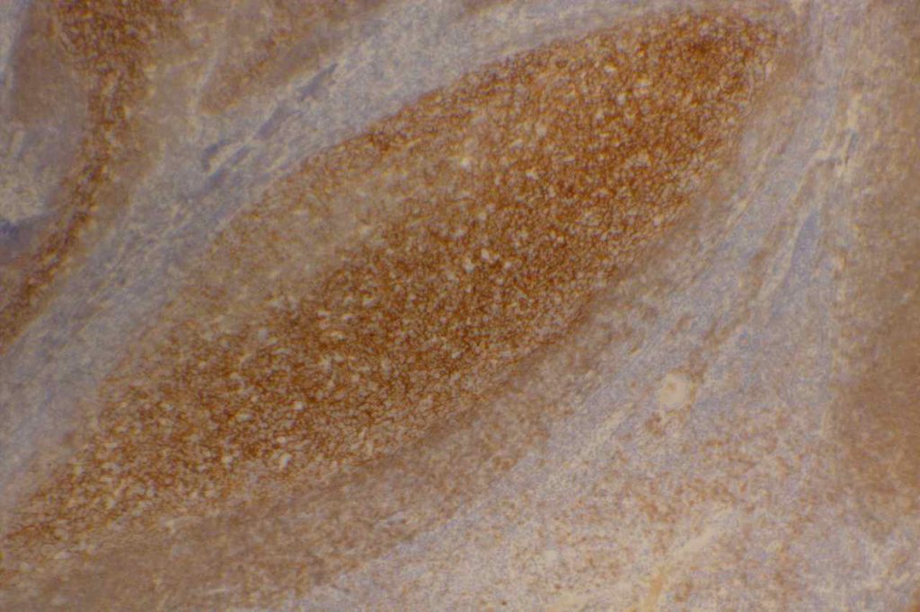
CD21 - Tonsil
CD21 highlighting the follicular dendritic meshwork in benign tonsil tissue.

References
Bone Marrow IHC.  Torlakovic, EE, et. al. American Society for Clinical Pathology Pathology Press © 2009.  pp. 64.
 
MD DY-PW, BacSc F.  A case of t (14; 18)-negative follicular lymphoma with atypical immunophenotype: usefulness of immunoarchitecture of Ki67, CD79a and follicular dendritic cell meshwork in making the diagnosis.  Malaysian Journal of Pathology. 2014. p. 125-129.
 
Harris NL, Swerdlow SH, Jaffe ES, et al. FollicularLymphoma. In: Swerdlow SH, Campo E, Harris NL,Jaffe ES, Pileri SA, Stein H, Thiele J, VardimanJW, editors. WHO classication of tumours of haematopoietic and lymphoid tissues. 4th ed. Lyon: IARC Press; 2008. p. 220–6.
 
Troxell ML, Schwartz EJ, van de Rijn M, Ross DT, Warnke RA, Higgins JP, et al. Follicular dendritic cell immunohistochemical markers in angioimmunoblastic T-cell lymphoma. Appl Immunohistochem Mol Morphol. 2005;13: 297–303.
 
Boyd SD, Natkunam Y, Allen JR, Warnke RA. Selective immunophenotyping for diagnosis of B-cell neoplasms: immunohistochemistry and flow cytometry strategies and results. Appl Immunohistochem Mol Morphol. 2013;21: 116–131. doi:10.1097/PAI.0b013e31825d550a

CD23

CD23 is a transmembrane glycoprotein expressed by different hematopoietic cells and is a low-affinity receptor for IgE.  It is also involved in promoting survival of B-cells in the germinal center.  CD23 is useful as a follicular cell dendritic cell marker and is classically expressed in cases of CLL/SLL.  CD23 has been identified in many types of lymphomas, but is most commonly used to differentiate between CLL/SLL (CD23+) and mantle cell lymphoma (CD23-).  This testing is typically performed by flow cytometry, but immunohistochemisty for CD23 is available.  Expression of CD23 has been associated with better prognosis (at least in limited published data) in follicular lymphoma, CLL/SLL, mantle cell lymphoma, and diffuse large B-cell lymphoma dependent upon expression characteristics.  CD23 is not commonly performed/used as a prognostic marker for B-cell lymphomas.
 
Rarely CD23 may be expressed in cases of Hairy cell leukemia (17%) and DLBCL (16%).  Approximately 70% of Mediastinal large B-cell lymphoma cases express CD23.  Practically, this IHC marker is used as a follicular dendritic cell marker and to help differentiate CLL/SLL from mantle cell lymphoma.  Follicular dendritic cell tumors will also express CD23 like CD21.  CD21 is more sensitive compared to CD23 as a follicular dendritic marker.
 
Follicular Lymphoma (FL) – CD23 has been found to be expressed in some cases of FL, especially from inguinal lymph nodes, and prognosis appears comparatively better.   Olteanuet. al found that 87% of inguinal lymph nodes expressed CD23, compared to 61% from other sites, and that survival was prolonged more in CD23+ cases. 
 
Diffuse Large B-Cell Lymphoma – A subset of DLBCLs may express CD23, which may have a better prognosis (CD23 is not commonly performed for this purpose).
 
Chronic Lymphocytic Leukemia/Small Lymphocytic Lymphoma (CLL/SLL) – CD23 expression is characteristic of CLL/SLL, particularly in comparison to another CD5+ lymphoma, mantle cell lymphoma.  Strong membrane expression has been associated with a better outcome.  DiRaimondo,et. al found ~6% of CLL cases to be CD23 negative (flow cytometry), and they had a worse prognosis.  Many of these cases may have been misdiagnosed mantle cell lymphomas.
 
Mantle Cell Lymphoma (MCL) – CD23 is characteristically negative in MCL, which helps to differentiate it from CLL/SLL.  However, ~21% of cases of MCL were found to be CD23+ by Gao,et. al, and other studies have shown CD23 expression in MCL ranging from 0% to 45% (most data appears to be based on flow cytometry).
CD23 Expression Pattern
  • CLL/SLL – characteristically expressed (6% may be negative, probably much lower)
  • Mantle cell lymphoma may be CD23+ (21%+, 0-45%)
  • B-cell Lymphomas (e.g. some DLBCL and follicular lymphomas may show expression)
  • Follicular Dendritc Cells (not as sensitive as CD21)
  • B-cells in mantle zone of lymphoid follicles

Photomicrographs

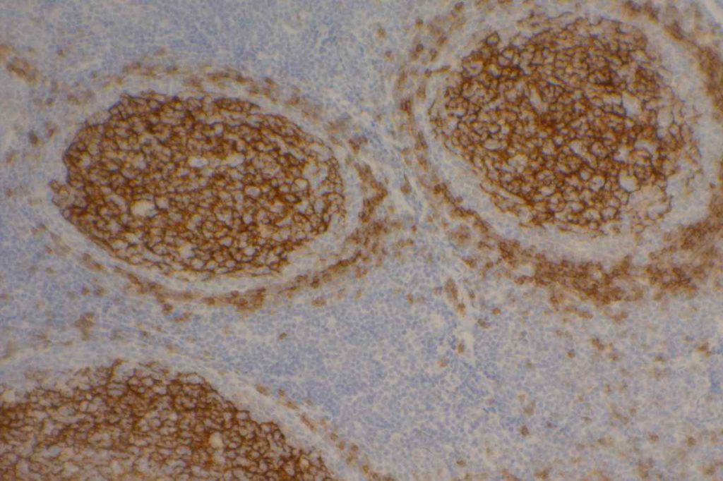
CD23 - Tonsil
CD23 expression highlighting follicular dendritic meshwork in a normal tonsil.


Reference
Bone Marrow IHC.  Torlakovic, EE, et. al. American Society for Clinical Pathology Pathology Press © 2009.  pp. 69.
 
Linderoth J, Jerkeman M, Cavallin-Stahl E, et al. Immunohistochemical expression of CD23 and CD40 may identify prognostically favorable subgroups of diffuse largeB-cell lymphoma: a Nordic Lymphoma Group Study.ClinCancer Res.2003;9:722-728.
 
Olteanu H, Fenske TS, Harrington AM, Szabo A, He P, Kroft SH. CD23 Expression in Follicular Lymphoma: Clinicopathologic Correlations. Am J Clin Pathol. 2011;135: 46–53. doi:10.1309/AJCP27YWLIQRAJPW
 
Gao J, Peterson L, Nelson B, Goolsby C, Chen Y-H. Immunophenotypic variations in mantle cell lymphoma. Am J Clin Pathol. 2009;132: 699–706. doi:10.1309/AJCPV8LN5ENMZOVY
 
Troxell ML, Schwartz EJ, van de Rijn M, Ross DT, Warnke RA, Higgins JP, et al. Follicular dendritic cell immunohistochemical markers in angioimmunoblastic T-cell lymphoma. Appl Immunohistochem Mol Morphol. 2005;13: 297–303.
 
Dalton RR, Admirand JH, Medeiros LJ. Small Lymphocytic Lymphoma. Pathology Case Reviews. 2004;9: 7.
 
DiRaimondo F, Albitar M, Huh Y, O’Brien S, Montillo M, Tedeschi A, et al. The clinical and diagnostic relevance of CD23 expression in the chronic lymphoproliferative disease. Cancer. 2002;94: 1721–1730. doi:10.1002/cncr.10401