Tag Archives: IHC

Prostate – IHC Stains

Prostate Adenocarcinoma
IHC Expression Characteristics
Negative
Typically negative, rarely positive.
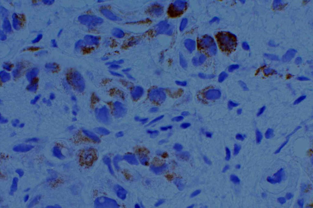
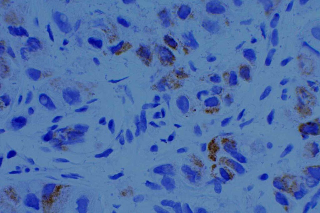
Racemase enzyme expressed by high grade PIN and Prostate adencoarcinoma (NOT a specific marker for prostate tissue).  Relatively good specificity (~90%), but may mark benign prostate lesions.  Often used in a cocktail with p63 and HMWKs (e.g. PIN4)
>95% sensitivity for prostate adenocarcinoma (highly specific)
PSAP
Earlier marker than PSA (now used occasionally as a backup) with less specificity compared to PSA
Combination stain containing a cocktail of AMACR (red), p63, and high molecular weight cytokeratins (CK5 & CK14)
34BetaE12
HMWK that is often used to highlight the basal layer of prostate glands (positivity means it is not invasive adenocarcinoma).
HMWK which has similar characteristics as 34betaE12
Nuclear marker which highlights the basal layer of benign prostate glands.
Prostatic Duct Carcinoma
 
IHC expression is the same as prostate adenocarcinoma.  Morphology is similar to breast DCIS.  Sometimes it may be confused with a primary colon adenocarcinoma (CDX-2+) or a primary urothelial carcinoma (PSA=).

References
Diagnostic Immunohistochemistry: Theranostic and Genomic Applications. [edited by] DJ Dabbs. 3rd Edition.  Elsevier, 2010.

Breast – Predictive Markers

One of the most important aspects of breast cancer diagnosis is the evaluation of therapeutic markers (ER, PR, and HER2).  Ki-67 is often included in the panel as a prognostic marker.  ER expression determines eligibility to receive hormonal therapy (Tamoxifin), PR expression is a prognostic marker, and HER-2 over-expression determines eligibility to receive Herceptin®.  Diagnostically, the challenge is to consistently and accurately perform and interpret these IHC markers.
 
Estrogen Receptor (ER):
  • Nuclear Marker
  • Stain is reported as PERCENT STAINING OF TUMOR CELLS and STAIN INTENSITY (1+, 2+, 3+).
  • 1% or greater nuclear expression in tumor cells is considered positive, and therefore eligible to receive hormonal therapy.
  • CAP-ASCO recommendations are for <1 hr. from time of excision/biopsy to having a cut edge of tumor in 10% neutral bufferedormalin fixative.  Fixation window of 6-72 hrs.  These times should be noted in the pathology report (time of excision, time in gross room, and time in fixative).
  • Negative staining results in biopsy material without an internal control should be repeated on the excisional specimen using blocks with both tumor and benign breast parenchyma.
Progesterone Receptor (PR):
  • Nuclear Marker
  • Stain is reported as PERCENT STAINING OF TUMOR CELLS and STAIN INTENSITY (1+, 2+, 3+).
  • 1% or greater nuclear expression in tumor cells is considered positive.
  • PR expression is a prognostic marker, and not directly used for eligibility to receive a specific treatment.
  • PR expression without ER expression should raise significant concern that the ER and PR slides have been mixed up, or there is a problem with the ER assay.  Many scientists believe that ER expression is required for PR expression.
HER-2 Overexpression (HER-2):
  • Membraneous stain
  • Stain is interpreted by combining stain intensity and percentage of tumor involvement to classify as (0, 1+, 2+, or 3+).
    • 0 (negative) = No staining or cell membrane staining in <10% of tumor cells.
    • 1+ (negative) =  Faint membrane staining (partial membrane staining) in >10% of tumor cells.
    • 2+ (equivocal) =  Weak to moderate complete membrane staining in >10% of tumor cells, or strong complete staining in <10% of invasive tumor cells.
    • 3+ (positive) =  Strong complete membrane staining in >10% of tumor cells.
  • CAP-ASCO recommendations are for <1 hr. from time of excision/biopsy to having a cut edge of tumor in 10% neutral buffered fomalin fixative.  Fixation window of 6-72 hrs.  Over-fixation is probably not a clinically significant issue practically, but given the absence of relevant IHC data and the highly regulated environment surrounding HER2 testing, f/u FISH testing for negative results (outside the fixative window) is necessary.
  • Equivocal (2+) results should be followed-up with FISH testing, if IHC is used as the initial testing modality (most common).  Less than 1/3rd of equivocal cases show Her2 over-expression by FISH analysis.

References
 
Hammond ME, et. al.  “ASCO-CAP Guideline Recommendations for IHC Testing of ER and PR in Breast Cancer”.  Arch Pathol Lab Med-Vol. 134, June 2010.
 
Wolff, A. C., Hammond, M. E. H., Hicks, D. G., Dowsett, M., McShane, L. M., Allison, K. H., et al. (2013). Recommendations for Human Epidermal Growth Factor Receptor 2 Testing in Breast Cancer: American Society of Clinical Oncology/College of American Pathologists Clinical Practice Guideline Update. Archives of pathology & laboratory medicine. doi:10.5858/arpa.2013-0953-SA 
 
Tafe, L. J., Janjigian, Y. Y., Zaidinski, M., Hedvat, C. V., Hameed, M. R., Tang, L. H., et al. (2011). Human epidermal growth factor receptor 2 testing in gastroesophageal cancer: correlation between immunohistochemistry and fluorescence in situ hybridization. Archives of pathology & laboratory medicine, 135(11), 1460–1465. doi:10.5858/arpa.2010-0541-OA 
 
Arch Pathol Lab Med. 2001;125:746.

Breast – IHC Specific Markers

IHC stain expression pattern for various IHC antibodies in breast carcinoma.
 
IHC Stain
Comments
50-70%
30-60%
>80%
<10%
10-25%
0%
0%
0%
Mesothelin
<10%
Clin Cancer Rs 2005;11(10) May 15, 2005.
 
When evaluating for a possible breast primary, it is usually part of a larger differential diagnosis.  CK7 and CK20 is the common starting point for carcinomas of unknown primary site (CUPS), and breast has a characteristic CK7+/CK20= profile.  Unfortunately, this pattern is not uncommon, and more specific markers need to be performed.  Two specific breast markers are GCDFP-15 and ER., but their sensitivity while good is limited.  GATA-3 is a relatively newer antibody, which shows excellent sensitivity with good specificity, and should be strongly considered to be part of an antibody panel in work-up of potential breast carcinoma cases. 
  • GCDFP-15 is very specific for breast carcinoma in the setting of CUPS, but limited sensitivity.  
  • ER expression may be highly suggestive of a breast primary (especially epidemiologically), but it is not as specific.
  • Other female organs (ovary/uterus) not uncommonly express ER, and practically any tissue can occasionally excess ER.
  • GATA-3 appears to have excellent sensitivity (>90%) with good (not perfect) specificity.
Liu, et al (Biocare Medical, Concord, CA)
Tumor
GATA-3
GCDFP-15
MGB
Breast Carcinoma
94%
35-55%
65-70%
ER-negative breast ca.
69%
15%
35%
Urothelial Carcinoma
86%


Breast Metastasis vs. Ovarian Ca. Primary
Tumor
WT-1
CA-125
GCDFP-15
Primary Ovarian Ca.
(N=41)
76%
73%
0%
Metastatic Breast Ca.
(N-40)
3%
10%
43%
P-Value
<0.001
<0.001
<0.001
Chen, USCAP, 2004

References
Liu, H., Shi, J., Wilkerson, M. L., & Lin, F. (2012). Immunohistochemical evaluation of GATA3 expression in tumors and normal tissues: a useful immunomarker for breast and urothelial carcinomas. American Journal of Clinical Pathology, 138(1), 57–64. doi:10.1309/AJCP5UAFMSA9ZQBZ
 
Miettinen, M., McCue, P. A., Sarlomo-Rikala, M., Rys, J., Czapiewski, P., Wazny, K., et al. (2014). GATA3: A Multispecific But Potentially Useful Marker in Surgical Pathology: A Systematic Analysis of 2500 Epithelial and Nonepithelial Tumors. The American Journal of Surgical Pathology, 38(1), 13–22. doi:10.1097/PAS.0b013e3182a0218f 
 
Clin Cancer Rs 2005;11(10) May 15, 2005.
 
Chen, USCAP, 2004

Breast – Normal IHC Expression

Normal breast ducts and lobules are lined by a 2-cell layer composed of luminal and myoepithelial cells.  There are also interspersed “basal” cells, which probably represent the epithelial progenitor cells.
 
IHC Marker
Luminal Cells
Myoepithelial Cells
LMWCKs
(CK7/8/18)
Positive
Negative
Variable Expression
Negative
HMWCKs
(CK5/14/17)
Negative
Positive
SMA
Negative
Positive
Negative
Positive
Negative
Positive
SMA=Smooth Muscle Actin, SMM-HC=Smooth Muscle Myosin Heavy Chain
 
An understanding of the normal IHC expression pattern in breast ductal tissue is important when considering IHC use in the differential diagnosis of breast pathology.

References
Hicks DG. Immunohistochemistry in the diagnostic evaluation of breast lesions. Appl Immunohistochem Mol Morphol. 2011;19(6):501–505. doi:10.1097/PAI.0b013e31822c8a48.
 
Liu H. Application of immunohistochemistry in breast pathology: a review and update. Arch Pathol Lab Med. 2014;138(12):1629–1642. doi:10.5858/arpa.2014-0094-RA.

HepPar-1

General
HepPar-1 is a mitochondrial antigen present in normal hepatocytes, and is a relatively specific marker for hepatic origin.  It also stains approximately 80-100% of hepatocellular carcinomas.  It is most commonly used to help identify tumors of primary hepatic origin, and exclude cholangiocarcinoma in the differential diagnosis. 
Interpretation
The staining pattern is granular and cytoplasmic in location.  The staining pattern can be heterogeneous ranging from focal (<5%) in poorly differentiated HCC to strong diffuse staining in well-differentiated HCC.  As with most issues in pathology and IHC, less differentiated lesions more often need IHC, which tend to perform less-optimally than described in the literature (case selection bias).  Also beware of focal staining of benign entrapped hepatocytes.
Specificity
HepPar-1 is not a pertfectly specific marker for HCC.  One study (Wee) notes specificity as 73% with non-hepatic tumors including adenocarcinomas from lung, gallbladder, pancreas, stomach, small intestine, adenoma of colon, adrenal gland carcinoma, paraganglioma, and melanoma showing expression.  Other studies show the specificity near 90% for HCC.
 
Up to 25% (probably <10% overall) of lung carcinomas (mostly adenocarcinomas) have been noted to have HepPar1 expression (cytoplasmic and granular).  The expression pattern may be focal/patchy, but these tumors also typically express TTF-1.
 
Use of HepPar-1 as part of a panel (and clinical-radiologic correlation) is recommended for optimal interpretation. 
Photomicrographs
HepPar-1 - Metastatic Hepatocellular Carcinoma
HepPar-1 – Metastatic Hepatocellular Carcinoma
HepPar-1 - Metastatic Hepatocellular Carcinoma
HepPar-1 – Metastatic Hepatocellular Carcinoma
References
Wee, A. (2006). Diagnostic utility of immunohistochemistry in hepatocellular carcinoma, its variants and their mimics. Applied Immunohistochemistry & Molecular Morphology : AIMM / Official Publication of the Society for Applied Immunohistochemistry, 14(3), 266–272.
 
Allende, D., & Yerian, L. (2009). Immunohistochemical Markers in the Diagnosis of Hepatocellular Carcinoma. Pathology Case Reviews, 14(1), 40–46.
 
Yousem, S. A., Lale, S., & Dacic, S. (2013). HepPar-1 expression in primary lung adenocarcinoma. American Journal of Clinical Pathology, 140(2), 225–230. doi:10.1309/AJCP4MXTNQRVOE2T
 
Minervi el al., 1997.  
 
Chu et al., Am J Surg Pathol 26:978-88, 2002.
 
L. Lamps. “The Differential Diagnosis of Hepatic Tumors.”  UAMS, Lecture, 2005.