Tag Archives: Lung

ROS-1

ROS-1 rearrangements with at least 12 different partner proteins have been identified in a small subset of lung non-small cell carcinomas (1–2%), which shows susceptibility to tyrosine kinase inhibitors (TKIs) similar to ALK rearranged tumors.  ROS-1 is considered a oncogene found on chromosome 6.  The exact mechanism of activation of this gene protein product with the various gene rearrangement partners is not understood.  The protein function is similar to that of the ALK family, which is why this mutation was studied for possible response to ALK inhibitors (crizotinib).
 
Recently, ROS-1 mutated tumors have been approved for TKI therapy with identification of a rearrangement by FISH analysis.  Like ALK, ROS-1 FISH utilizes a break apart probe to identify the presence of a gene rearrangement.  Other successful modalities for identification of ROS-1 rearrangements include ‘next generation’ sequencing (NGS) and immunohistochemistry.
 
Immunohistochemistry (IHC) has been studied as an alternative to FISH as a screening modality.  Based on multiple studies, the sensitivity of IHC appears to be near 100% with the specificity of at least 92%.  These studies were performed using the D4D6 rabbit monoclonal antibody clone (Cell Signaling Technology, Danvers, Massachusetts).

Stain Interpretation
Unlike ALK, there is no known normal tissue counterpart which can be used as a control.  Therefore, known ROS-1 positive tumors or cell lines  (HCC78 cell line with the SLC34A2-ROS1 rearrangement) are generally used.  ROS-1 expression is cytoplasmic with described expression ranging from finely granular to globular cytoplasmic staining and membranous staining.  No consensuses has been established as to the minimal threshold of positivity.
 
Possible interpretation pitfalls include weak staining of type II pneumocytes and alveolar macrophages along with osteoclast-type giant cells in bone biopsies.  Like any immunostain, contextual evaluation is critical.

References
Thunnissen E, Allen TC, Adam J, Aisner DL, Beasley MB, Borczuk AC, et al. Immunohistochemistry of Pulmonary Biomarkers: A Perspective From Members of the Pulmonary Pathology Society. Arch Pathol Lab Med. 2018;142: 408–419. doi:10.5858/arpa.2017-0106-SA
 
Shaw AT, Ou S-HI, Bang Y-J, Camidge DR, Solomon BJ, Salgia R, et al. Crizotinib in ROS1-rearranged non-small-cell lung cancer. N Engl J Med. 2014;371: 1963–1971. doi:10.1056/NEJMoa1406766
 
Bubendorf L, Büttner R, Al-Dayel F, Dietel M, Elmberger G, Kerr K, et al. Testing for ROS1 in non-small cell lung cancer: a review with recommendations. Virchows Arch. 2016;469: 489–503. doi:10.1007/s00428-016-2000-3
 
Boyle TA, Masago K, Ellison KE, Yatabe Y, Hirsch FR (2015) ROS1 immunohistochemistry among major genotypes of non- small-cell lung cancer. Clin Lung Cancer 16(2):106–111. doi:10.1016/j.cllc.2014.10.003 
 
CaoB,WeiP,LiuZ,BiR,LuY,ZhangL,ZhangJ,YangY,Shen C, Du X, Zhou X (2016) Detection of lung adenocarcinoma with ROS1 rearrangement by IHC, FISH, and RT-PCR and analysis of its clinicopathologic features. Onco Targets Ther 9:131–138. doi:10.2147/OTT.S94997 
 
Sholl LM, Sun H, Butaney M, Zhang C, Lee C, Janne PA, Rodig SJ (2013) ROS1 immunohistochemistry for detection of ROS1-rearranged lung adenocarcinomas. Am J Surg Pathol 37(9):14411449. doi:10.1097/PAS.0b013e3182960fa7
 
Yoshida A, Tsuta K, Wakai S, Arai Y, Asamura H, Shibata T, Furuta, K, Kohno T, Kushima R (2014) Immunohistochemical detection of ROS1 is useful for identifying ROS1 rearrangements in lung can- cers. Mod Pathol 27(5):711720. doi:10.1038/modpathol.2013.192
 
Rogers TM, Russell PA, Wright G, Wainer Z, Pang JM, Henricksen LA, Singh S, Stanislaw S, Grille J, Roberts E, Solomon B, Fox SB (2015) Comparison of methods in the detection of ALK and ROS1 rearrangements in lung cancer. J Thorac Oncol 10(4):611618. doi:10.1097/JTO.0000000000000465
 
Rimkunas VM, Crosby KE, Li D, Hu Y, Kelly ME, Gu TL, Mack JS, Silver MR, Zhou X, Haack H (2012) Analysis of receptor tyro- sine kinase ROS1-positive tumors in non-small cell lung cancer: identification of a FIG-ROS1 fusion. Clin Cancer Res 18(16): 44494457. doi:10.1158/1078-0432.CCR-11-3351
 
Mescam-Mancini L, Lantuejoul S, Moro-Sibilot D, Rouquette I, Souquet PJ, Audigier-Valette C, Sabourin JC, Decroisette C, Sakhri L, Brambilla E, McLeer-Florin A (2014) On the relevance of a testing algorithm for the detection of ROS1-rearranged lung adenocarcinomas. Lung Cancer 83(2):168–173. doi:10.1016/j. lungcan.2013.11.019 
 
Shan L, Lian F, Guo L, Qiu T, Ling Y, Ying J, Lin D (2015) Detection of ROS1 gene rearrangement in lung adenocarcinoma: comparison of IHC, FISH and real-time RT-PCR. PLoS One 10(3): e0120422. doi:10.1371/journal.pone.0120422 

ALK

ALK (Anaplastic Lymphoma Kinase) is a transmembrane molecule that is only normally expressed in some neural tissues.  It has characteristic expression in a significant proportion of Anaplastic Large Cell Lymphoma (ALCL) cases.  It has also been expressed in cases of pleomorphic liposarcoma, inflammatory myofibroblastic tumor, Merkel cell carcinoma, and a small subset of diffuse large B-cell lymphomas.
 
A subset (1-5%) of non-small cell lung carcinomas (usually adenocarcinomas) have an ELM4-ALK mutation, which is often sensitive to the tyrosine kinase inhibitor (TKI) crizotinib (Pfizer).  Newer TKIs, including ceritinib (Novartis) and alectinib (Hoffmann-La Roche), have also been found to be effective.  
 
Historically, ALK translocations have been identified by FISH analysis.  IHC is also an accepted method with an FDA approved test (IHC CDx Assay).  PCR is being studied as an alternative.  ALK antibody clones 5A4 (Novocastra, Leica Biosystems, Buffalo Grove, Illinois), ALK1 (Dako, Santa Clara, California), 1A4 (Origene, Rockville, Maryland) and D5F3 (Cell signaling Technology, Danvers, Massachusetts) have been successfully used to identify ALK mutated lung tumors, with the 5A4 & D5F3 having equivalent sensitivity. 
 
The ALK1 clone is not as sensitive and the 1A4 clone lacks specificity compared to other antibodies.  Please consult the current medical literature for FDA approved tests for ALK translocation identification in non-small cell lung carcinomas.
Normal Expression
  • Neural Tissue
Abnormal Expression

Interpretation
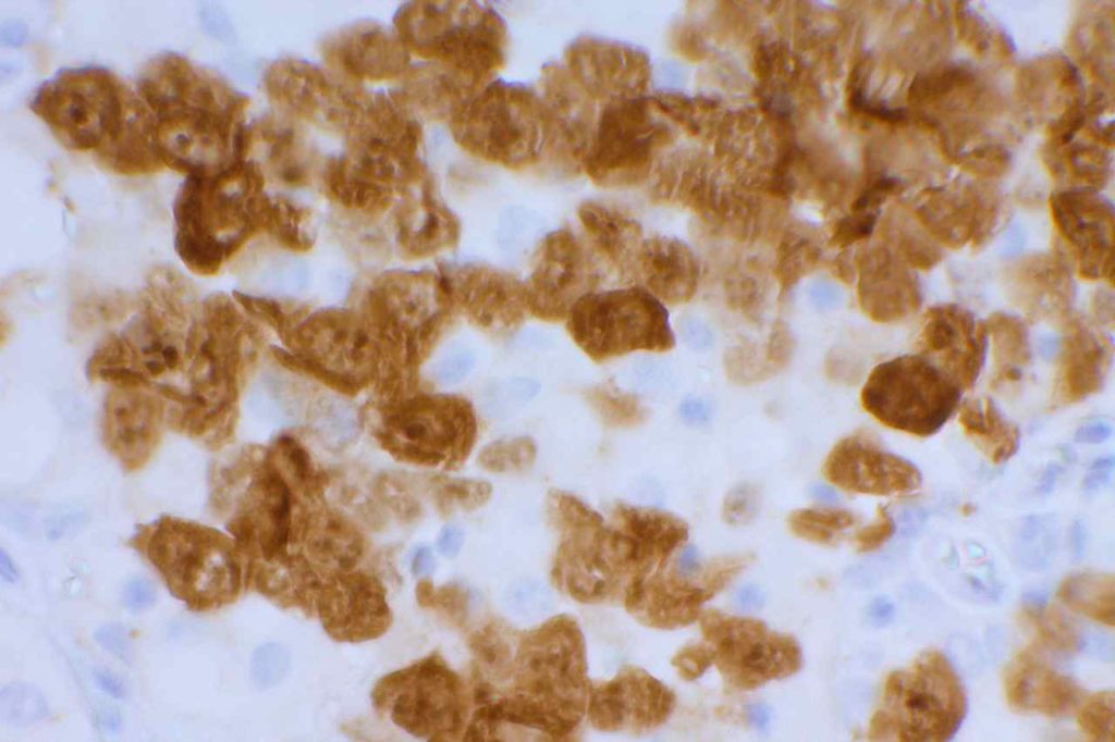
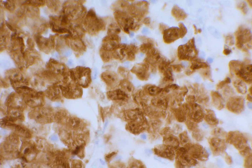
ALK may stain in a cytoplasmic and/or nuclear pattern.  In ALCL the combined pattern of cytoplasmic and nuclear staining is associated with the t(2;5).
 

In lung ALK staining/expression is cytoplasmic.  Like other markers (e.g. Napsin A), staining may be present in macrophages.  Necrotic tumor, extracellular mucin, and cells of neural origin may also stain.


Photo Gallery
ALK - ALCL
ALK expression in anaplastic large cell lymphoma
ALK - ALCL
ALK expression in anaplastic large cell lymphoma

References
Thunnissen E, Allen TC, Adam J, Aisner DL, Beasley MB, Borczuk AC, et al. Immunohistochemistry of Pulmonary Biomarkers: A Perspective From Members of the Pulmonary Pathology Society. Arch Pathol Lab Med. 2018;142: 408–419. doi:10.5858/arpa.2017-0106-SA
 
Chan, J. K. C., Ip, Y. T., & Cheuk, W. (2013). The Utility of Immunohistochemistry for Providing Genetic Information on Tumors. International Journal of Surgical Pathology, 21(5), 455–475. doi:10.1177/1066896913502529
 
Chan, J. K. C. (2013). Newly Available Antibodies With Practical Applications in Surgical Pathology. International Journal of Surgical Pathology, 21(6), 553–572. doi:10.1177/1066896913507601
 
Paik, J. H., Choe, G., Kim, H., Choe, J.-Y., Lee, H. J., Lee, C.-T., et al. (2011). Screening of Anaplastic Lymphoma Kinase Rearrangement by Immunohistochemistry in Non-small Cell Lung Cancer: Correlation with Fluorescence In Situ Hybridization. Journal of Thoracic Oncology : Official Publication of the International Association for the Study of Lung Cancer, 6(3), 466–472. doi:10.1097/JTO.0b013e31820b82e8
 
Yi, E. S., Boland, J. M., Maleszewski, J. J., Roden, A. C., Oliveira, A. M., Aubry, M.-C., et al. (2011). Correlation of IHC and FISH for ALK Gene Rearrangement in Non-small Cell Lung Carcinoma: IHC Score Algorithm for FISH. Journal of Thoracic Oncology : Official Publication of the International Association for the Study of Lung Cancer, 6(3), 459–465. doi:10.1097/JTO.0b013e318209edb9
 
 Mino-Kenudson, M., Chirieac, L. R., Law, K., Hornick, J. L., Lindeman, N., Mark, E. J., et al. (2010). A novel, highly sensitive antibody allows for the routine detection of ALK-rearranged lung adenocarcinomas by standard immunohistochemistry. Clinical Cancer Research : an Official Journal of the American Association for Cancer Research, 16(5), 1561–1571. doi:10.1158/1078-0432.CCR-09-2845
 
Reichard, K. K., McKenna, R. W., & Kroft, S. H. (2007). ALK-positive diffuse large B-cell lymphoma: report of four cases and review of the literature. Modern Pathology : an Official Journal of the United States and Canadian Academy of Pathology, Inc, 20(3), 310–319. doi:10.1038/modpathol.3800742
 
Medeiros, L. J., & Elenitoba-Johnson, K. S. J. (2007). Anaplastic Large Cell Lymphoma. American Journal of Clinical Pathology, 127(5), 707–722.  
 
Takeuchi K, Choi YL, Togashi Y, et al. KIF5B-ALK, a novel fusion oncokinase identified by an immunohistochemistry-based diagnostic system for ALK-positive lung cancer. Clin Cancer Res. 2009;15(9):3143–3149. doi:10.1158/ 1078-0432.CCR-08-3248.