Tag Archives: MDS

Myelofibrosis – Etiologies

Myelofibrosis is characterized by (typically) increased reticulin fibrosis or (less commonly) collagen fibrosis (trichrome stain). Hematologic malignancies are often leading culprits, but consideration of other etiologies should be considered.

The following categories and entities should be considered with the finding of myelofibrosis.


Infectious diseases
  • Tuberculosis
Autoimmune disorders
  • Systemic lupus erythematosus (SLE)
  • Sjogren’s syndrome (SS)
  • Systemic sclerosis
  • Primary autoimmune myelofibrosis
  • Connective tissue disease
Drug associated conditions
  • Thrombopoietin receptor agonist toxicity
Endocrine disorders
  • Hyperparathyroidism (primary or secondary)
  • Vitamin D deficiency (nutritional or rickets)
  • Osteomalacia
Hematologic malignancies
Other hematologic malignancies
  • Paroxysmal nocturnal hemoglobinuria (PNH)
  • Gray platelets syndrome
Other
  • Primary hypertrophic osteoarthropathy
  • Paget disease
  • Metastatic solid tumor malignancies

Myelofibrosis Grading
Grade
Comment
Scattered linear fibers without intersections.  Normal bone marrow.
MF1
Loose network of reticulin fibers with intersections (particularly perivascular)
Diffuse increase of reticulin fibers with increased density and numerous intersections.  Focal bundles of thick fibers.
Diffuse increase of reticulin fibers with increased density and numerous intersections.  Increased thick bundles of fibers consistent with collagen fibrosis.  Osteosclerosis usually present.

In cases of MF2 or MF3, it is recommended to perform trichrome stain to evaluate for collagen fibrosis.


References

Marcellino B, Jamal El SM, Mascarenhas JO. Distinguishing autoimmune myelofibrosis from primary myelofibrosis. Clin Adv Hematol Oncol. 2018;16: 619–626.

MDS Cytogenetics

The following are cytogenetic abnormalities that are considered as presumptive evidence of MDS in the setting of persistent cytopenias:
 
-7 or del (7q)
-5 or del(5q)
i(17q) or t(17p)
-13 or del(13q)
del(11q)
del(12p) or t(12p)
del(9q)
idic(X)(q13)
t(11;16)(q23;p13.3)
t(3;21)(q26.2;q22.1)
t(1;3)(p36.3;q21.2)
t(2;11)p21;q23)
inv(3)(q21q26.2)
t(6;9)(p23;q34)

Continue reading MDS Cytogenetics

SB3F1 Mutation

  • SB3F1 is a RNA splicesome, mutated SB3F1 may result in an alternative function proteins instead of a loss of function mutation (Obeng, et al.)  
  • Point mutations associated with MDS are in the regions of exons 14 to 16
  • ~25% of all cases of MDS have a SB3F1 mutation
  • ~80% of MDS-RS SLD have SBF3F1 mutation
  • 30-70% of MDS-RS MLD have SB3F1 mutation
  • 20% of MDS/MPN cases have SB3F1 mutation
  • Heterozygous mutation of SB3F1 mutation is associated with disease
  • Obeng et al. demonstrated in mice that an isolated SB3F1 mutation is sufficient to cause MDS-type findings
  • The presence of a SB3F1 mutation has a positive predictive value (PPV) of finding ring sideroblasts of 97.7%.

Continue reading SB3F1 Mutation

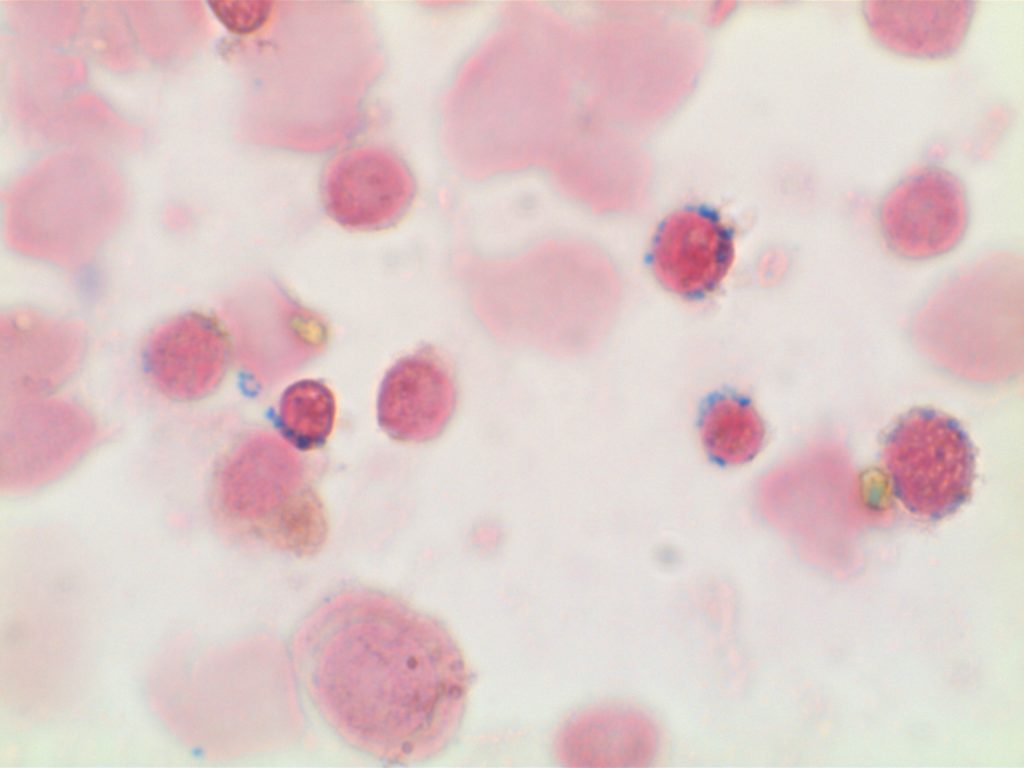
Ring Sideroblasts

General
  • Many cases of myelodysplasia (MDS-RS) and MDS/MPN with ring sideroblasts and thrombocytosis (MDS/MPN-RS-T) are associated with SF3B1 mutations.
    • SF3B1 – splicosome gene, often comuted with JAK2 in cases of MDS/MPN-RS-T (less frequently CALR and MPL)
    • Only 5% RS is required to diagnosis MDS-RS if a SF3B1 mutation is identified.  If no mutation, then the 15% RS threshold remains.
Photomicrographs
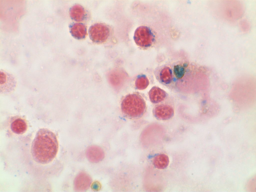
Ringed Sideroblasts in MDS

MDS-SLD

Myelodysplastic Syndrome with Single Lineage Dysplasia (MDS-SLD)

2016 WHO revision that effectively replaces the 2008 category of refractory cytopenia with unilineage dysplasia (RCUD).


Diagnostic Criteria

Immunohistochemistry
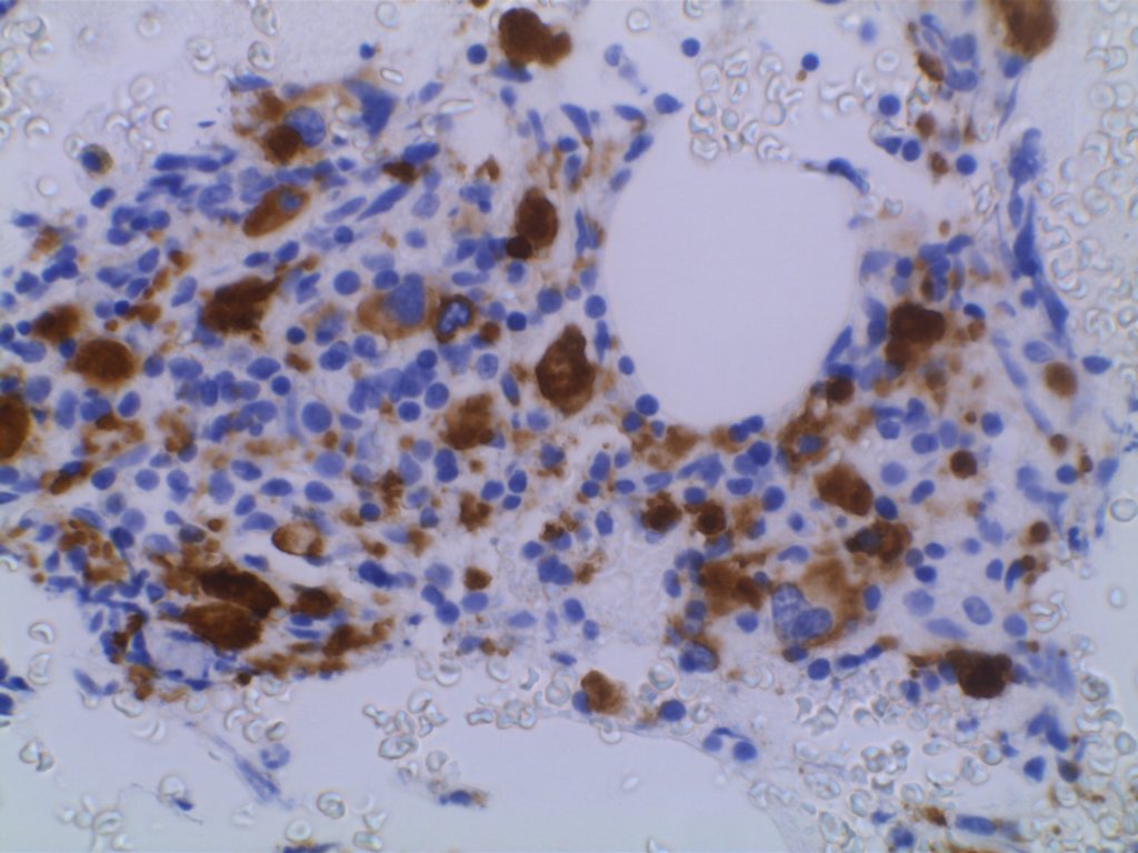
The use of immunohistochemistry (IHC) in many of the MDS syndromes is limited.  Identifying an increased blast population is one of the most useful, and may indicate a more aggressive course or transformation to acute myelogenous leukemia (AML).  Helpful IHC markers may include:
 
Stain
Comment
CD34 marks immature cells including myeloblasts.  In the setting of AML, it is ~70% sensitive.  A subset of lymphoblasts may express CD34.
CD117 is a specific myeloid marker, and marks a subset of myeloblasts.  The expression is dim, and one often must look at 20-40X to clearly see expression.  Mast cells (fried egg looking cell) will have very strong expression.
CD71 marks nucleated erythroid cells.  This may be helpful in quantitating and differentiating erythroid cells from myeloid cells.  This marker may be set-up as a double stain with CD34.
In the setting of hematopoietic cells, E-Cadherin marks immature erythroid cells.  Like CD71, E-Cadherin may be useful to differentiate immature erythroid cells from immature myeloid cells.
TdT is a sensitive lymphoblast (~95%) marker.  It is not entirely specific for lymphoblasts, but other markers can help clarify diagnostic difficulties (B and T-cell markers).  
References
Swerdlow SH, Campo E, Harris, NL, Jaffe ES, Pileri SA, Stein H, Thiele J (Eds):  WHO Classification of Tumours of Haematopoietic and Lymphoid Tissues (Revised 4th edition). IARC: Lyon 2017
 
Hematopathology. [edited by] Jaffe, ES. 1st. ed. Elsevier, Inc. © 2011.

CD61

CD61 is a marker of megakaryocytes/platelets.  The antigen is glycoprotein IIIa, and any cellular proliferation with megakaryocytic differentiation may express CD61.  This marker has been found useful in bone marrow biopsies looking for identifying highlighting abnormal and micromegakaryocytes, which is found in some cases of myelodysplasia (MDS) and myeloproliferative neoplasms (MPN).  
 
Some data suggests that at least 25% of the staining megakaryocytes should be micromegakaryocytes to have specificity for MDS (normal bone marrow <10% micromegakaryocytes).  The opinion of some expert hematopathologists is that a significant proportion for MDS is >50% micromegakaryocytes.
Photomicrographs
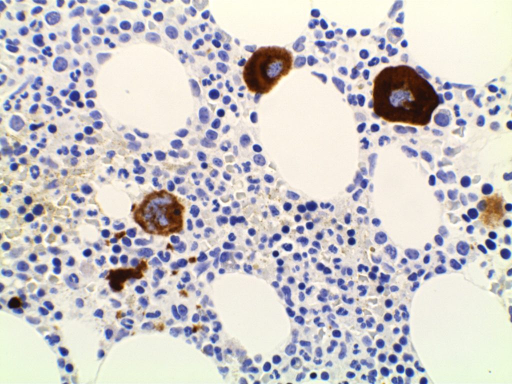
CD61 - Megakaryocytes
CD61 expression in a normal bone marrow highlighting megakaryocytes.
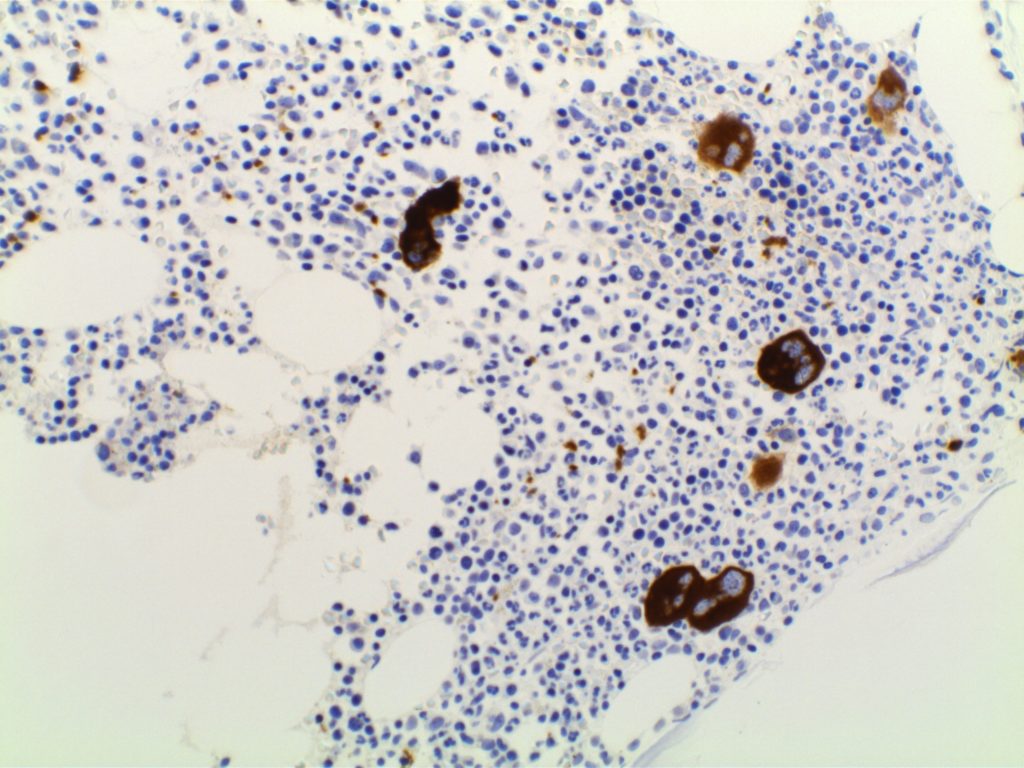
CD61 - Megakaryocytes
CD61 expression in a normal bone marrow highlighting megakaryocytes.
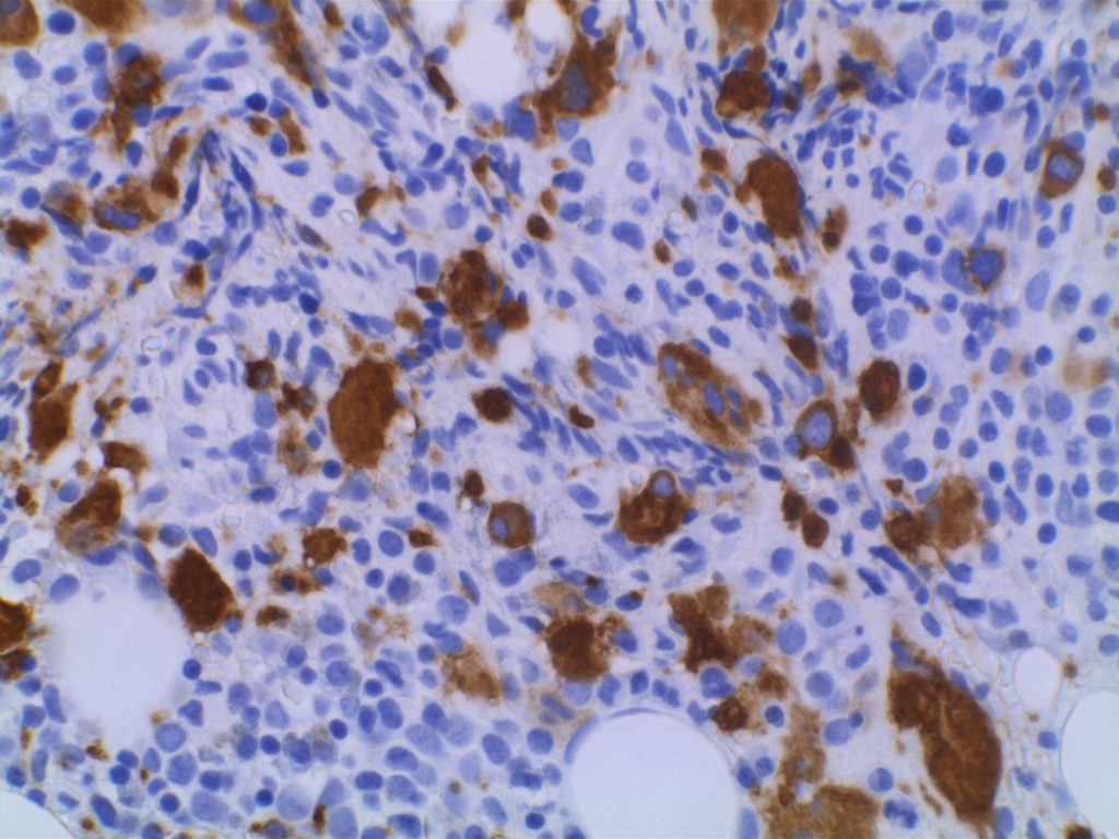
CD61 - RAEB
CD61 expression in MDS (RAEB) with increased numbers of small and hypolobated megakaryocytes.
CD61 - RAEB
CD61 expression in MDS (RAEB) with increased numbers of small and hypolobated megakaryocytes.

References
Quitmann H, Wagner S, Fischer R. Dysmegakaryopoiesis in myelodysplastic syndromes (MDS): an immunomorphometric study of bone marrow trephine biopsy specimens. Journal of Clinical Pathology. 1991.
 
Chuang SS, Li CY. Useful Panel of Antibodies for the Classification of Acute Leukemia by lmmunohistochemical Methods in Bone Marrow Trephine Biopsy Specimens. Am J Clin Pathol. 1997.
 
Jawad MD, Go RS, Reichard KK, Shi M. Increased Multinucleated Megakaryocytes as an Isolated Finding in Bone Marrow:  A Rare Finding and Its Clinical Significance. Am J Clin Pathol. 2016;146: 561–566. doi:10.1093/ajcp/aqw144
 
FOX, S.B., LORENZEN, J., HERYET, A., JONES, M., GATTER, K.C. and MASON, D.Y. (1990), Megakaryocytes in myelodysplasia: an immunohistochemical study on bone marrow trephines. Histopathology, 17: 69–74. doi:10.1111/j.1365-2559.1990.tb00665.x
 
Bone Marrow IHC.  Torlakovic, EE, et. al. American Society for Clinical Pathology Pathology Press © 2009.  pp. 109-113.