Tag Archives: mesothelioma

WT-1 (Wilms tumor gene product)

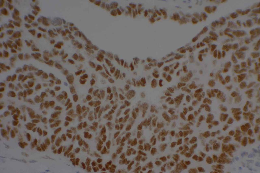
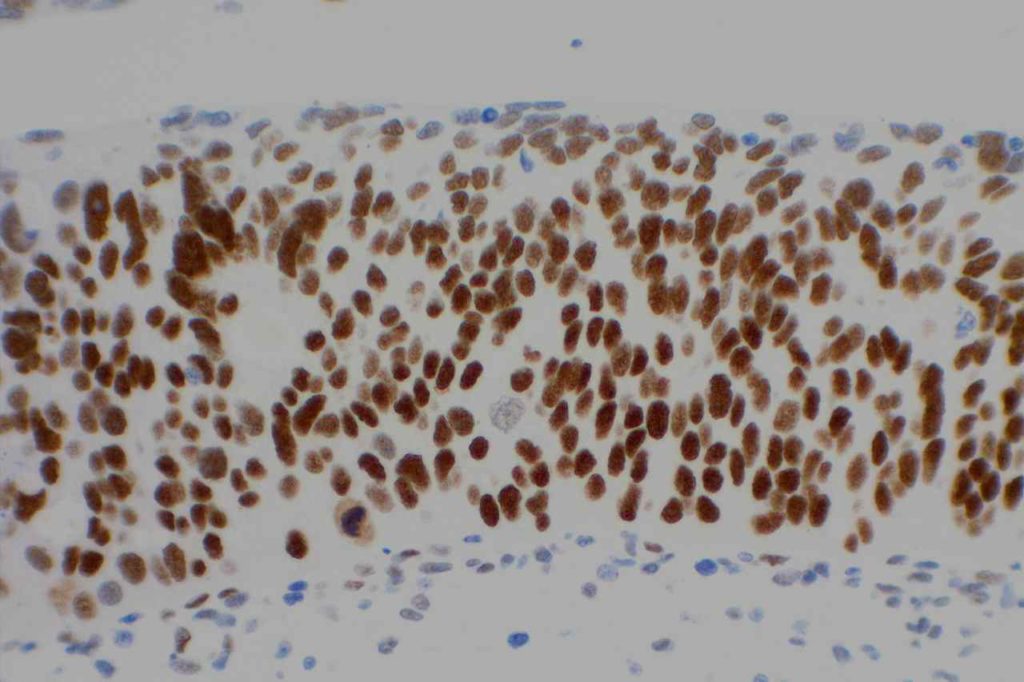
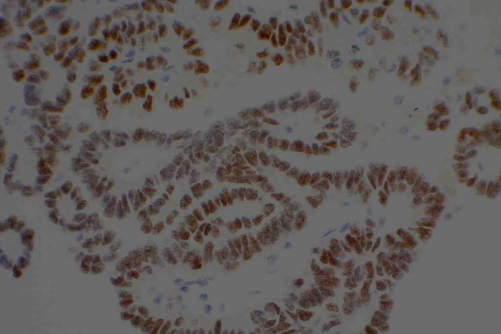
WT-1 (Wilms tumor gene product) is a marker most commonly used to identify OVARIAN SEROUS CARCINOMAS.  It is also commonly used in a panel to differentiate mesothelioma (positive) from adenocarcinoma (negative).  WT-1 has a normal expression distribution in adult tissues, which includes Sertoli cells, ovarian stromal and surface epithelium, and mesothelium.  The 6F-H2 clone has shown better sensitivity compared to other WT-1 antibodies (Ordonez).
 
Tumors / lesions expressing WT-1:
  • Mesothelioma (>75% of epithelial mesotheliomas, usually negative in sarcomatoid variants)
  • Ovarian Serous Carcinomas
  • Wilms tumor
  • Desmoplastic Small Round Cell Tumors
  • Metanephric Adenomas
 
Tumor
Expression (%)
Ovarian Serous Carcinoma
93%
Ovarian Mucinous Carcinoma
0%
Pancreatobiliary Carcinoma
0%
Breast Carcinoma
0%
Lung Carcinoma
0%
Colon Adenocarcinoma
0%
Renal Cell Carcinoma
0%
Thyroid Carcinoma
0%
Prostate Adenocarcinoma
0%
 
WT-1 expression in differentiating mesothelioma from adenocarcinoma (Marchevsky).
Tumor
Expression (%)
Epithelioid Mesothelioma
77%
Sarcomatoid Mesothelioma
13%
Adenocarcnioma
4%
Photomicrographs
WT-1 - Wilms Tumor
WT-1 – Wilms Tumor
WT-1 - Mesothelioma
WT-1 – Mesothelioma
WT-1 - Ovarian Serous Carcinoma
WT-1 – Ovarian Serous Carcinoma
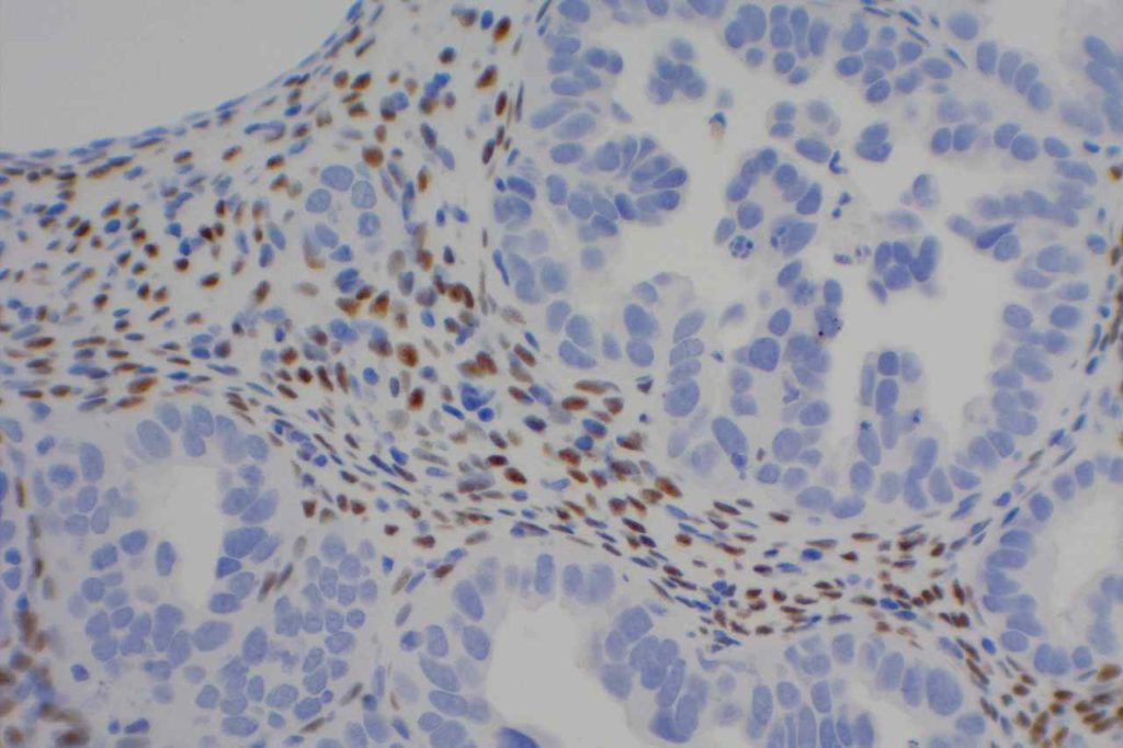
WT-1 - Stromal Cells
WT-1 expression in stromal cells.

 
References:
Muir, T. E., Cheville, J. C., & Lager, D. J. (2001). Metanephric adenoma, nephrogenic rests, and Wilms’ tumor: a histologic and immunophenotypic comparison. The American Journal of Surgical Pathology, 25(10), 1290–1296.
 
Marchevsky, A. M. (2008). Application of immunohistochemistry to the diagnosis of malignant mesothelioma. Archives of Pathology & Laboratory Medicine, 132(3), 397–401.
 
Ordóñez, N. G. (2005). Immunohistochemical diagnosis of epithelioid mesothelioma: an update. Archives of Pathology & Laboratory Medicine, 129(11), 1407–1414.  
  
Hadi, AIMM Annual Meeting, “Carcinomas of Unknown Primary”, presentation, 2011.

CD15 (Leu-M1)

CD15 (Leu-M1) is a hematopoietic antigen expressed by granulocytes, monocytes, Reed-Sternberg cells, and subsets of T and B-cells.  CD15 may also be expressed in glandular or neuroendocrine epithelial cells as well as part of a panel to distinguish between metastatic adenocarcinoma (positive) and mesothelioma (negative).
CD15 Expression
  • Neutrophils (~90%)
  • Monocytes (30-60%)
  • Promyelocytes
  • Adenocarcinoma (72%)
  • Classical Hodgkin Lymphoma cells
  • T-cell Lymphomas (subset, MF)
  • Acute Myelogenous Leukemia (particularly with monocytic differentiation)
Classical Hodgkin Lymphoma
CD15 stains strongly the cytoplasm of granulocytes.  CD15 is most commonly used as a diagnostic marker for classical Hodgkin lymphoma (CHL).  The combined staining of CD15 and CD30 in the appropriate morphologic setting is very specific for Hodgkin Lymphoma.  The biggest issue with this marker in the diagnosis of CHL is sensitivity.  In a well-optimized setting using a preferred clone, the sensitivity can be fairly high, ~70%.  In general when CHL cases are positive for CD15, it will stain a subset of the CD30 positive cells.
 
When this marker is not well optimized and/or using a suboptimal clone, then the negative results can not be trusted (even when expressed in background granulocytes).  Comparison analysis with a reference lab to compare IHC expression in validation cases is recommended for this antibody.  PAX-5 is also a helpful marker, when CD15 is negative and CHL is suspected, as it will be dimly expressed (compared to background B-cells) in ~90% of CHL cases.
Bone Marrow
CD15 will stain at least a subset of normal granulocytes(~90%), monocytes (30-60%), promyelocytes in the bone marrow.  A subset of cases of acute myelogenous leukemia (AML) will express CD15.  CD15 is a myeloid marker, and appears to be preferentially expressed in AML cases with monocytic differentiation (not entirely sensitive or specific).  Flow cytometry is more sensitive in detected CD15 expression than immunohistochemistry (IHC).  A subset of neoplastic T and B-cells may also express CD15.
Mesothelioma vs. Adenocarcinoma
CD15 has been used as part of a panel to differentiate between mesothelioma and adenocarcinoma.  CD15 is expressed in ~7% of epithelioid mesotheliomas and is not known to be expressed in sarcomatous mesothelioma while being expressed in a majority of adenocarcinomas (~72%).  CD15 is not typically one of the main markers used in the mesothelioma vs. adenocarcinoma panel (there are other markers with better sensitivity/specificity profiles), but may be helpful in certain circumstances.
CD15 Expression (Marchevsky)
  • Epithelial mesothelioma – 7%
  • Sarcomatous mesothelioma – 0%
  • Adenocarcinoma – 72%
  • Squamous cell carcinoma – 30%
  • Renal cell carcinoma – 25-100%
Photomicrographs
CD15 - Classical Hodgkin Lymphoma
CD15 – Classical Hodgkin Lymphoma.  Note dark staining granulocytes in the background.
CD15 - Classical Hodgkin Lymphoma
CD15 – Classical Hodgkin Lymphoma

References
Wick, MR. “Immunohistochemical approaches to the diagnosis of undifferentiated malignant tumor.”Annals of Diagnostic Pathology12(2008):72-84.
 
Bone Marrow IHC.  Torlakovic, EE, et. al. American Society for Clinical Pathology Pathology Press © 2009.  pp. 51.
 
Marchevsky, A. M. (2008). Application of immunohistochemistry to the diagnosis of malignant mesothelioma. Archives of Pathology & Laboratory Medicine, 132(3), 397–401. 
 
Dunphy CH, Polski JM, Evans HL, Gardner LJ. Evaluation of bone marrow specimens with acute myelogenous leukemia for CD34, CD15, CD117, and myeloperoxidase. Arch Pathol Lab Med. 2001;125: 1063–1069.