Tag Archives: T-Cell

CD8

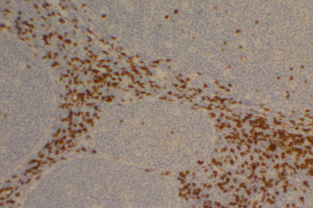
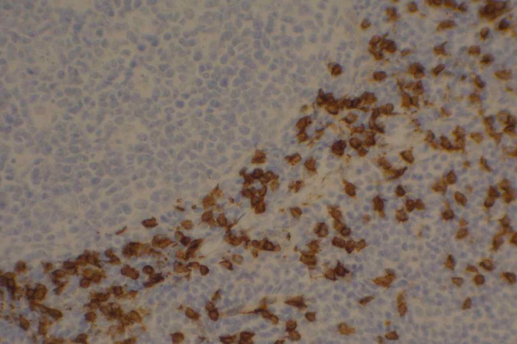
CD8 is aT-cell marker that is expressed by cytotoxic T-cells (contains perforin and granzyme, which are directly toxic), and stains a sub-set of CD3 positive T-cells.  CD3 positive T-cells express either CD4 or CD8.  Only thymocytes should express both CD4 and CD8 during a phase of their maturation process.  CD8 can be a helpful marker in characterizing T-cell disorders.
 
T-Cell antigens (usually CD5) can be expressed in B-cell lymphomas, but occasionally other T-cell markers (including CD8) may also be expressed (CD8, up to 3% but flow cytometry).  Use of panels (particularly when dealing with T-cell differentiation) is generally more helpful.
 
Quantification (absolute or relative to CD3 and/or CD4) of CD8+ lymphocytes associated with tumors (stromal infiltration, tumor infiltrating lymphocytes, etc.) is an active area of research.  Quantification of such findings has shown prognostic significance (varies based on tumor), and will likely become even more important as treatment regimen begin to more frequently incorporate “immunotherapy” (e.g. PD-L1 inhibitors).  
 
Evaluation of T-cells subsets is also an area of interest in medical conditions such as gluten-sensitive enteropathy and colitis.  However, no standard use of T-cell markers is  currently a common practice.  Though some advocate use in detection of early cases of gluten-sensitive enteropathy (controversial).
 
Other cytotoxic markers, such as granzyme, TIA-1, and perforin, may be helpful to further characterize CD8 expression lesions/neoplasms to confirm the cytotoxic nature of the cells vs. aberrant expression in a non-cytotoxic lesion.
CD8 Expression
  • Dendritic cells of lymphoid origin (dendritic cells of myeloid origin do not express CD8)
  • T-cell Lymphomas (CD8+ less common than CD4+)
  • Mature T-cells (subset CD4-)
  • T-ALL
  • T-cell Large Granular Lymphocyte Leukemia
  • Dim expression may be seen in endothelial cells

Photomicrographs
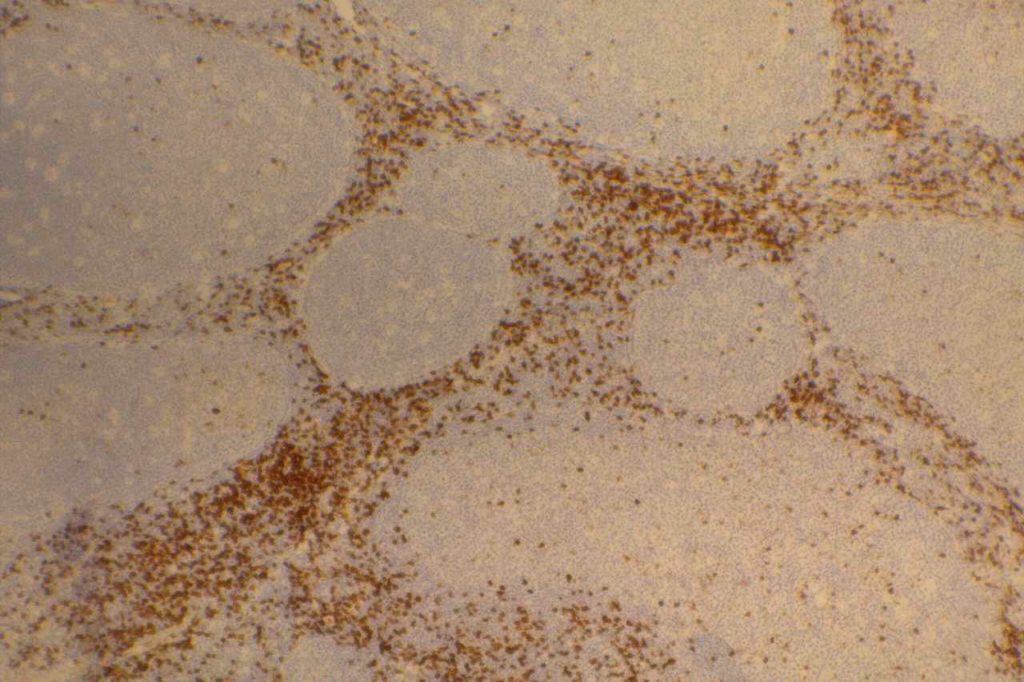
CD8 - Benign Tonsil
CD8 – Benign Tonsil
CD8 - Benign Tonsil
CD8 – Benign Tonsil
CD8 - Benign Tonsil
CD8 – Benign Tonsil

References
Carulli G, Stacchini A, Marini A, Ciriello MM, Zucca A, Cannizzo E, et al. Aberrant expression of CD8 in B-cell non-Hodgkin lymphoma: a multicenter study of 951 bone marrow samples with lymphomatous infiltration. Am J Clin Pathol. 2009;132: 186–90; quiz 306. doi:10.1309/AJCPNCOHS92ARWRQ
 
Paulson KG, Iyer JG, Simonson WT, Blom A, Thibodeau RM, Schmidt M, et al. CD8+ Lymphocyte Intratumoral Infiltration as a Stage-Independent Predictor of Merkel Cell Carcinoma Survival: A Population-Based Study. Am J Clin Pathol. 2014;142: 452–458. doi:10.1309/AJCPIKDZM39CRPNC
 
An JL, Ji QH, An JJ, Masuda S, Tsuneyama K. Clinicopathological analysis of CD8-positive lymphocytes in the tumor parenchyma and stroma of hepatocellular carcinoma. Oncol Lett. 2014;8: 2284–2290. doi:10.3892/ol.2014.2516
 
Ortonne N, Buyukbabani N, Delfau-Larue M-H, Bagot M, Wechsler J. Value of the CD8-CD3 ratio for the diagnosis of mycosis fungoides. Mod Pathol. 2003;16: 857–862. doi:10.1097/01.MP.0000084112.81779.BB
 
Hudacko R, Kathy Zhou X, Yantiss RK. Immunohistochemical stains for CD3 and CD8 do not improve detection of gluten-sensitive enteropathy in duodenal biopsies. Mod Pathol. 2013;26: 1241–1245. doi:10.1038/modpathol.2013.57
 
Bone Marrow IHC.  Torlakovic, EE, et. al. American Society for Clinical Pathology Pathology Press © 2009.  pp. 32-33.

CD7

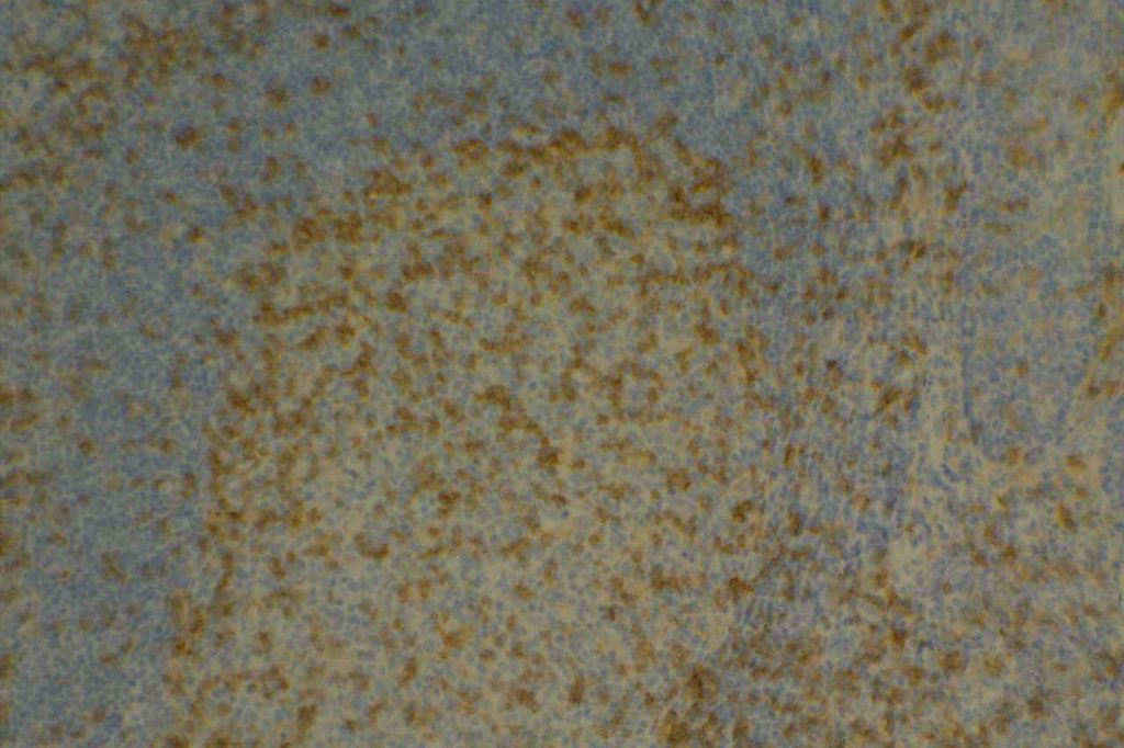
CD7 is a T-cell marker.  It is a specific T-cell marker first expressed early in T-cell development, but often one of the first to be dropped in cases of T-cell lymphoma.  However, loss of CD7 expression is not SPECIFIC for lymphoma, as normal benign T-cells may also lose expression.  Therefore the IHC findings must always be combined with the morphology when making a diagnosis of T-cell lymphoma.  CD7 is typically used as part of a larger T-cell panel including CD2, CD3, CD4, CD5, CD7, and CD8.  In challenging cases TCR gene rearrangement studies may also be helpful (specificity is not as good as B-cell gene rearrangement studies).
 
Aberrant expression of CD7 (and other T- and B-Cell markers) may be seen in acute leukemias (sometimes a helpful finding as a strategy for detecting minimal residual disease by flow cytometry).
CD7 Expression
  • Thymocytes
  • Mature T-cells
  • T-ALL
  • Mycosis Fungoides (often drops CD7 expression)

Photomicrographs
CD7 - Benign Tonsil
CD7 – Benign Tonsil

References
Bone Marrow IHC.  Torlakovic, EE, et. al. American Society for Clinical Pathology Pathology Press © 2009.  pp. 31-32.
 
Campbell SM, Peters SB, Zirwas MJ, Wong HK. Immunophenotypic diagnosis of primary cutaneous lymphomas: a review for the practicing dermatologist. J Clin Aesthet Dermatol. 2010;3: 21–25.
 
Jaffe ES, Nicolae A, Pittaluga S. Peripheral T-cell and NK-cell lymphomas in the WHO classification: pearls and pitfalls. Mod Pathol. 2013;26 Suppl 1: S71–87. doi:10.1038/modpathol.2012.181

CD4

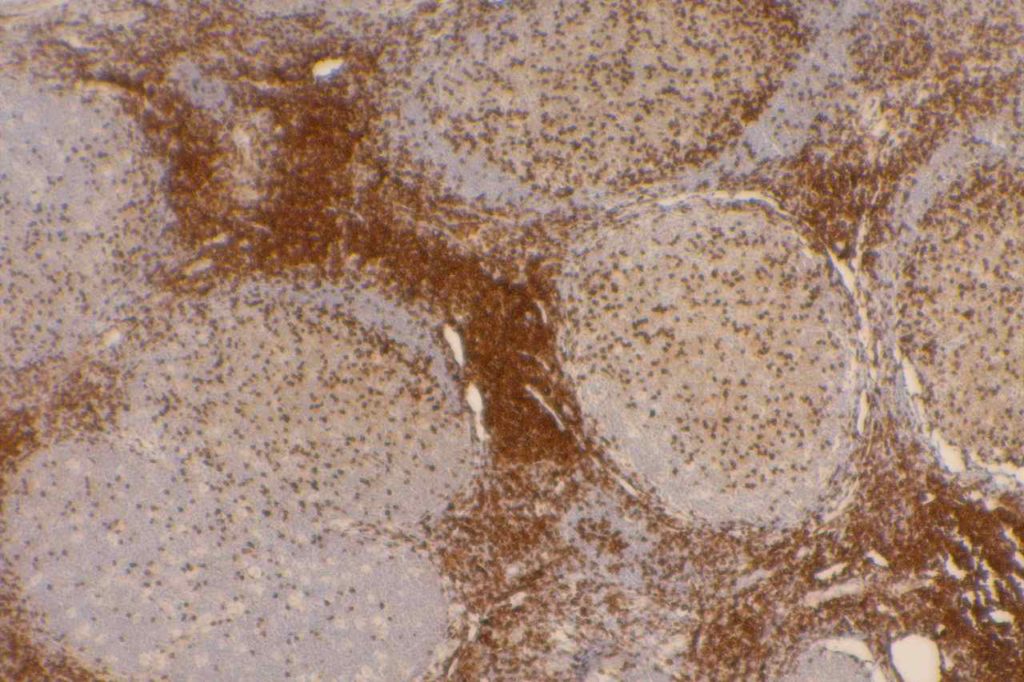
CD4 is a glycoprotein on the surface of a subset of T-cells, which defines the T-helper subset.  T-helper cells help to activate T-cell and B-cell immune response/proliferation in response to a foreign antigen.  CD4 is the target/receptor of the HIV virus.
 
CD4 stains a subset of CD3 positive T-cells (T-helper cells).  CD4 is expressed by thymocytes (80-90%), macrophages, Langerhans cells, and dendritic cells.  Many T-cell leukemia/lymphomas, blastic plasmacytoid dendritic cell neoplasms, and a sub-set of AMLs will express CD4.
 
Negative Staining:  NK cells, B-cell lymphoma, enteropathy associated T-cell lymphoma, epidermotropic cutaneous T-cell lymphoma, hepatosplenic alpha/beta and gamma/delta lymphoma, Hodgkin lymphoma, and T cell lymphoma with cytotoxic phenotype.
CD4 Expression
  • Thymoctye subsets
  • T-helper cells
  • Monocytes
  • Macrophages
  • CD4+/CD56+ hematodermic neoplasm
  • Dendritic cells (can express CD4 and CD8)
  • T-cell Lymphomas (CD4+ more common than CD8+)
  • Histiocytic disorders
  • Subset of AMLs
CD4 is typically used as part of a T-cell panel to identify aberrant expression or loss of expression in cell populations of interest.  Given the somewhat lack of specificity of the marker, it should be only interpreted within the context of the differential diagnosis and expected reactivity pattern(s).
 
By flow cytometry, low levels of CD4 expression has been demonstrated on erythroid precursor cells.  This probably lacks diagnostic significance (particularly IHC), but may have implications in HIV where CD4 is the receptor the virus is mediated through.

Photomicrographs
CD4 - Benign Tonsil
CD4 – Benign Tonsil
CD4 - Benign Tonsil
CD4 – Benign Tonsil
CD4 - Benign Tonsil
CD4 – Benign Tonsil

References
Bone Marrow IHC.  Torlakovic, EE, et. al. American Society for Clinical Pathology Pathology Press © 2009.  pp. 24-25.
 
Cleveland RP, Liu YC. CD4 Expression by erythroid precursor cells in human bone marrow. Blood. 1996;87: 2275–2282.
 
Herling M, Jones D. CD4+/CD56+ hematodermic tumor: the features of an evolving entity and its relationship to dendritic cells. Am J Clin Pathol. 2007;127: 687–700. doi:10.1309/FY6PK436NBK0RYD4